1933
Alterations of cortical and subcortical structures in mild cognitive impairment1Nanjing Drum Tower Hospital, Nanjing, China
Synopsis
Keywords: Neurodegeneration, Alzheimer's Disease
In this study, we investigated alterations of cortical morphology, subcortical nuclei volume and morphology in MCI using multiple morphological analysis methods. Moreover, we explored these imaging features relationship with cognitive performances and their efficiency in classification of MCI using support vector machine (SVM). We speculate that combining volumetric and morphological analysis methods will outperform than single analysis method in MCI identification.Abstract
Purpose: Grey matter changes are thought to be closely related to cognitive decline in mild cognitive impairment (MCI) patients but no consensus has yet emerged. The study aimed to explore the alterations of cortical and subcortical structures in MCI patients, and to investigate the association with cognitive assessment.Methods: A total 24 MCI individuals and 22 normal controls (NCs) were included. Voxel-based morphometry (VBM) analysis and vertex-based shape analysis were used to analyze the volume and shape of subcortical nuclei, respectively. We used surface-based morphometry (SBM) analysis through the Computational Anatomy Toolbox (CAT12) to compare cortical morphological changes between MCI and NCs. Relationships between abnormal changes of cortical and subcortical structures and cognitive assessment were investigated with spearman correlation analysis. Classification via support vector machine (SVM) was performed to evaluate whether MRI features could differentiate the MCI group with NCs accurately.
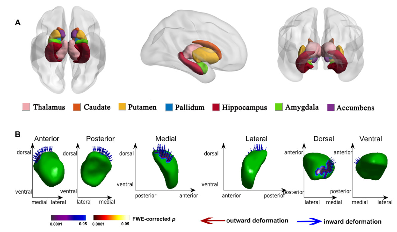
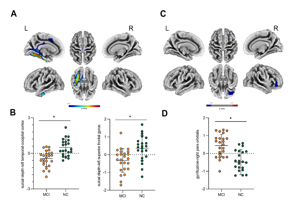
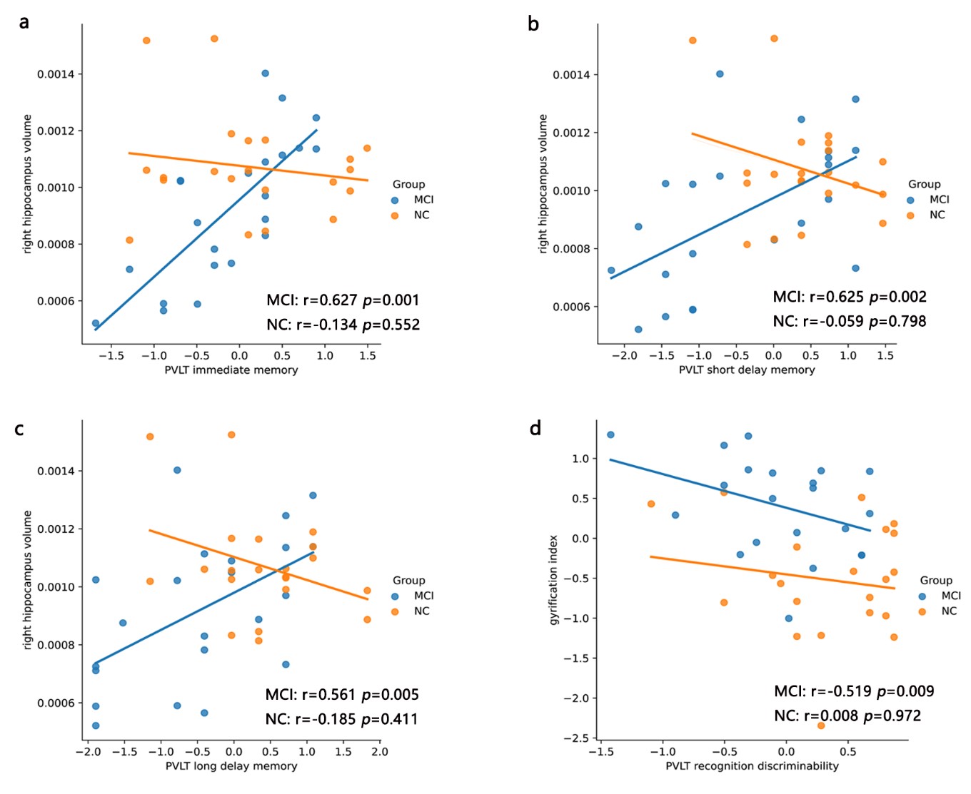
Results: Compared with NCs, MCI patients showed significant atrophy in the volume of left thalamus (p = 0.011), left hippocampus (p = 0.006), left amygdala (p = 0.031), right pallidum (p = 0.009) and right hippocampus (p = 0.008). Meanwhile, vertex-wise shape analysis showed inward deformation in the left amygdala in MCI group compared with NC (p < 0.05, FWE corrected). The SBM analysis revealed that MCI group exhibited significantly shallower sulci depth in the left hemisphere, predominantly in the fusiform, isthmus cingulate and superior frontal regions, and increased cortical gyrification index (GI) in the right frontal gyrus. Correlation analysis showed the positive correlation between adjusted right hippocampus volume and episode memory, while negative correlation between the altered GI and memory performance in MCI group. The SVM result showed that sulci depth and gyrification index derived from SBM outperformed subcortical imaging features in MCI identification. Particularly, when combing the cortical and subcortical morphological metrics, SVM reached the best performance with an accuracy of 89% to classify the MCI from NC.
Conclusion: The study indicates that obvious grey matter structural changes occur in MCI patients. These morphological alterations may be the basis of the brain functional differences underlying MCI and could be helpful for the early identification and clinical diagnosis of MCI. These findings contribute to our understanding of the neural mechanisms underlying memory impairment in MCI.
Acknowledgements
We sincerely thank all the subjects in this study.References
Wang, M.L., et al., Subcortical nuclei in Alzheimer's disease: a volumetric and diffusion kurtosis imaging study. Acta Radiol, 2018. 59(11): p. 1365-1371.
Huang, S., et al., Applications of Support Vector Machine (SVM) Learning in Cancer Genomics. Cancer genomics & proteomics, 2018. 15(1): p. 41-51.
Zhang, J., et al., Gray Matter Atrophy in Amnestic Mild Cognitive Impairment: A Voxel-Based Meta-Analysis. Front Aging Neurosci, 2021. 13: p. 627919.
Minkova, L., et al., Gray matter asymmetries in aging and neurodegeneration: A review and meta-analysis. Hum Brain Mapp, 2017. 38(12): p. 5890-5904. 8.
Patenaude, B., et al., A Bayesian model of shape and appearance for subcortical brain segmentation. Neuroimage, 2011. 56(3): p. 907-22.
Grijalva, C., et al., Dual-task performance is associated with brain MRI Morphometry in individuals with mild cognitive impairment. J Neuroimaging, 2021. 31(3): p. 588-601.
Figures