1928
Multiparametric MRI assessment of botulinum-toxin treatment for a patient with cervical dystonia
Xubin Chai1,2, Le He1, Changhao Zhu3, Wanting Hu3, Rong Xue2, Xiaolei Song1, and Li Wang4
1Center for Biomedical Imaging Research, Department of Biomedical Engineering, School of Medicine, Tsinghua University, Beijing, Beijing, China, 2State Key Laboratory of Brain and Cognitive Science, Institute of Biophysics, Chinese Academy of Sciences, Beijing, China, 3China School of Information Sciences and Technology, Northwest University, Xi'an, China, 4Department of Neurology, The First Hospital of Tsinghua University, Beijing, China
1Center for Biomedical Imaging Research, Department of Biomedical Engineering, School of Medicine, Tsinghua University, Beijing, Beijing, China, 2State Key Laboratory of Brain and Cognitive Science, Institute of Biophysics, Chinese Academy of Sciences, Beijing, China, 3China School of Information Sciences and Technology, Northwest University, Xi'an, China, 4Department of Neurology, The First Hospital of Tsinghua University, Beijing, China
Synopsis
Keywords: Neurodegeneration, Molecular Imaging
Cervical dystonia (CD) has been regarded as the most common form of focal dystonia, the affected neck muscles always in sustained situation lead to an abnormal rotation of the head[1]. The mechanisms of CD have not yet been thoroughly illustrated. Botulinum toxin (BT)is considered as the recommended first-line therapeutic method for the focal dystonia[2].Traditional MRI Scan has been used to evaluate any pathophysiological changes of brain anatomy or the affected neck muscles[3].However, seldom MRI have provided the cervical muscles metabolic messages[4].The CEST MRI could provide the metabolic aspects of the cervical muscles before and after the treatment of the BTIntroduction:
Multiparametric MRI for skeletal muscles have being employed in the clinical management of cervical dystonia (CD) patient. We compared multiparametric MRI acquired before and after the botulinum-toxin (BT) treatment, for a typical CD patient. Anatomy and metabolic assessment were achieved using m-Dixon and APT[5], respectively. We expect the imaging evaluations may be helpful for clinical decision-making and treatment assessment of CD patients in clinic.Materials and Methods:
Patient clinical information:The patient was a 72-year-old female. An abnormal cervical posture initially appeared 7 years before (left twisting of the head). She often used her hand to keep her head still (Sensory tricks). Six years ago, the patient was diagnosed with Cervical dystonia (CD). Symptoms improved with botulinum toxin injection every 3-6 months until the summer of 2019. She did not return to visit the clinic due to the COVID-19 epidemic. In the past 2 years, her symptoms worsened, the patient developed left neck stiffness and pain with involuntary shoulder shrug, and left subcostal twitch and dragging sensation, which could be relieved by clamping a bag. The above discomfort was significantly aggravated nearly one year. Tsui score was 6. We performed botulinum injection under EMG guidance. The total dosing of BTX-A was 300 U (including right sternocleidomastoideus 62.5U, right Splenius capitis 37.5U, left Splenius capitis 62.5U, left trapezius 50U, left semispinalis capit 12.5U, left levator scapulae 50U, left scalenus 25U). The patient’s symptoms improved gradually. One months after the injection, the patient felt her left neck stiffness and pain remission with involuntary shoulder shrug disappear. Two months later she can do housework and take part in physical exercise. Tsui score decreased to 1. MRI imaging protocol: One patient diagnosed with the cervical dystonia (female, 72 years) was enrolled with written informed consent signed before participation.MR scans were performed on a 3T scanner (Ingenia CX 3.0T; Philips Medical Systems, Best, The Netherlands), using a 16-channel torso coil and a 12-channel posterior coil as the receivers. The transverse and coronal planes crossing the cervical muscles (from C1-C7 areas) were chosen for CEST imaging. As listed in Table1, three B1 saturation offsets 0.6uT,0.9 uT and 2uT of the CEST images were acquired, with in-plane resolution of 2.5 mm X 2.5 mm respectively. Three series of CEST Z-spectral data were acquired, two with saturation power of 0.6 uT and 0.9uT, with 33 offsets distributed from -10 ppm to 10ppm, one with 2 uT saturation and 9 offsets from -4.3 ppm to 4.3 ppm with 0.8 ppm step size. The rest parameters of all datasets are shown in Table1. The areas of the three muscles before and after the BT treatment were shown in Table 2. All data were processed using custom-written MATLAB scripts, with APTw at 3.5 ppm for CEST quantification.Results and Discussion:
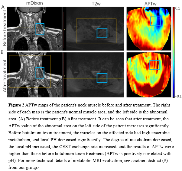
Figure 1: Area measurement for three muscles of the Splenius capitis, Levator scapulae and the Trapezius, based on m-Dixon images before and after BT treatment. The reduced area could reflect the relaxation of muscle and therapy effectiveness 45 days post BD treatment. Figure 2: APTw maps of the patient's neck muscle before and after treatment. The right side of each map is the patient's normal muscle area, and the left side is the abnormal area. (A) Before treatment ;(B) After treatment. It can be seen that after treatment, the APTw value of the abnormal area on the left side of the patient increases significantly. Before botulinum toxin treatment, the muscles on the affected side had high anaerobic metabolism, and local PH decreased significantly. The degree of metabolism decreased, the local pH increased, the CEST exchange rate increased, and the results of APTw were higher than those before botulinum toxin treatment (APTw is positively correlated with pH).For more technical details of metabolic MRI evaluation, see another abstract (#) from our group. Figure 3 Areas of the Corresponding five slices of three muscles of Splenius capitis, Levator scapulae and Trapezius based on them coronal results of mDixon sequence before and after BT treatment. The results showed that the areas of the three muscles decreased after BT treatment. Statistical analysis showed that the shrinkage of trapezius muscles had statistical significance, p-value ≤0.05,while the area of the muscles of Splenius capitis and levator scapularis had no statistical significance,p-value>0.05Conclusion:
CEST contrast maps and quantitative curves suggested the obviously changes before and after the BT treatment of the CD and could be used to assess the dynamic metabolic transform of the cervical muscles, which has potential for cervical dystonia CEST imaging in clinic.Acknowledgements
The authors acknowledged funding from National Natural Science Foundation of China (82071914). and the startup package from Tsinghua University to Dr. Song.References
1. Stacy M. Idiopathic cervical dystonia: an overview. Neurology 2000; 55(12 Suppl 5):S2-8. 2. Jankovic J. Treatment of cervical dystonia with botulinum toxin. Mov Disord 2004; 19 Suppl 8:S109-115. 3. Nevrly M, Hlustik P, Hok P, Otruba P, Tudos Z, Kanovsky P. Changes in sensorimotor network activation after botulinum toxin type A injections in patients with cervical dystonia: a functional MRI study. Exp Brain Res 2018; 236(10):2627-2637. 4. Rosales R, Dressler D. On muscle spindles, dystonia and botulinum toxin. European journal of neurology 2010; 17:71-80. 5. Chen Y, Dang X, Zhao B, Zheng Z, He X, Song X. B(0) Correction for 3T Amide Proton Transfer (APT) MRI Using a Simplified Two-Pool Lorentzian Model of Symmetric Water and Asymmetric Solutes. Tomography 2022; 8(4):1974-1986.Figures
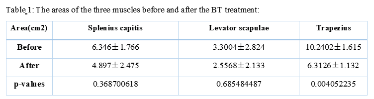
Table 1: The areas of the three muscles before and after the BT
treatment:
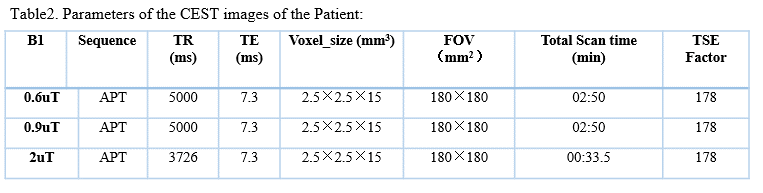
Table2. Parameters of the CEST images of the Patient:
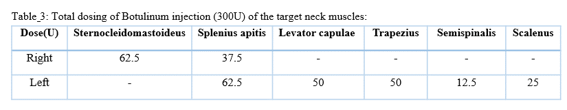
Table 3: Total dosing
of Botulinum injection (300U) of the target neck muscles:

Figure 1 The
areas of the three muscles of the Splenius capitis, Levator scapulae and the
Trapezius,before and after the treatment of the BT.
The areas of the three muscles have decreased while compare with the before and
after the treatment of the BT.
Figure
2 APTw maps of the
patient's neck muscle before and after treatment.(A)
Before treatment ;(B) After treatment.The
APTw value of the abnormal area on the left side of the patient increases
significantly. Before botulinum toxin treatment, the muscles on the affected
side had high anaerobic metabolism, and local PH decreased significantly. The
degree of metabolism decreased, the local pH increased.For more technical
details of metabolic MRI evaluation, see another abstract (#) from our group.
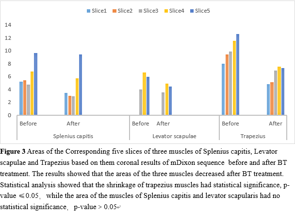
Figure
3 Areas of the
Corresponding five slices of three muscles of Splenius capitis, Levator
scapulae and Trapezius based on them coronal results of mDixon sequence before and after BT treatment. The results
showed that the areas of the three muscles decreased after BT treatment.
Statistical analysis showed that the shrinkage of trapezius muscles had
statistical significance, p-value ≤0.05,while the area of
the muscles of Splenius capitis and levator scapularis had no statistical
significance,p-value>0.05
DOI: https://doi.org/10.58530/2023/1928