1926
Olfactory Oddball Detection Task Activates the Left Temporoparietal Junction1Pennsylvania State University College of Medicine, Hershey, PA, United States
Synopsis
Keywords: Neurodegeneration, Neuro, Olfactory fMRI
Both the medial and inferior temporal lobes have been previously implicated in odor-identification. However, the precise neural substrate remains unclear. We used a novel oddball detection olfactory fMRI task to probe the neural basis of odor identification. fMRI activation was detected in the left temporoparietal junction along with known olfactory brain structures. Given the presence of odor identification deficits in early Alzheimer’s disease (AD), our paradigm has the potential to establish relationships between olfactory deficits, neurodegeneration, and memory impairment.Introduction
Olfactory function, particularly, odor-identification, is negatively affected in mild cognitive impairment (MCI) and preclinical Alzheimer’s Disease (AD)1. Unfortunately, we do not yet know the neurobiological basis of odor-identification that can potentially relate AD neurodegeneration to specific functional deficits in olfaction. In this study, we propose the development of an olfactory functional magnetic resonance imaging (fMRI) oddball detection task for healthy adults in order to identify the neural substrate that supports odor-identification.Materials and Methods
Eighteen subjects (n=18, mean age=28.55; 10 females) took part in the imaging study. Subjects completed two pseudorandomized and counterbalanced fMRI runs of the oddball detection olfactory task (Figure 1) on a Siemens 3.0 Tesla clinical MRI system (Prisma Fit, Siemens Medical Solutions, Inc., Erlangen, Germany) with a 64-channel head coil. The oddball task required subjects to press a button with their right index finger to identify the oddball, coffee. A T2*-weighted EPI sequence was used for fMRI image acquisition (TR/TE/FA = 2000 ms/30 ms/70°, resolution = 2.75 mm × 2.75 mm; 32 slices with 4 mm thickness). A T1-weighted MPRAGE image was acquired for anatomical overlay.fMRI data was pre-processed and analyzed using SPM12 software. Images were normalized to Montreal Neurological Institute (MNI) space and smoothed using an 8 × 8 × 8 mm3 kernel.
Results and Discussion
Our oddball olfactory fMRI paradigm identified a network consisting of the primary olfactory cortex, insula and hippocampus (Figure 2), which are known to be affected by neurodegeneration2,3. The odorants used in this study individually activated these brain regions. The oddball vs. non-oddball contrast activated the left temporoparietal junction (TPJ)4 (Figure 3).Our paradigm is novel because it has a dynamic range in odor-identification for testing people with normal cognition and MCI. Since our paradigm is modeled after well-established behavioral olfactory tests such as UPSIT and OLFACT, the fMRI activation during this task can be directly correlated to UPSIT/OLFACT scores5,6. Many studies have focused on the right TPJ7, but our study has shown that the left TPJ also plays a role in odor oddball detection which may be related to attentional control and salience/novel detection. The TPJ has been linked to the Ventral Attention Network (VAN) and Wernicke’s area. The VAN region seems to be responsible for supramodal ventral alerting4,8. The specific TPJ region found in our study matches those of the coordinates related to the VAN and left TPJ4,9.
The novelty of this study lies in the finding that the left TPJ plays a role in olfactory oddball detection. Since odor identification is also significantly reduced in early AD patients3 relative to age matched controls, our paradigm provides the basis for the development of a neurocognitive model for odor-identification, which can be applied to broader translational studies on olfactory function, aging, and neurological disease.
Acknowledgements
We thank the Center for NMR Research and Center for Aging and Neurodegenerative Diseases of Penn State University College of Medicine for all the staffs' kind help and suggestion. This research is supported by the National Institute on Aging grants (1R01AG070088-01A1 and1R21AG064486)References
1. Murphy, C. Olfactory and other sensory impairments in Alzheimer disease. Nat Rev Neurol 15, 11–24 (2019).
2. Price, J. L. CHAPTER 32 - Olfaction. in The Human Nervous System (Second Edition) (eds. Paxinos, G. & Mai, J. K.) 1197–1211 (Academic Press, 2004). doi:10.1016/B978-012547626-3/50033-8.
3. Vasavada, M. M. et al. Olfactory cortex degeneration in Alzheimer’s disease and mild cognitive impairment. J Alzheimers Dis 45, 947–958 (2015).
4. Kim, H. Involvement of the dorsal and ventral attention networks in oddball stimulus processing: A meta-analysis. Human Brain Mapping 35, 2265–2284 (2014).
5. Doty, R. L., Shaman, P., Kimmelman, C. P. & Dann, M. S. University of Pennsylvania Smell Identification Test: a rapid quantitative olfactory function test for the clinic. Laryngoscope 94, 176–178 (1984).
6. Zhang, Z. et al. Altered Odor-Induced Brain Activity as an Early Manifestation of Cognitive Decline in Patients With Type 2 Diabetes. Diabetes 67, 994–1006 (2018).
7. Dugué, L., Merriam, E. P., Heeger, D. J. & Carrasco, M. Specific Visual Subregions of TPJ Mediate Reorienting of Spatial Attention. Cerebral Cortex 28, 2375–2390 (2018).
8. Vossel, S., Geng, J. J. & Fink, G. R. Dorsal and Ventral Attention Systems: Distinct Neural Circuits but Collaborative Roles. Neuroscientist 20, 150–159 (2014).
9. Pride, N. A., Korgaonkar, M. S., North, K. N. & Payne, J. M. Impaired engagement of the ventral attention system in neurofibromatosis type 1. Brain Imaging and Behavior 12, 499–508 (2018).
Figures
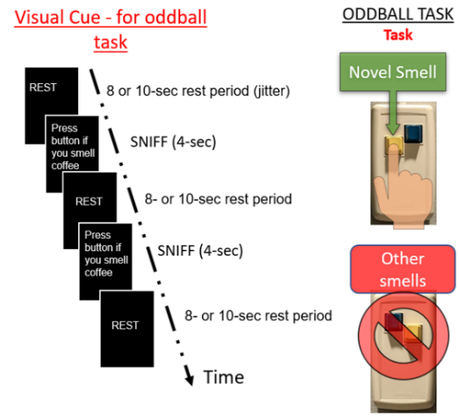
Figure 1. fMRI oddball paradigm presentation structure for coffee, along with oddball task instructions. Participants are allowed to answer from the moment the novel stimulus presents itself until the time the next odor is presented, giving subjects 12-14s to think and answer. Rest periods varied between 8-10s, at irregular intervals, and were counterbalanced. Odorants used were bubblegum, lemon, rose, and coffee (oddball).
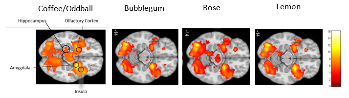
Figure 2. The oddball stimulus alone activated the Olfactory cortex, Hippocampus, Insula, and Amygdala. (n=18, GLM Level 2 ANOVA, p < 0.05 FWE, extent threshold = 20). Z= -14
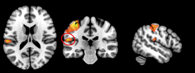
Figure 3. The oddball minus non-oddball contrast activated the left TPJ (temporoparietal junction): circled in red, which includes the left Superior temporal and Supramarginal gyri., (n=18, GLM Level 2 ANOVA, p < 0.05 FWE, extent threshold = 20)