1924
Hippocampal subfields atrophy based on automated volumetry analysis in end-stage-renal disease patients1the First Affiliated Hospital of Dalian Medical University, Dalian, China
Synopsis
Keywords: Neurodegeneration, Neurodegeneration
Cognitive impairment is very common in patients with end-stage renal disease (ESRD). Hippocampal atrophy has been proven to be an effective marker for distinguishing the normal population from the cognitively impaired patients. However, there are almost no studies on changes in hippocampal structure, especially the volume of hippocampal subfields, in ESRD patients. In this study, automated volumetry of hippocampal subfields in ESRD patients was performed and compared with healthy controls, and found that ESRD patients have hippocampal subfield atrophy, especially in bilateral fimbria and right hippocampal tail.Introduction
Chronic kidney disease (CKD) has become a worldwide public health problem due to its progressive course and the risk of adverse outcomes [1]. Cognitive impairment (CI) is very common in patients with end-stage renal disease (ESRD)2. Although some studies have found that when the estimated glomerular filtration rate (eGFR) is lower than 30 mL/min/1.73 m2, the decline in executive function is the most obvious [2]. Especially, ESRD patients also have a certain degree of memory impairment [3]. Hippocampal atrophy has been proven to be an effective marker for distinguishing the normal population from the cognitively impaired patients, and studies have found the relationship between the volume of the hippocampus subfields (DG/CA3, CA1, subiculum) and memory [4]. However, there are almost no studies on changes in hippocampal structure, especially the volume of hippocampal subfields, in ESRD patients. In this study, we aim to use an automatic hippocampal segmentation method to determine if there was a change in whole or subfield hippocampal volume in ESRD patients.Methods
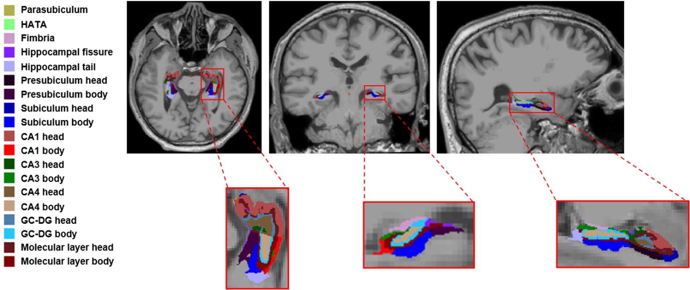
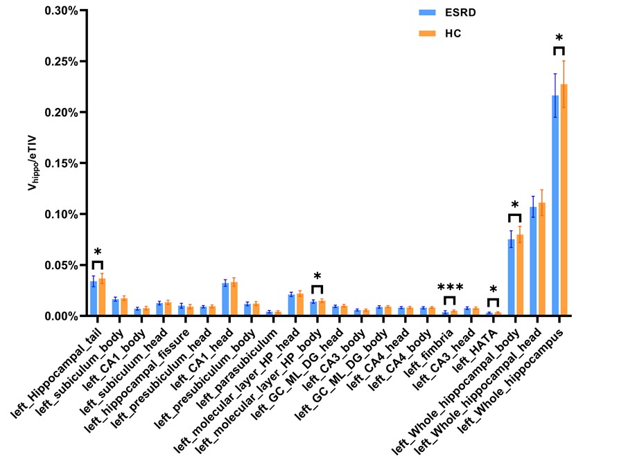
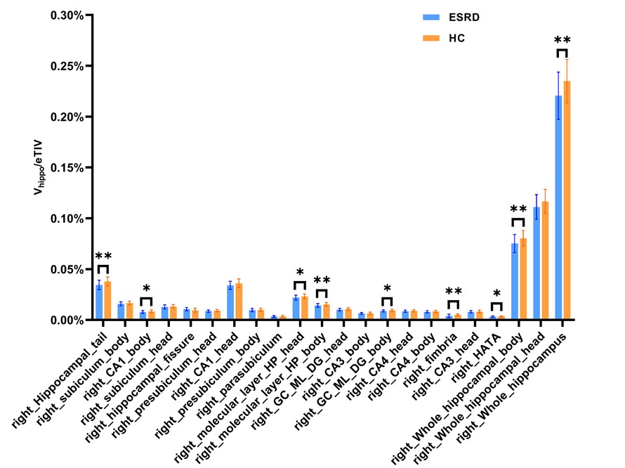
Fifty-two patients with clinically confirmed ESRD (mean age: 59.71 ± 9.06, range from 33 to 79 years; 26 females, 26 males) and 28 healthy control subjects (mean age: 60.25 ± 6.37, range from 44 to 69 years; 18 females, 10 males) were recruited and all were right-handed. Informed consent was acquired from each subject. Participants were scanned using a 3.0 T MR scanner (Ingenia CX, Philips Healthcare, the Netherlands) with a 32-channel receive-only head coil. On whole-brain, T1-weighted multishot turbo field echo (MS-TFE) was acquired: repetition time (TR)/echo time (TE) = 6.6/3.0 ms, field of view (FOV) = 256 × 256 mm, flip = 12°, 188 sagittal slices, and 1 mm3 spatial resolution. Automated volumetric measurement of the hippocampal subfields was done using FreeSurfer 7.0 hippocampal subfields module on the T1 image. The hippocampus was segmented, bilaterally, into the hippocampal tail, subiculum, CA1, hippocampal fissure, presubiculum, parasubiculum, molecular layer, granule cell layers of the dentate gyrus (GC-ML-DG), CA3, CA4, fimbria, and the hippocampus-amygdala-transition-area (HATA) (Figure 1). The estimated total intracranial volume (eTIV) volumes were also extracted. In order to reduce the influence of individual variation, we used a proportion method which expresses the brain volume (v) as a proportion of eTIV. The normalized volume (ṽ) is computed as ṽ = v/TIV [5]. Data analyses were performed using SPSS 22.0, independent-samples t test was used to compare the subfield hippocampal volume between ESRD group and HC group. P < 0.05 was considered statistically significant. FDR correction was applied to reduce the false-positive errors.Results
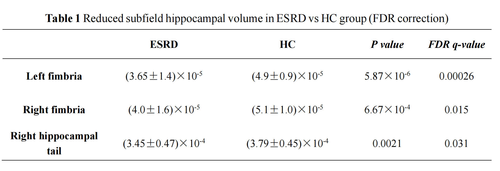
The subfield hippocampal volume had a reduction trend in ESRD group, especially for the right side (P < 0.05, Figure 2-3). After FDR correction, the volumes of left fimbria (P < 0.001), right fimbria (P = 0.015) and right hippocampal tail (P = 0.031) decreased significantly compared to HCs (Table 1). The whole volumes of the bilateral hippocampi of ESRD group were decreased than that of HC group, but there was no significant difference after FDR correction.Discussion
This study investigated the volumes hippocampal substructures in patients with ESRD for the first time. We observed the reduced volumes of left fimbria, right fimbria and right hippocampal tail in patients with ESRD while compared with HCs. The fimbria is a white matter tract that begins at the posterior end of the hippocampus and transitions into the fornix which is the main connecting tract of the limbic system [6]. The fimbria-fornix pathway plays a vital role in spatial memory [7]. Therefore, we have reason to suspect that the hippocampal atrophy of ESRD patients is dominated by the white matter regions. The hippocampal tail is a well-connected area of the hippocampus, especially with the prefrontal cortex8, and the prefrontal cortex is closely related to memory function. In addition, a study showed that hippocampal tail volume was related to visuospatial memory performance [8]. Therefore, we believe that hippocampal subregion (mainly bilateral fimbria and right hippocampal tail) atrophy in ESRD patients may mainly affect visuospatial memory function, but it is only speculation at present, and further research is needed to confirm it.Conclusion
Patients with ESRD have hippocampal subfield atrophy, especially in bilateral fimbria and right hippocampal tail.Acknowledgements
No acknowledgement found.References
[1]. Zhang L, Wang F, Wang L, et al. Prevalence of chronic kidney disease in China: a cross-sectional survey. Lancet 2012;379:815-822
[2]. Berger I, Wu S, Masson P, et al. Cognition in chronic kidney disease: a systematic review and meta-analysis. BMC Med 2016;14:206
[3]. Chang CY, Lin CC, Tsai CF, et al. Cognitive impairment and hippocampal atrophy in chronic kidney disease. Acta Neurol Scand 2017;136:477-485
[4]. Bennett IJ, Stark SM, Stark CEL. Recognition Memory Dysfunction Relates to Hippocampal Subfield Volume: A Study of Cognitively Normal and Mildly Impaired Older Adults. J Gerontol B Psychol Sci Soc Sci 2019;74:1132-1141
[5]. Gabere M, Thu Pham NT, Graff-Radford J, et al. Automated Hippocampal Subfield Volumetric Analyses in Atypical Alzheimer's Disease. J Alzheimers Dis 2020;78:927-937
[6]. Powell TP, Guillery RW, Cowan WM. A quantitative study of the fornixmamillo-thalamic system. J Anat 1957;91:419-437
[7]. Dahmani L, Courcot B, Near J, et al. Fimbria-Fornix Volume Is Associated With Spatial Memory and Olfactory Identification in Humans. Front Syst Neurosci 2019;13:87
[8]. De Meo E, Portaccio E, Prestipino E, et al. Effect of BDNF Val66Met polymorphism on hippocampal subfields in multiple sclerosis patients. Mol Psychiatry 2021
Figures