1869
Improving Image Quality While Reducing Geometric Distortion of Diffusion-weighted MRI in prostate: Comparison of FOCUS-MUSE, MUSE, and SS DWI1Department of Radiology, The First Affiliated Hospital, Sun Yat-Sen University, Guangzhou, China
Synopsis
Keywords: Prostate, Diffusion/other diffusion imaging techniques
The current study investigated the qualitative and quantitative image quality of three DWI sequences of the prostate: SS DWI, MUSE DWI and a new DWI sequence named FOCUS-MUSE DWI. Twenty-three healthy volunteers were enrolled. The diagnostic image quality score was assessed by two radiologists on the three DWI sequences. And the ADC values in the peripheral and transitional zone were measured together with the Dice Coefficient calculated for quantitative comparison. Our results indicated that FOCUS-MUSE DWI had a superior image quality and more stable ADC measurement compared with SS DWI and MUSE DWI. The geometric distortion level was also reduced.Synopsis
The prospective study aims to investigate the qualitative and quantitative image quality of three magnetic resonance diffusion-weighted imaging (DWI) sequences of the prostate: single-shot (SS) DWI, multiplexed sensitivity encoding (MUSE) DWI and a new DWI sequence named field-of-view optimized and constrained undistorted single-shot (FOCUS)-MUSE DWI. A total of 23 healthy volunteers were enrolled. The diagnostic image quality score was assessed by two radiologists on the above three DWI sequences. And the apparent diffusion coefficient (ADC) values in the peripheral and transitional zone were measured as well as the Dice Coefficient were calculated for quantitative comparison. Our results indicated that FOCUS-MUSE DWI had a superior image quality as well as more stable ADC measurement compared with SS DWI and MUSE DWI. The geometric distortion level was also reduced (0.9224 vs. 0.8927 vs. 0.8989, p < 0.001).Introduction
Diffusion-weighted imaging (DWI) is an integral part of multiparametric magnetic resonance imaging (mpMRI) for detecting, localizing, and staging prostate cancer[1]. However, the single-shot (SS) DWI was limited by severe geometric distortion and low spatial resolution[2]. In magnetic resonance diffusion-weighted imaging (DWI), multiplexed sensitivity encoding (MUSE) was reported to possess a high signal-to-noise ratio (SNR) and field-of-view optimized and constrained undistorted single-shot (FOCUS) was designed to improve the spatial resolution[3-5]. A new DWI sequence named FOCUS-MUSE DWI which combined the strengths of MUSE and FOCUS was developed to better delineate the prostatic diseases. To the best of our knowledge, the clinical utility of FOCUS-MUSE DWI in prostate MRI has not yet been reported. Therefore, the purpose of the current study is to investigate the qualitative and quantitative image quality of FOCUS-MUSE DWI and compared them with that of MUSE DWI and SS DWI.Methods and Materials
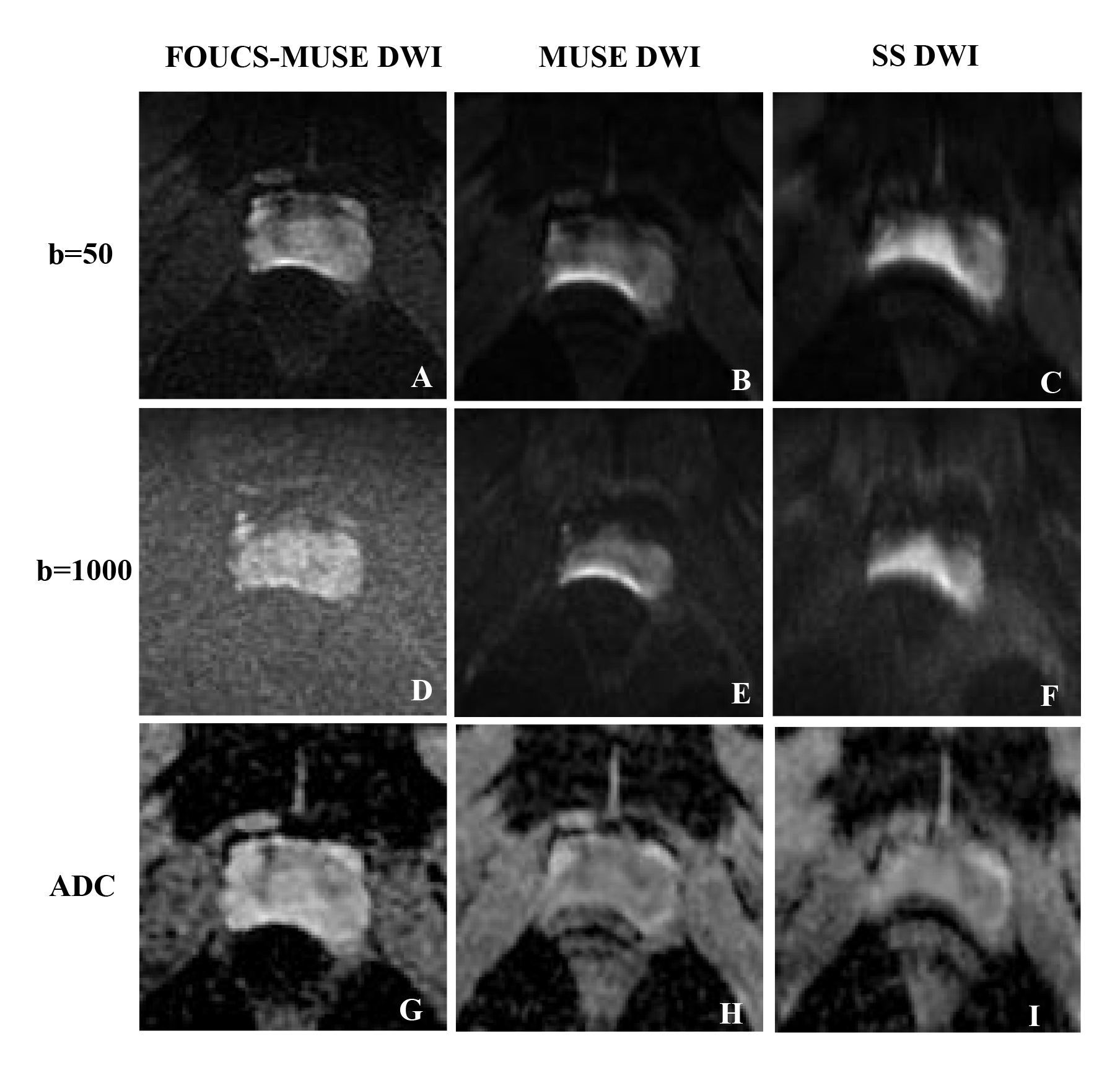
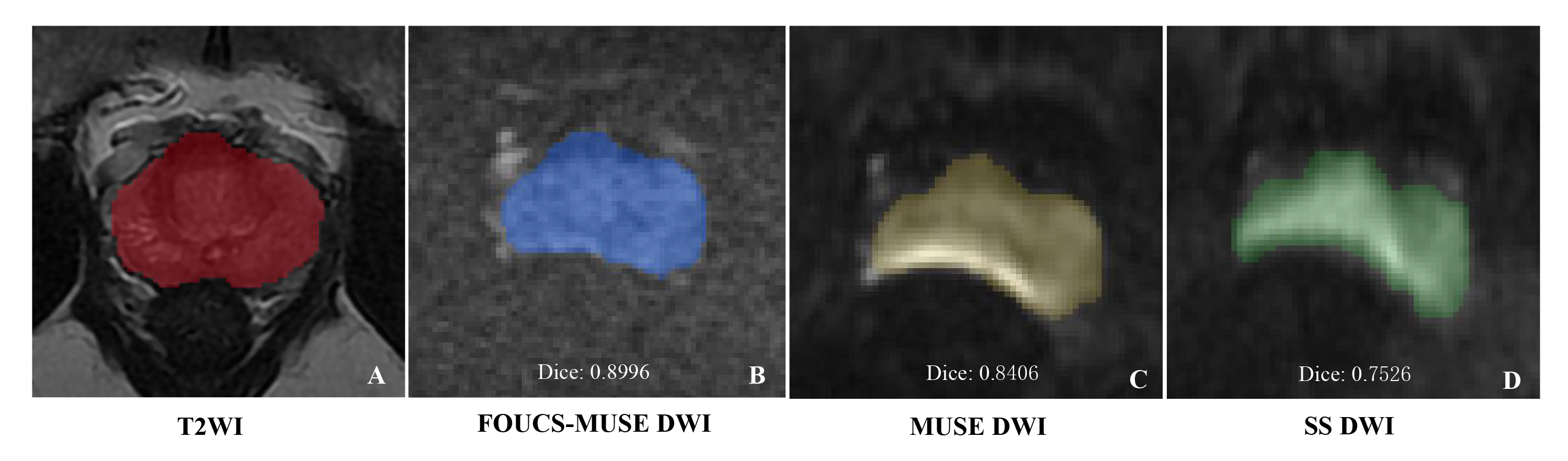
A total of 23 healthy volunteers (mean age, 28.96 ± 6.00 years) were prospectively enrolled and underwent prostate magnetic resonance imaging at a 3 T scanner. The axial and sagittal T2-weighted turbo spin echo images and the three DWI sequences, including FOCUS-MUSE DWI, MUSE DWI, and SS DWI were acquired. The subjective qualitative image features (resolution, capsule demarcation, zonal anatomy, artifacts, geometric distortion, and overall image quality) were rated by two radiologists who were blinded to the type of DWI on a 5-point Likert scale. The ADC values in the peripheral and transitional zone were measured by two radiologists. For analyzing the geometric distortion levels, the outlines of prostates were manually drawn and the Dice Coefficient[6] were calculated, with T2-weighted images as reference. Statistical analysis was performed using paired Wilcoxon signed-rank tests. Interobserver agreement was evaluated using the intraclass correlation coefficient (ICC). A P value <0.05 was considered to be of statistical significance.Results
Interobserver agreement for qualitative features was excellent (ICC 0.757–0.914). The resolution, capsule demarcation, zonal anatomy, artifacts, geometric distortion, and overall image quality were significantly better for FOCUS-MUSE DWI than SS DWI and MUSE DWI at b50, b1000, and ADC images. There were no statistically significant differences between SS DWI and MUSE DWI concerning these subjective image features (all p>0.05). FOCUS-MUSE DWI maintained the best intraobserver and interobserver reliability of ADC measurements [peripheral zone: 0.953(0.892,0.980) and 0.889(0.731,0.954); transitional zone: 0.837(0.487,0.940) and 0.778(0.550,0.899)] among the three DWI sequences. The Dice Coefficient were higher for FOCUS-MUSE DWI compared to SS DWI [0.9224(0.9114,0.9435) vs. 0.8927(0.8749,0.9289), p < 0.001] and MUSE DWI [(0.9114,0.9435) vs. 0.8989(0.8406,0.9197), p < 0.001] at b1000 images.Discussion
Both qualitative and quantitative evaluations indicated that FOCUS-MUSE DWI had better image quality and more subtle Geometric Distortion, which might attribute to its combination of FOCUS and MUSE technique. It was reported that a decreased field-of-view (FOV) in the phase-encoding direction can provide better image quality, anatomic details, and lesion conspicuity than conventional full FOV[7]. Meanwhile, the reduced SNR caused by reduced FOV can be compensated by the MUSE technique, which employs multi-shot excitation together with segment k-space filling in the phase encoding direction[8-10]. In addition, a more stable ADC measurement was observed in the FOCUS-MUSE DWI among the three DWI sequences, which may be the consequence of fewer susceptibility artifacts of the FOCUS method as well as the shorter T2 signal decay of the MUSE technique[8].Conclusions
FOCUS-MUSE DWI had a superior image quality compared with SS-DWI and MUSE DWI and outperformed the other two DWI sequences in achieving stable ADC measurements. In addition, the geometric distortion level was significantly reduced.Summary of main finding
The FOCUS-MUSE DWI facilitates the diagnosis of prostatic diseases due to its high image quality and less geometric distortion.Acknowledgements
No acknowledgement found.References
[1] WILLIAMS I S, MCVEY A, PERERA S, et al. Modern paradigms for prostate cancer detection and management [J]. The Medical journal of Australia, 2022, 217(8): 424-33.
[2] FARZANEH F, RIEDERER S J, PELC N J. Analysis of T2 limitations and off-resonance effects on spatial resolution and artifacts in echo-planar imaging [J]. Magnetic resonance in medicine, 1990, 14(1): 123-39.
[3] DONG H, LI Y, YU K, et al. Comparison of image quality and application values on different field-of-view diffusion-weighted imaging of breast cancer [J]. Acta radiologica (Stockholm, Sweden : 1987), 2016, 57(1): 19-24.
[4] CHANG H C, CHEN G, CHUNG H W, et al. Multi-shot Diffusion-Weighted MRI With Multiplexed Sensitivity Encoding (MUSE) in the Assessment of Active Inflammation in Crohn's Disease [J]. Journal of magnetic resonance imaging : JMRI, 2022, 55(1): 126-37.
[5] KIM H, LEE J M, YOON J H, et al. Reduced Field-of-View Diffusion-Weighted Magnetic Resonance Imaging of the Pancreas: Comparison with Conventional Single-Shot Echo-Planar Imaging [J]. Korean journal of radiology, 2015, 16(6): 1216-25.
[6] JOHANSSON J, LAGERSTRAND K, IVARSSON L, et al. Brain diffusion MRI with multiplexed sensitivity encoding for reduced distortion in a pediatric patient population [J]. Magnetic resonance imaging, 2022, 87: 97-103.
[7] MANNELLI L, MONTI S, CORRIAS G, et al. Comparison of Navigator Triggering Reduced Field of View and Large Field of View Diffusion-Weighted Imaging of the Pancreas [J]. Journal of computer assisted tomography, 2019, 43(1): 143-8.
[8]CHEN N K, GUIDON A, CHANG H C, et al. A robust multi-shot scan strategy for high-resolution diffusion weighted MRI enabled by multiplexed sensitivity-encoding (MUSE) [J]. NeuroImage, 2013, 72: 41-7. [
9]CHEN X, ZHANG Y, CAO Y, et al. A feasible study on using multiplexed sensitivity-encoding to reduce geometric distortion in diffusion-weighted echo planar imaging [J]. Magnetic resonance imaging, 2018, 54: 153-9.
[10]BAXTER G C, PATTERSON A J, WOITEK R, et al. Improving the image quality of DWI in breast cancer: comparison of multi-shot DWI using multiplexed sensitivity encoding to conventional single-shot echo-planar imaging DWI [J]. The British journal of radiology, 2021, 94(1119): 20200427.
Figures