1829
Pilot Tone-navigated motion estimation for liver DW-MRI1Radiology, Boston Children's Hospital and Harvard Medical School, Boston, MA, United States, 2MR R&D Collaborations, Siemens Medical Solutions USA, Inc., Los Angeles, CA, United States, 3MR R&D Collaborations, Siemens Medical Solutions USA, Inc., Austin, TX, United States, 4MR R&D Collaborations, Siemens Medical Solutions USA, Inc., New York, NY, United States
Synopsis
Keywords: Motion Correction, Diffusion/other diffusion imaging techniques
Diffusion-weighted MRI (DW-MRI) is capable of detecting and characterizing liver tumors and following-up treatments. Unfortunately, respiratory motion during DW-MRI scan causes misalignments between slices and reduces image quality. 3D slice-to-volume registration (SVR) can be employed to correct for motion. However, motion estimates may be inaccurate for high b-value images where SNR decreases. In this work, we propose to use PT estimated motion correction, which is calibrated on 3D SVR motion parameters obtained from low b-value images. We showed misalignments between slices are reduced by the proposed PT-based motion correction compared to SVR-based motion correction and no correction.Introduction
Abdominal diffusion-weighted imaging MRI (DWI-MRI) is a powerful clinical tool to detect and characterize liver tumors and follow-up treatments. However, misalignments between slices are inevitable due to respiration-induced motion, particularly in axial imaging, reducing image quality and robustness of quantitative disease markers. Breath-holding can be used to reduce motion in abdominal imaging but it is challenging for some patients, especially children. Prospective gating is another alternative, but it reduces the efficiency of the acquisition by discarding a large portion of acquired data and increases acquisition time. Therefore, several image-based retrospective motion measurement and correction techniques based on registration for motion parameter estimation have been introduced1. Methods based on 3D slice-to-volume registration (SVR) have been shown to be effective in motion correction2,3. However, registration is ill-posed and may especially be challenging for axial abdominal DW-MRI due to poor resolution in the direction of the largest motion (superior-inferior) and lack of motion-free template volume to use as reference. In addition, registration may be less reliable for high b-value images where SNR decreases4. Recently, a small wireless RF transmitter that generates a reference signal called pilot tone (PT) was shown to be capable of measuring physiological motion changes5-7. The PT signal can be detected simultaneously with the MRI signal and can be used as an alternative navigator. However, the pilot tone signal does not directly provide motion parameters. In this work, we propose to use PT estimated motion correction which is calibrated on 3D SVR motion parameters obtained from low b-value images, by building a motion model. Such a motion model has been previously proposed to estimate motion parameters from the FID navigator signal8. This motion model and the pilot tone signal are then used to predict motion parameters for each slice and can correct for motion in higher b-value images, and for slices where SVR may be less reliable due to the limitations of registration.Methods
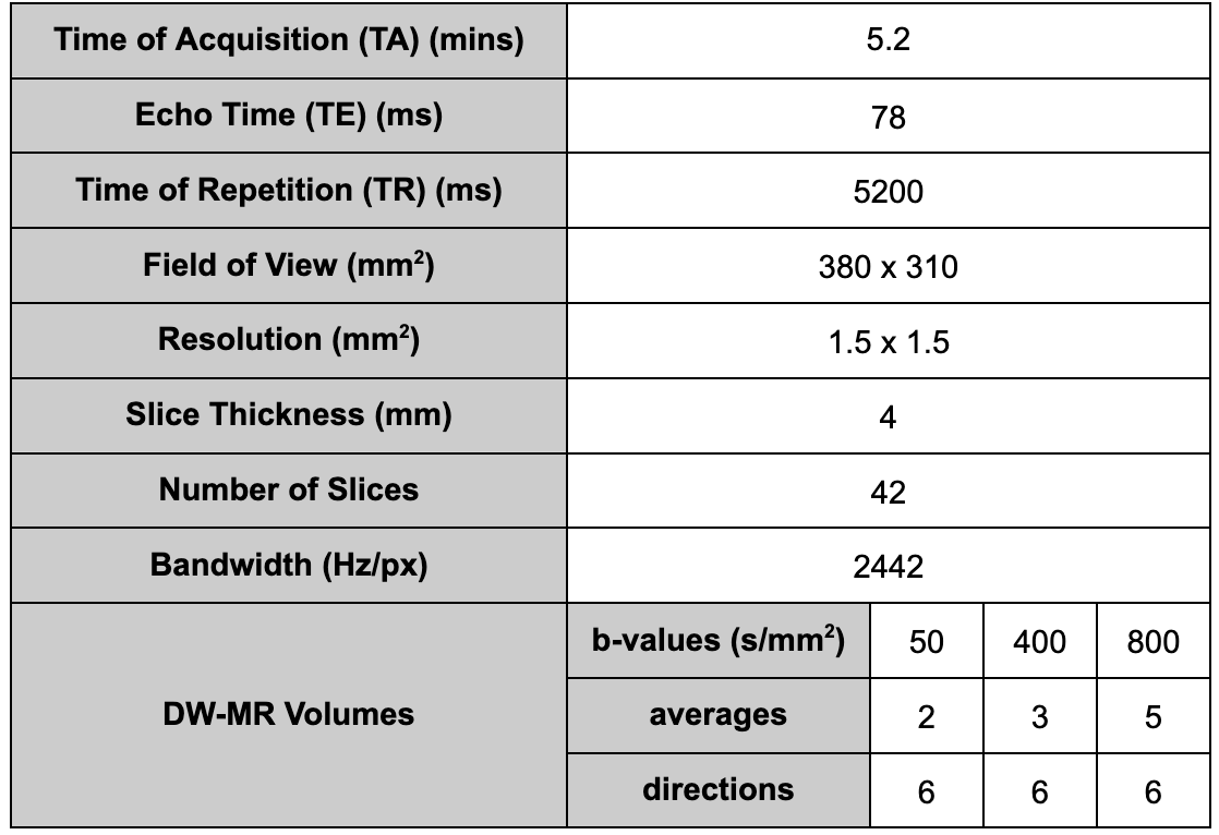
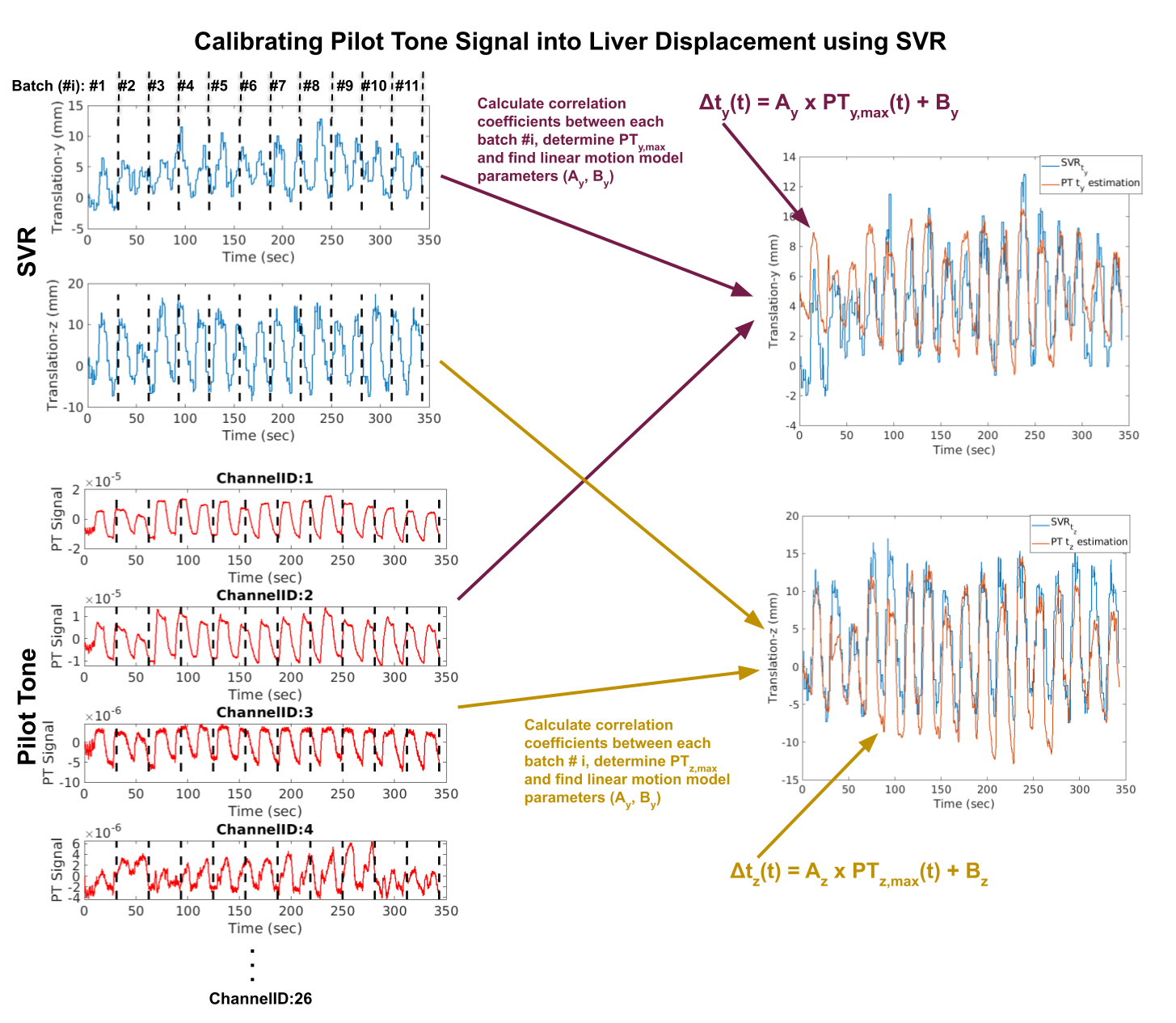
A liver diffusion-weighted MRI clinical protocol was acquired on 3 volunteers (2 female, age=37.3±4.0 years) with 60 DW-MR volumes using a 3T scanner (MAGNETOM Prisma, Siemens Healthcare, Erlangen, Germany). Imaging parameters are given in Table 1. A PT transmitter box was placed on the patient table next to the hip of the volunteer. Volunteers were asked to perform deep/irregular free-breathing. Raw data were saved for offline processing. PT navigator signals were extracted from the raw data for each channel (PTcha). Motion parameters (3 translation: tx, ty, tz and 3 rotation: rx, ry, rz) were estimated using SVR. Each PT channel measurement, PTcha, as well as y- and z-translation,tk (k=y,z) were divided into number of slices (Nsli) x 6 batches as PTcha,i, and tk,i, where i is the batch index. Only two largest parameters (ty, tz) were selected for correction based on PT because the other four parameters were smaller and set to zero (i.e., no correction applied for tx, rx, ry and rz ). Correlation coefficients between each PTcha,i and tk,i batch were calculated for each b50 acquisition. Linear motion model was fitted between the pair of PTcha,i (called, PTk,max) and tk,i (tk,max) that has the highest correlation coefficient, hence the estimated translation is $$$\Delta\hat{t_k}(t) = A_k x PT_{k,max}(t)+B_k$$$ [Eqn. 1]. The linear motion model parameters (A, B) were used to estimate ty, tz based on translated PT signal into displacement using Eqn. 1 for the entire acquisition, and subsequently used for motion correction for each slice. The overview of estimating y- and z- translations by calibrating PT signal is shown in Figure 1. After motion correction, each slice is transformed to the correct position and a point cloud is obtained from motion corrected slices in a volume. Kernel interpolation is used to resample the points in the point cloud back into a regular grid to generate a motion-corrected volume2,3. Single exponential model was fitted to estimate ADC maps.Results
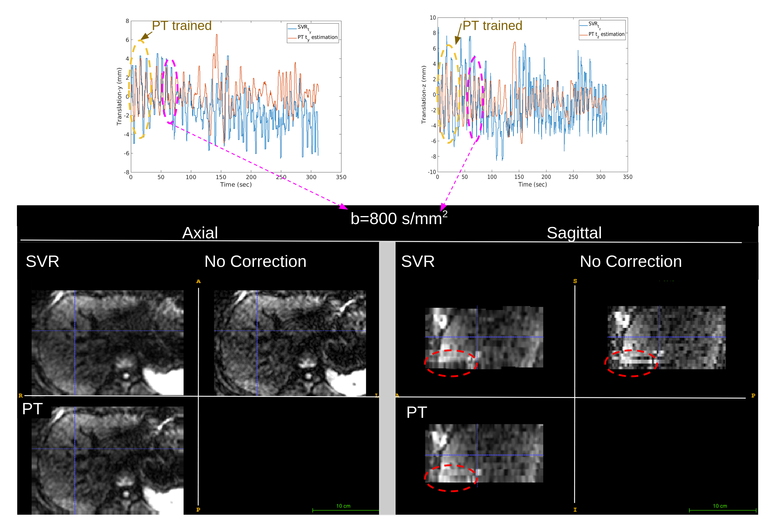
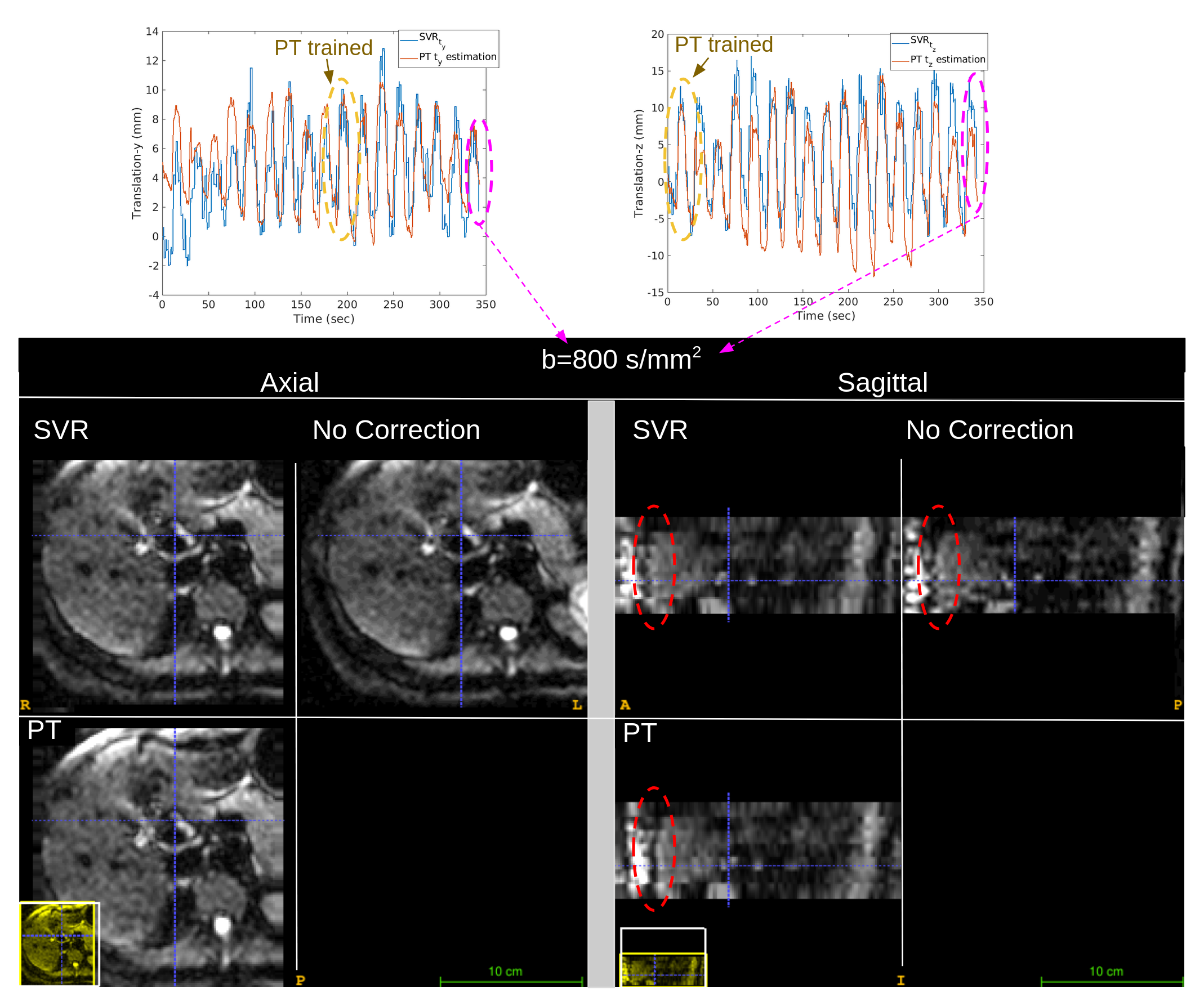
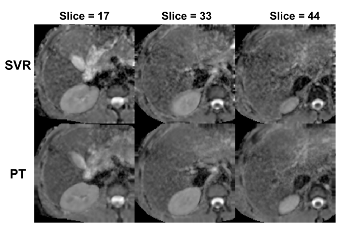
b=800 s/mm2 images comparing PT, SVR-based motion correction and no correction are depicted for volunteer #1 and #2 in Figure 2 and 3, respectively. It can be observed that misalignments of slices are eliminated with the proposed PT-based motion correction technique. In Figure 4, estimated ADC maps from PT and SVR -based motion corrected DWI are shown for volunteer #3. ADC maps estimated from PT-based motion corrected DWI are less noisy and sharper compared to ones estimated from SVR-based motion corrected.Discussion and Conclusion
The PT signal from multiple channels provides successful estimation of translation parameters of rigid motion for each slice using the motion model learned from a small portion of data used for training the model. The predicted PT-based motion estimations from SVR motion parameters obtained at low b-values can be used to correct for the position of each slice for realignment to a reference volume. Pilot tone navigator combined with SVR method can potentially improve the motion parameters compared to directly estimating the motion parameters from SVR, especially for high b-value images where the registration accuracy may reduce due to lower SNR and poor quality of the images.Acknowledgements
This work was supported in part by NIH grants R01 EB019483, R01 NS121657, R01 DK125561, R21 DK123569, R21 EB02962, S10OD025111. We thank the Center for Advanced Imaging Innovation and Research (CAI2R) at NYU for supplying the 3T Pilot Tone device used in this work.References
1. S. Kurugol, M. Freiman, L. Domachevsky, O. Afacan, J.M. Perez-Rossello, M.J. Callahan S.K. Warfield, “Motion-robust parameter estimation in abdominal diffusion-weighted MRI by simultaneous image registration and model estimation”, Medical Image Analysis, 39, 124-132, 2017.
2.S. Kurugol, B. Marami, O. Afacan, S.K. Warfield, A. Gholipour,”Motion-Robust Spatially Constrained Parameter Estimation in Renal Diffusion-Weighted MRI by 3D Motion Tracking and Correction of Sequential Slices”, In: Molecular Imaging, Reconstruction and Analysis of Moving Body Organs, and Stroke Imaging and Treatment. RAMBO 2017. Lecture Notes in Computer Science, vol 10555. Springer.
3. J. Coll-Font, J., O. Afacan, S. Hoge, S., H. Garg, K. Shashi, B. Marami, A. Gholipour, J. Chow, S. K. Warfield, S. Kurugol. Retrospective Distortion and Motion Correction for Free-Breathing DW-MRI of the Kidneys Using Dual-Echo EPI and Slice-to-Volume Registration. Journal of Magnetic Resonance Imaging, 2020.
4. Y. Mazaheri, R. K. G. Do, A. Shukla-Dave, J. O. Deasy, Y. Lu, and O. Akin: Motion Correction of Multi-b-value Diffusion-weighted Imaging in the Liver. Academic Radiology 19(12), 1573 (2012).
5. Ludwig, Juliane, et al. "Pilot tone–based motion correction for prospective respiratory compensated cardiac cine MRI." Magnetic Resonance in Medicine 85.5 (2021): 2403-2416.
6. Solomon, Eddy, et al. "Free-breathing radial imaging using a pilot-tone radiofrequency transmitter for detection of respiratory motion." Magnetic resonance in medicine 85.5 (2021): 2672-2685.
7. Falcão, M. B. L. et al. Pilot tone navigation for respiratory and cardiac motion-resolved free-running 5D flow MRI. Magn Reson Med 87, 718–732 (2022).
8. Ariyurek C, Wallace TE, Kober T, Kurugol S, Afacan O. Prospective motion correction in kidney MRI using FID navigators. Magn Reson Med. 2023;89:276-285.
Figures