1825
Inline Retrospective Motion Correction and Dynamical Alignment Using an Optical Markerless Motion Tracker
Ulrich Lindberg1, Stefan Glimberg2, Gerard Crelier3, Martin Buehrer3, and Henrik Bo Wiberg Larsson1
1Functional Imaging Unit, Department of Clinical Physiology and Nuclear Medicine, Rigshospitalet, Copenhagen, Denmark, 2TracInnovations, Ballerup, Denmark, 3GyroTools, Zurich, Switzerland
1Functional Imaging Unit, Department of Clinical Physiology and Nuclear Medicine, Rigshospitalet, Copenhagen, Denmark, 2TracInnovations, Ballerup, Denmark, 3GyroTools, Zurich, Switzerland
Synopsis
Keywords: Image Reconstruction, Motion Correction
Using an external motion tracking system for recording head movement during MR-acquisition and a modified version of the Philips reconstruction software (Recon2.0) that enables phase re-alignment and re-gridding of acquisition profiles during the standard immediate reconstruction process, we present a fully integrated retrospective motion correction solution that deliver both corrected and non-corrected images to the user and allows for the immediate visual assessment of the correction quality upon acquisition.Introduction
Head motion during data acquisition may lead to motion artefacts on the resulting MR images degrading the diagnostic value1. Furthermore, dynamical studies or a set of series used in combination for e.g., T1-mapping may suffer from misalignment due to motion. With the addition of an external motion tracking system for recording head movement during MR-acquisition we here present a fully integrated solution for retrospective motion correction (RMC) applied directly after data acquisition inline on the scanner host computer as well as an image-to-image alignment.Methods
Motion estimates for retrospective correction were obtained in real time during scanning with an optical markerless motion tracker (Tracoline TCL3.1, TracInnovations, Ballerup, Denmark), including the software TracSuite v3.2.0. The motion tracker computes motion estimates in real time at 30 Hz by continuously aligning a surface reconstruction of the subjects face to a reference surface acquired at the beginning of the scan (Fig. 1). All scans were acquired on a 3T Philips (Philips Medical Systems, Best, The Netherlands) Achieva dStream (software release 5.7.1.2) equipped with a 32-channel phase-array receive head coil. A variety of scans including both T1- and T2-weighted acquisitions as well as fast dynamical imaging was acquired in healthy volunteers performing controlled head motion. A custom software module running on the scanner host system received motion updates in real time from the motion tracker and stored it within the raw data of the image acquisition, therefore allowing immediate image reconstruction both with and without motion correction. A modified version of the Philips reconstruction software (Recon2.0) performed phase re-alignment and re-gridding of acquisition profiles during the standard immediate reconstruction process.Results
Visually improved image quality was seen in all cases (Fig. 2) In general, the retrospective correction performs very well on true rigid-body motions, as demonstrated also with phantom experiments. In the in-vivo experiments however, depending on the motion pattern and the presence of non-rigid body motion some reminiscent motion artefacts were still visible in the resulting reconstructed images.Image-to-image alignment yielded comparable and sometimes superior registration compared to image-based registration algorithms that depend on image contrast.
The inline reconstruction works well and provides a lean workflow for the user as the reconstructed images are readily available on the scanner host computer directly after data acquisition.
Discussion
The fully integrated retrospective motion correction solution delivers both corrected and non-corrected images to the user and allows for the immediate visual assessment of the correction quality. The limits of the used correction scheme are apparent especially in cases when the motion of the brain is not rigid compared to the head motion and when the acquisition coverage is insufficient for a precise re-gridding of acquisition profiles. The latter can be addressed by applying also prospective correction methods during acquisition2.Conclusion
We have implemented a fully integrated retrospective motion correction application in Philips Recon2.0 that carries out motion correction and image reconstruction directly on the scanner resulting in markedly improved image quality.Acknowledgements
No acknowledgement found.References
- Andre JB, Bresnahan BW, Mossa-Basha M, Hoff MN, Smith CP, Anzai Y, Cohen WA. Toward Quantifying the Prevalence, Severity, and Cost Associated With Patient Motion During Clinical MR Examinations. J Am Coll Radiol. 2015 Jul;12(7):689-95. doi: 10.1016/j.jacr.2015.03.007. Epub 2015 May 9.
- Slipsager JM, Glimberg SL, Højgaard L, Paulsen RR, Wighton P, Tisdall MD, Jaimes C, Gagoski BA, Grant PE, van der Kouwe A, Olesen OV, Frost R. Comparison of prospective and retrospective motion correction in 3D-encoded neuroanatomical MRI. Magn Reson Med. 2022 Feb;87(2):629-645. doi: 10.1002/mrm.28991. Epub 2021 Sep 7.
Figures
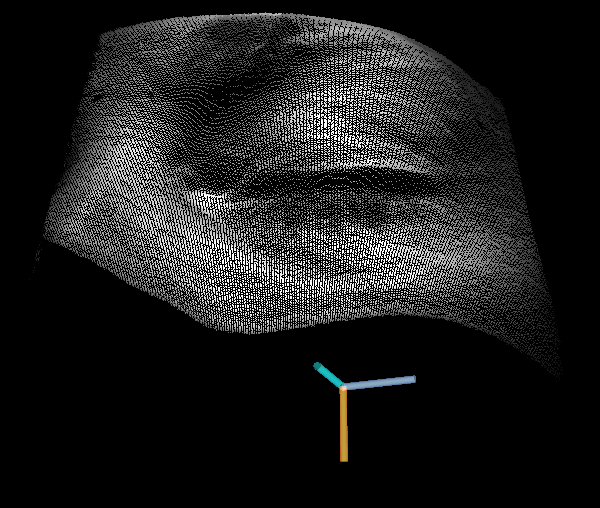
Surface point cloud image from the tracking system. The surface point cloud image is updated with 30 Hz and aligned to a reference surface point cloud.
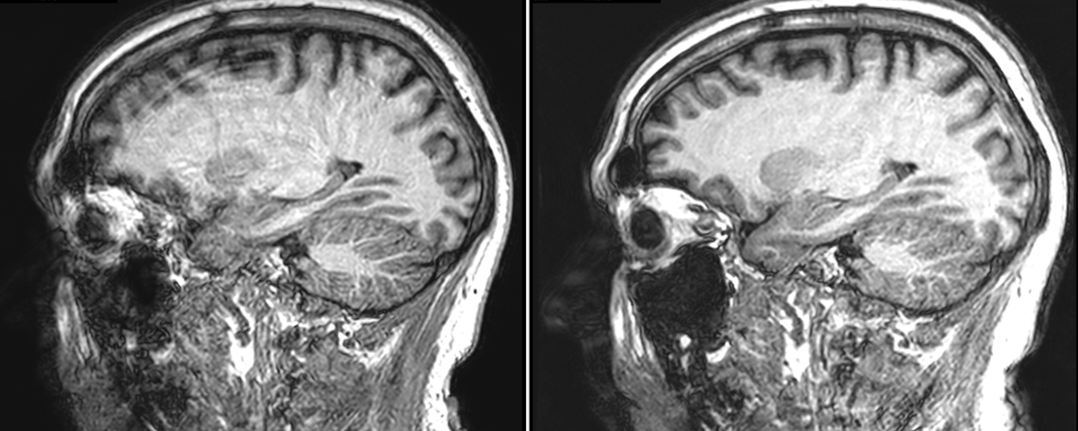
Retrospective motion correction of 3D T1w structural scan (acquisition matrix 256x256x170, acquisition time 5:35 min) with continuous heavy breathing. Left: without correction. Right: Corrected using data from the external tracking device send to the scanner in real time and corrected during reconstruction.
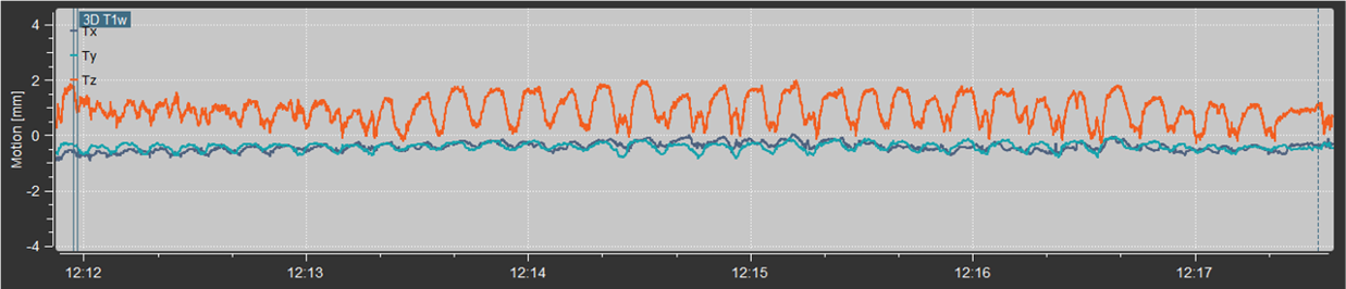
Motion measurements during the 5:35 min 3D T1w structural scan. The volunteer was asked to perform exaggerated respiratory motions. Top: absolute motion of the average point on the tracked surface. Middle: X-, Y-, and Z-translations. Bottom: X-, Y-, and Z-rotations.
DOI: https://doi.org/10.58530/2023/1825