1819
Effectiveness and Reliability Assessment on Head Motion Capturing and Correction (MoCAP)
Zhuoyang Gu1,2, Lianghu Guo1, Qing Yang1, Xinyi Cai1, Tianli Tao1, Sifan He1, Hua Jiang1, Haifeng Tang1, Qian Wang1, Xiaopeng Zong1, Dinggang Shen1, Qiang He2, and Han Zhang1
1School of Biomedical Engineering, ShanghaiTech University, Shanghai, China, 2United Imaging Healthcare Co., Ltd., Shanghai, China
1School of Biomedical Engineering, ShanghaiTech University, Shanghai, China, 2United Imaging Healthcare Co., Ltd., Shanghai, China
Synopsis
Keywords: Motion Correction, Brain, MRI acquisition
Head motion monitoring and compensation during MRI is essential to imaging quality and success rates of acquisition. MoCAP is a novel real-time head motion monitoring and correction technique utilizing structured light to perform prospective motion correction by adjusting MR gradient. We perform effectiveness and reliability assessment on MoCAP in structural and functional MRI. MoCAP can significantly improve quality of the two modalities, reducing motion artifacts and enhancing validity and reliability of post-processing results. MoCAP is promising in the MRI field for special populations like children and patients with difficulty in keeping still during scan.Introduction
Recent MRI studies often require prolonged scan time, posing difficulty for certain cohorts such as children and patients who cannot keep still. Motion artifacts reduce image quality and may lead to biases in the post-processing results. In structural MRI (sMRI), head motion could reduce gray matter volume and thickness estimates1. In functional MRI (fMRI), head motion could significantly confound BOLD signals and resultant functional connectivity (FC) estimates2. Head motion artifact reduction has been extensively studied with various post-processing techniques3, however, real-time head motion monitoring and correction for sMRI and fMRI has not been well studied.MoCAP is such a technique that utilizes structured light to perform prospective motion correction during acquisition. A 3D structured-light camera continuously monitors subject's face during MRI scan at a frequency of 30Hz and feeds back 6 degree-of-freedom head position information to MR system to upgrade the gradients. It ensures that the relative position of the patient's head and the acquired FOV remain fixed during the entire scan. Images will be re-acquired if motion exceeds a certain threshold.
In this work, we systematically evaluate the effectiveness of MoCAP for sMRI and fMRI. The results show that MoCAP substantially improves the quality of T1w sMRI and derived surface-based measurement, as well as the FC results of resting-state fMRI.
Methods
Eleven healthy adults aged 22–33 years (4 females) were scanned using a research-dedicated, United Imaging 3.0T uMR890 scanner equipped with MoCAP. The experiment includes 8 runs with randomized order. In case of large head motion, subjects were asked to move their head slowly following a figure “8” trajectory. For small head motion, the subjects stayed as still as possible, and two runs were acquired immediately for test-retest reliability assessment. MoCAP was turning on or off in different runs (see Fig. 1 for an example).For quality assessment on sMRI, various image quality metrics including CNR, SNR, and FWHM were calculated4; region-wise surface geometric features were extracted5; a motion score was calculated according to 6 summarizing x/y/z transition and rotation recorded by MoCAP. These features were compared among different runs using paired t-tests. For region-wise analysis, p threshold was set to 0.05 after false discovery rate correction.
For fMRI, a deep learning-based brain parcellation model7 was used to generate each subject’s brain regions of interest for region-averaged fMRI signal extraction. Two different parcellation schemes were used to generate whole-brain FC matrices. Seed-based correlation was also used to generate primary visual network and default mode network for quality evaluation.
Results
MoCAP substantially reduced motion artifacts in T1w images with large head motion (Fig. 2). CNR, SNR, and FWHM significantly improve with MoCAP when head motion is large (Fig. 3). Comparisons on region-wise surface geometric measures show significant differences with large head motion between MoCAP on and off (Fig. 4a, cortical thickness, Fig. 4b, all metrics). For fMRI, qualitative comparison shows FC results are significantly improved when MoCAP turns on, even only with small head motion (Fig. 5).Discussion and Conclusions
MoCAP considerably improves the quality of sMRI and fMRI, especially when there are large head motions. The reliability of MoCAP is good as revealed by the test-retest experiments. Results suggested that, for sMRI, there are little differences in image quality w/ and w/o MoCAP when subjects are instructed to stay still. This is probably because the subjects in the experiment are healthy young adults who controlled head motion quite well during scan. MoCAP may improve fMRI quality in certain case even when head motion is small. Collectively, we recommend keeping MoCAP on during sMRI and fMRI scan for improved data quality and success rate.Acknowledgements
This work is partially supported by the National Key Technology R&D Program (Nos. 2022ZD0209000, 2021ZD0200516), Shanghai Pilot Program for Basic Research - Chinese Academy of Science, Shanghai Branch (No. JCYJ-SHFY-2022-014), Open Research Fund Program of National Innovation Center for Advanced Medical Devices (No. NMED2021ZD-01-001), Shenzhen Science and Technology Program (No. KCXFZ20211020163408012), and Shanghai Pujiang Program (No. 21PJ1421400).References
1. Reuter, M.; Tisdall, M. D.; Qureshi, A.; Buckner, R. L.; van der Kouwe, A. J.; Fischl, B., Head motion during MRI acquisition reduces gray matter volume and thickness estimates. Neuroimage 2015, 107, 107-115.2. Power, J. D.; Barnes, K. A.; Snyder, A. Z.; Schlaggar, B. L.; Petersen, S. E., Spurious but systematic correlations in functional connectivity MRI networks arise from subject motion. Neuroimage 2012, 59 (3), 2142-2154.
3. Zaitsev, M.; Maclaren, J.; Herbst, M., Motion artifacts in MRI: A complex problem with many partial solutions. Journal of Magnetic Resonance Imaging 2015, 42 (4), 887-901.
4. Esteban, O.; Birman, D.; Schaer, M.; Koyejo, O. O.; Poldrack, R. A.; Gorgolewski, K. J., MRIQC: Advancing the automatic prediction of image quality in MRI from unseen sites. PloS one 2017, 12 (9), e0184661.
5. Fischl, B., FreeSurfer. Neuroimage 2012, 62 (2), 774-781.
6. Moore, J.; Jimenez, J.; Lin, W.; Powers, W.; Zong, X., Prospective Motion Correction and Automatic Segmentation of Penetrating Arteries in Phase Contrast MRI at 7 T. bioRxiv 2022.
7. Wei, J.; Shi, F.; Cui, Z.; Pan, Y.; Xia, Y.; Shen, D. In Consistent Segmentation of Longitudinal Brain MR Images with Spatio-Temporal Constrained Networks, International Conference on Medical Image Computing and Computer-Assisted Intervention, Springer: 2021; pp 89-98.
Figures
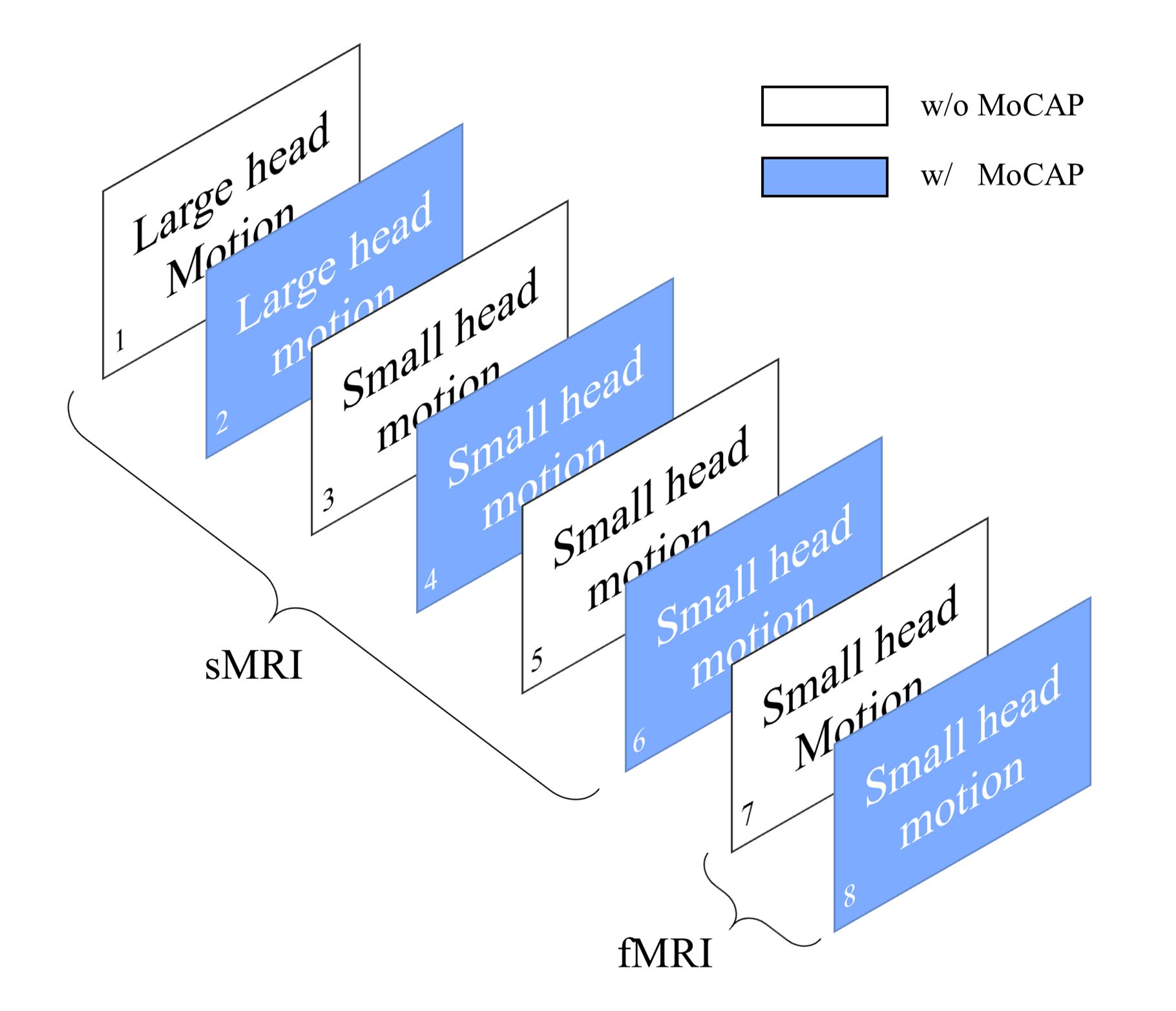
Fig. 1 Experiment design of sMRI/fMRI acquisition w and w/o MoCAP
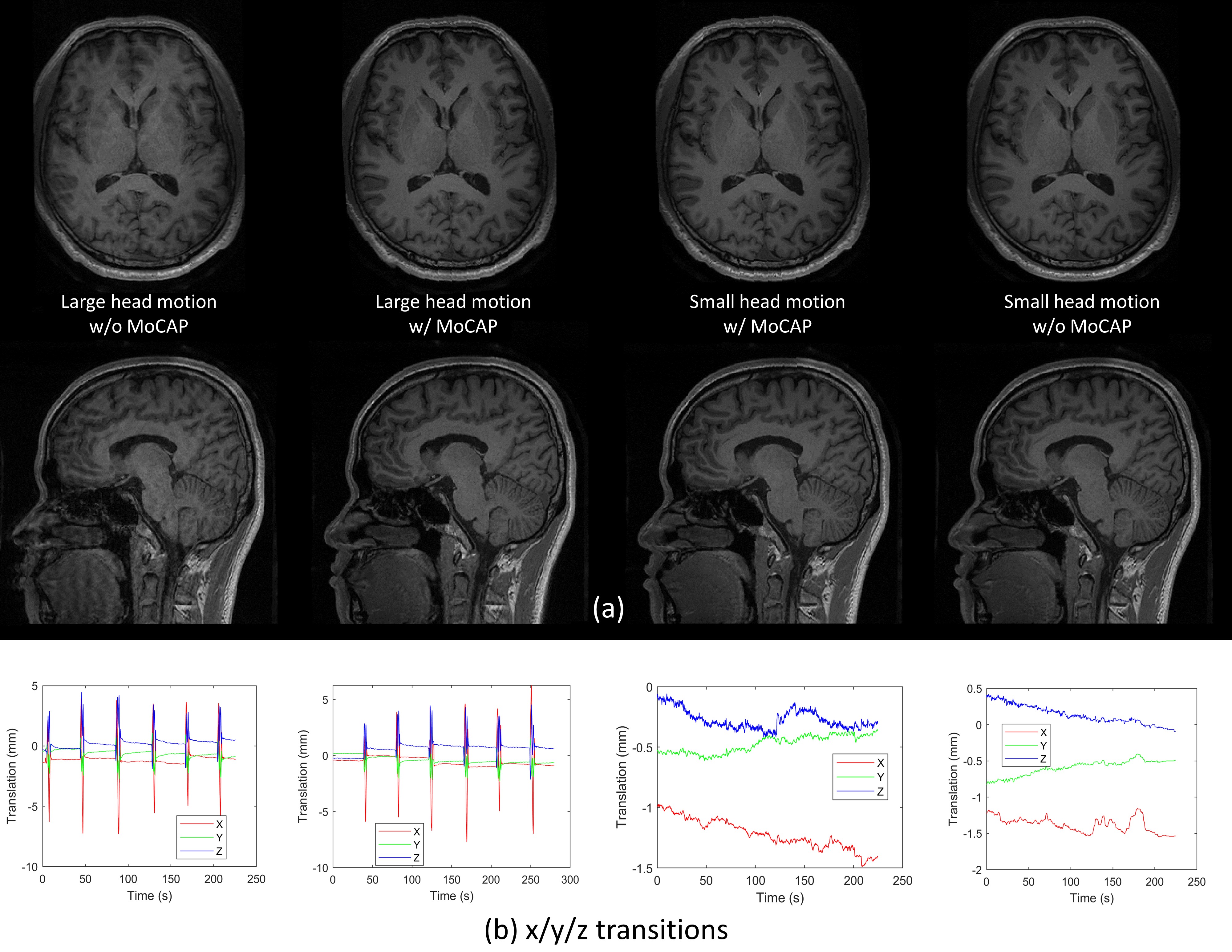
Fig. 2 (a) T1w image quality comparison with large and small head motion, w/ and w/o MoCAP. (b) head motion curves (show x/y/z transitions only, x/y/z rotations are not shown) recorded by MoCAP during T1w scans with large and small head motion, w/ and w/o MoCAP.
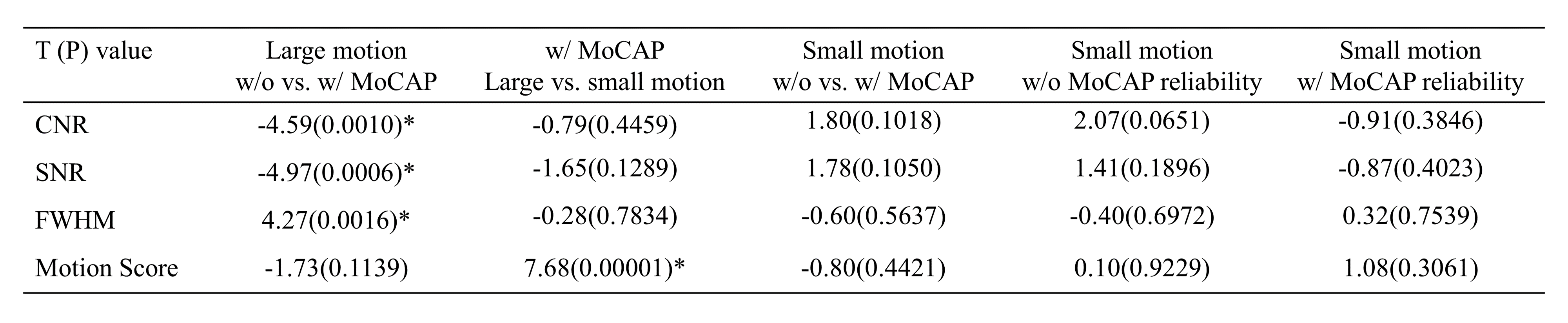
Fig.3 Comparisons of image quality metrics across different runs (* indicate significant).
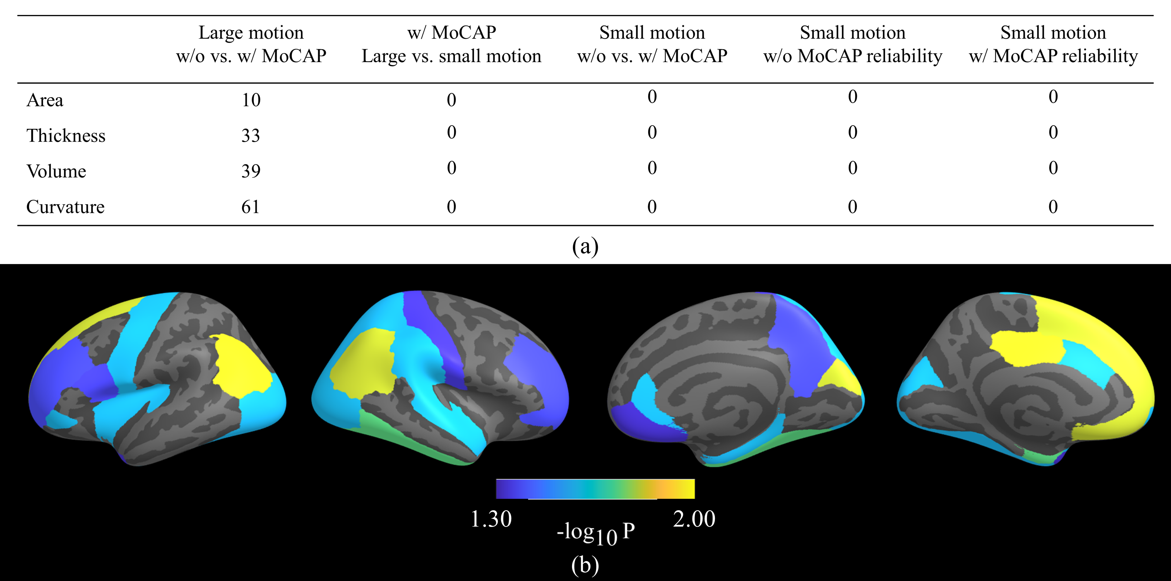
Fig. 4 (a) Comparisons on region-wise surface geometric features across different runs. Figures indicate the total number of brain regions with significant difference (p<0.05, FDR correction). (b) Comparing region-wise surface geometric measures between w/ and w/o MoCAP in large head motion scenario. Regions with colors are those with significant differences using paired t-test (p<0.05, FDR corrected).
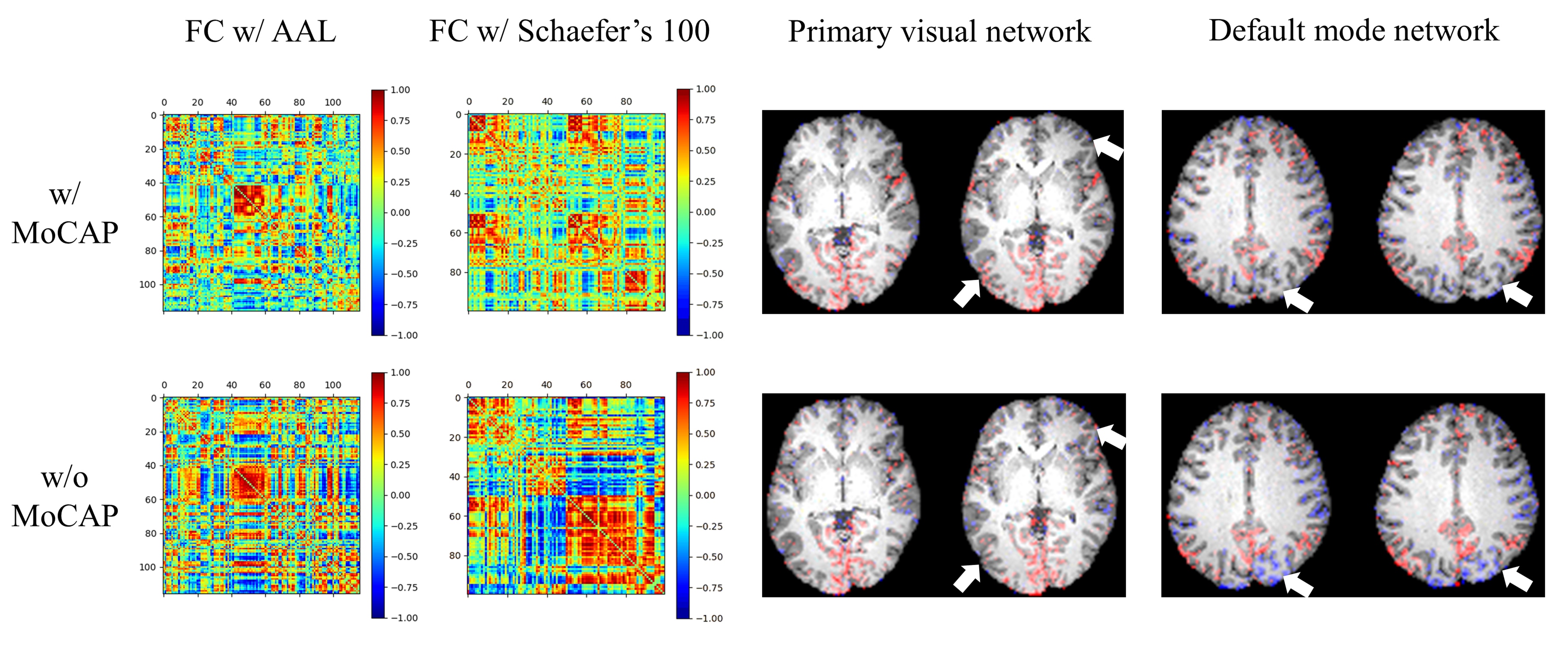
Fig. 5 Comparisons on functional connectivity matrices with two parcellation schemes, as well as the seed-based correlation maps (primary visual network and default mode network) derived from fMRI, between w/ and w/o MoCAP. Results are from one exemplary subject during resting-state scan with small head motion. Arrows show suspicious motion artifacts on two resting-state network maps.
DOI: https://doi.org/10.58530/2023/1819