1805
Quantitative DWI in patients with hepatocellular carcinoma: effects of simultaneous multi-slice acquisition and gadoxetic acid injection1West China Hospital, Chengdu, China, 2MR Collaborations, Siemens Healthineers Ltd., Shanghai, China
Synopsis
Keywords: Quantitative Imaging, Liver, simultaneous multi-slice diffusion weighted imaging
Diffusion weighted imaging (DWI) and gadoxetic acid-enhanced magnetic resonance imaging (MRI) are important in diagnosing hepatocellular carcinoma (HCC). Previous studies have validated that gadoxetic acid administration during enhanced MRI improves signal-to-noise ratio without influencing apparent diffusion coefficient (ADC), while the impact of simultaneous multi-slice (SMS) technique on the measurement of ADC is still unknown. Our study assessed the influence of gadoxetic acid administration on ADC values of liver with and without SMS acceleration. We found that SMS technique and gadoxetic acid administration did not affect the ADC values of liver parenchyma and HCC.
Background and purpose
Diffusion weighted imaging (DWI) is sensitive to hepatocellular carcinoma (HCC) by reflecting the diffusion of water molecules1,2, and the degree of diffusion restriction can be quantified by apparent diffusion coefficient (ADC)3. In addition, gadoxetic acid-enhanced magnetic resonance imaging (MRI) allows for functional information of liver cell membrane, thus improving the diagnostic capabilities of HCC4. Although these sequences provide considerable diagnostic value, the relative long acquisition time is still a matter of concern. Fortunately, simultaneous multi-slice (SMS) technique enables accelerated acquisition process by exciting multiple slices simultaneously5. A previous study has found that gadoxetic acid injection during enhanced MRI can improve image signal-to-noise ratio without influencing ADC derived from DWI6. Therefore, this study aimed to investigate whether SMS acceleration influence quantitative parameter of breath-hold and free-breathing (FB) DWI in the patients with HCC.Methods
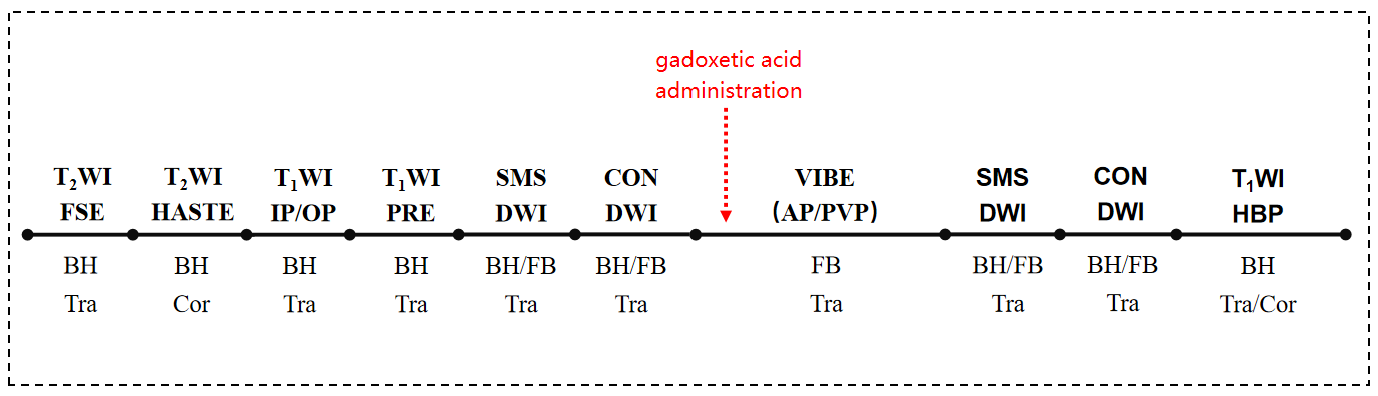
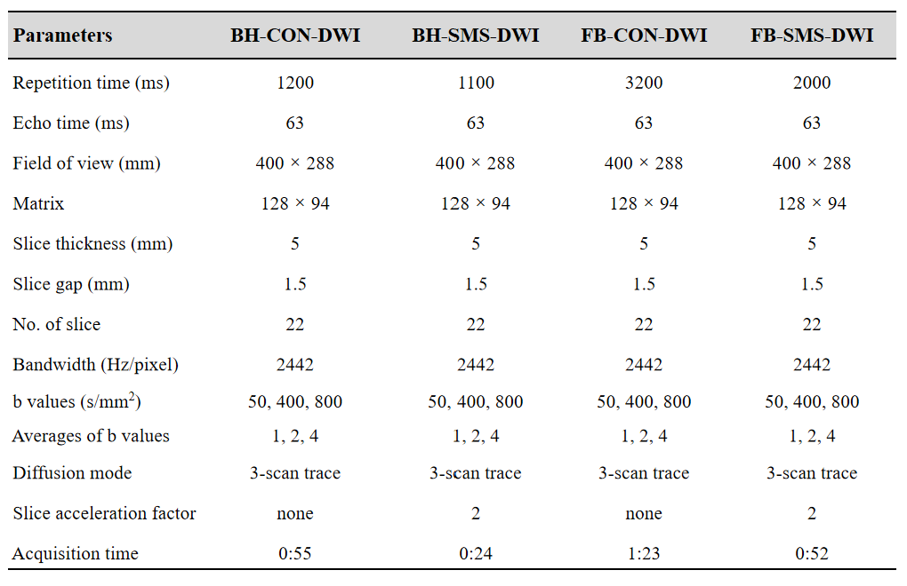
A total of 38 patients (32 men and 6 women; mean age ± standard deviation, 53.63±12.65 years) with histologically proven HCC were enrolled between November 2020 and January 2021. Each patient underwent BH and FB DWI with and without SMS technique using a 3T MRI system (MAGNETOM Skyra, Siemens Healthcare, Erlangen, Germany) before and after the injection of gadoxetic acid, the sequence of the scanning protocol is presented in Figure 1. The detailed imaging acquisition parameters are shown in Figure 2. ADC maps were automatically generated on MR system’s console and its values of the normal liver parenchyma and HCC lesions were measured by positioning region of interest (ROI) with an average area of 1.5 cm2 in the right liver lobe at similar slice positions between scans, while excluding major vessels and artifacts. The Student’s t-test or Mann-Whitney U test was used to compare the ADC values between conventional DWI (CON-DWI) and SMS-DWI, as well as ADC values before and after gadoxetic acid injection.Results
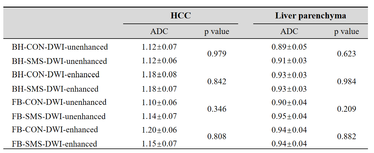
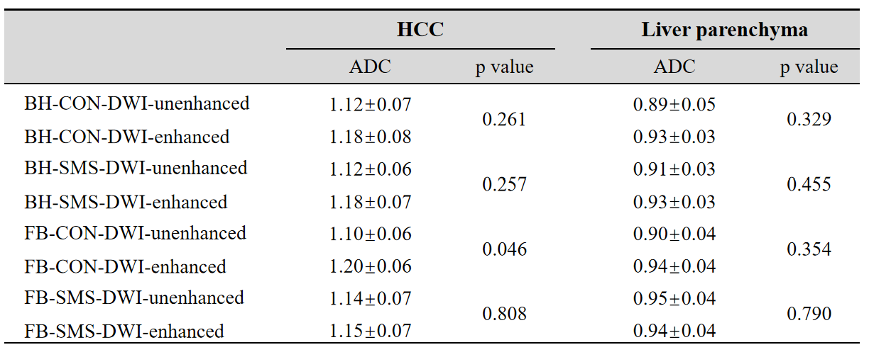
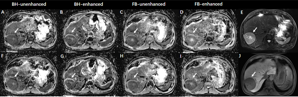
Both SMS and gadoxetic acid administration showed no statistical difference in the ADC values of liver parenchyma and HCC lesions (P=0.257-0.984) in BH-DWI. As for FB-DWI, except that the ADC values of HCC lesions on the enhanced CON-DWI were significantly higher than the unenhanced CON-DWI (P=0.046), others were observed no difference (P=0.209-0.882). The ADC values of liver parenchyma and HCC with and without SMS acceleration are detailed in Figure 3, and those before and after gadoxetic acid administration are presented in Figure 4. Besides, an example of a 59-year-old male HCC patient is shown in Figure 5.Discussion
For BH-DWI scheme, SMS and gadoxetic acid administration have no impact on the ADC values of liver parenchyma and HCC lesions. For FB-DWI scheme, similar results were observed except that gadoxetic acid administration influenced the ADC values of HCC lesions on CON-DWI. However, according to Chen et al., they reported no significant difference between the influence of different breathing schemes, including BH, FB, respiratory-triggered (RT), and navigator-triggered (NT), on liver ADC derived from CON-DWI7. Another study conducted by Tang et al. investigated the impact of gadoxetic acid administration on ADC values of hepatic lesions generated from RT CON-DWI and concluded that they would not be affected8. Therefore, we speculated that the significantly higher HCC ADC values of CON-DWI after gadoxetic acid injection may attribute to relatively small sample size in current study or the joint influence of FB and gadoxetic acid administration on CON-DWI.Conclusion
SMS technique and gadoxetic acid administration have no impact on ADC values of liver and HCC. Moreover, FB scheme is recommended to patients if they have difficulty in breath-holding.Acknowledgements
NoneReferences
1. Malayeri AA, El Khouli RH, Zaheer A, Jacobs MA, Corona-Villalobos CP, Kamel IR, et al. Principles and Applications of Diffusion-weighted Imaging in Cancer Detection, Staging, and Treatment Follow-up. Radiographics 2011;31:1773-1791.
2. Gluskin JS, Chegai F, Monti S, Squillaci E, Mannelli L. Hepatocellular Carcinoma and Diffusion-Weighted MRI: Detection and Evaluation of Treatment Response. J Cancer. 2016;7(11):1565-1570.
3. Vermoolen MA, Kwee TC, Nievelstein RAJ. Apparent diffusion coefficient measurements in the differentiation between benign and malignant lesions: a systematic review. Insights into Imaging 2012;3:395-409.
4. Ringe KI, Husarik DB, Sirlin CB, Merkle EM. Gadoxetate disodium-enhanced MRI of the liver: part 1, protocol optimization and lesion appearance in the noncirrhotic liver. AJR Am J Roentgenol. 2010;195(1):13-28.
5. Taron J, Weiß J, Martirosian P, et al. Clinical Robustness of Accelerated and Optimized Abdominal Diffusion-Weighted Imaging. Invest Radiol. 2017;52(10):590-595.
6. Choi JS, Kim MJ, Choi JY, Park MS, Lim JS, Kim KW. Diffusion-weighted MR imaging of liver on 3.0-Tesla system: effect of intravenous administration of gadoxetic acid disodium. Eur Radiol. 2010;20(5):1052-1060.
7. Chen X, Qin L, Pan D, et al. Liver diffusion-weighted MR imaging: reproducibility comparison of ADC measurements obtained with multiple breath-hold, free-breathing, respiratory-triggered, and navigator-triggered techniques. Radiology. 2014;271(1):113-125.
8. Tang H, Yuan Y, Deng L, et al. Identification of diffusion weighted imaging would be affected before and after Gd-EOB-DTPA in patients with focal hepatic lesions: an observational study. Ann Transl Med. 2022;10(6):346.
Figures