1758
Whole-tumor ADC Histogram Analysis in evaluating Tongue Squamous Cell Carcinoma heterogeneity and predicting Cervical Lymph Node Metastases1Shanghai Ninth People’s Hospital, affiliated to Shanghai Jiao Tong University, School of Medicine, Shanghai, China, 2MR Collaboration, Central Research Institute, Shanghai United Imaging Healthcare, Shanghai, China, 3Shanghai Pulmonary Hospital, Shanghai, China
Synopsis
Keywords: Head & Neck/ENT, Diffusion/other diffusion imaging techniques
Tumor heterogeneity occurred frequently in patients with SCC and associated with poor prognosis. Compared to clinic TNM stage or the histological grade, the histological heterogeneity has not been well addressed yet. Our results clearly demonstrated that higher incidence of CNM was observed in histological hetero-group than that in homo-group. whole-lesion ADC histogram metrics presented lower values in hetero-group and in CNM+ group. ADC75th and kurtosis were two independent prognostic factors for evaluating the CNM status. Whole-tumor ADC histogram offered an approach for detecting intra-tumoral heterogeneity and predicting cervical lymph node metastases status.Introduction
Tongue squamous cell carcinoma (SCCT) accounts for 40-50% of all oral cancers1, which is characterized by frequent lymphoid metastasis, a high rate of regional recurrence, and a poor prognosis. One major reason for the poor prognosis maybe the intra-tumoral heterogeneity of SCCT2. An understanding of tumor heterogeneity would help to predict the eventual prognosis and provide the basis of effective therapeutic strategies for patients with advanced SCCT. Diffusion weighted imaging(DWI)could assess microscopic thermal motion of water in biologic tissues by quantify the apparent diffusion coefficient(ADC)values3,4. However, mean ADC values obtained by placing a localized ROI on several representative sections of the tumor might have a limited ability to reflect the actual whole-tumor characteristics5. In this regard, voxel-based whole-tumor histogram analysis is more spatial- and texture-oriented, with which the spatial intra-tumoral heterogeneity can be better characterized5. Thus we aimed to determine the feasibility of quantitative ADC metrics from whole-lesion histogram analysis to access the intra-tumoral heterogeneity and the prognostic value for predicting cervical node metastases (CNM) in patients with SCCT.Methods
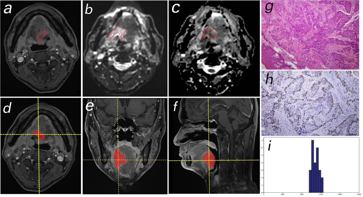
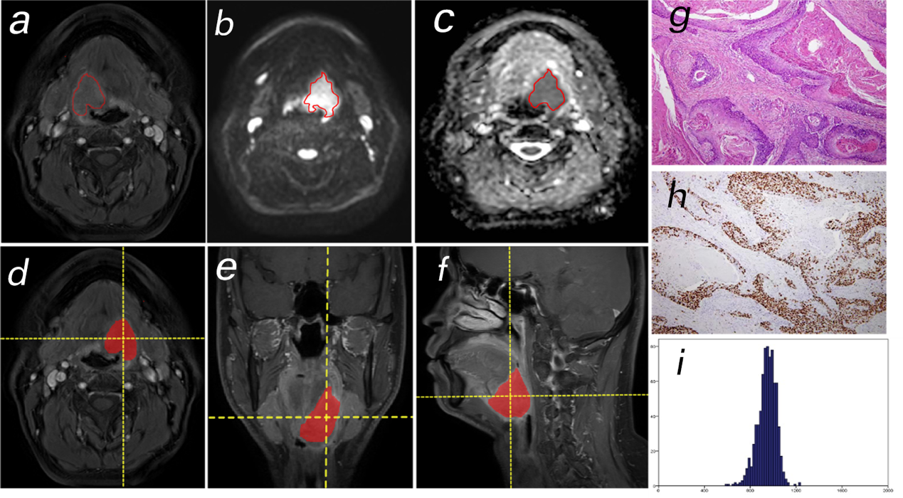
A total of 45 patients (mean age 54 ± 10 years, 23 patients with cervical lymph node metastases) with pathologically proven SCC were included in the study. Uneven histological grade such as grade I-II or grade II-III were defined as histological heterogeneous-group (hetero-group, 24 patients). All magnetic resonance imaging (MRI) examinations including T2-weighted imaging (T2WI), DWI and dynamic contrast-enhanced MRI (DCE-MRI) were performed on a 1.5T scanner (uMR560, United Imaging Healthcare) with a twelve-channel head-neck coil. ADC maps were generated by a voxel-by-voxel fitting based on the mono-exponential diffusion model. Volumes of interest (VOIs) analysis was performed by manually delineated on multiple slices of DWI images to cover the whole tumor and exclude the part of necrotic, cystic or hemorrhagic regions with reference to the T2WI and DCE images. Volumetric parameters including tumor volume, ADC histograms (mean, standard deviation (SD), 10th, 25th, 50th, 75th, and 90th percentiles, variance, skewness and kurtosis) were measured. The differences of metrics between different groups were evaluated using the independent student’s t-test or Mann-Whitney U test. Sensitivity, specificity, and area under the curve (AUC) were calculated for the diagnostic procedures. A p-value <0.05 was considered statistically significant.Results
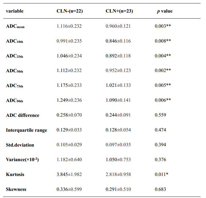
Whole-lesion ADC histogram metrics significantly lower in hetero-group than homo-group were mean ADC, percentiles from 10th to 90th, SD and variance (Table 1, Figure 1 and 2). Among all valuable ADC histogram parameters, ADCmean had the best discriminative powers to differentiate SCCT heterogeneity, yielding the sensitivity and specificity of 66.7% and 81.0%, respectively and the AUC of 0.731. Whole-lesion ADC histogram metrics significantly lower in CNM+ group were mean ADC, percentiles from 10th to 90th and kurtosis (Table 2). Higher incidence of CNM was observed in histological hetero-group than that in homo-group. ADC75th (Odds ratio [OR], 24.72; 95% CI, 2.62, 233.24; p = 0.005) and kurtosis (OR, 18.48; 95% CI, 2.089 163.68, p = 0.009) were two independent predictors of cervical lymph node metastasis.Discussion
This study investigated the capability of volumetric ADC histogram metrics in the preoperative evaluation of intra-tumoral heterogeneity and predicting cervical node metastases in patients with SCCT. Although the mean histological grade between two subgroups were balanced, the values of ADCmean and all ADC percentiles were significantly lower in high-heterogenous group than those in low heterogeneity group. Lower ADC values usually represented higher malignancy, possibly resulting from the limited extracellular and extravascular space with microstructural changes in the tumor such as the increasing cell densities. Although the driving factors for the tumoral histological heterogeneity is not well understood, the association between lower ADC histogram metrics and high histological heterogeneity strongly implicated its clinical relevance and potential therapeutic strategies. A decrease in kurtosis could be an indicator for high intra-tumoral heterogeneous. Our data indicated that lower kurtosis and lower ADC75th may be potentially used for predicting CNM status in patients with SCCT although further studies are required for both internal and external validation.Conclusion
In summary, whole-tumor ADC histogram metrics may serve as non-invasive biomarkers of tumoral heterogeneity of SCC. ADC histogram metrics, kurtosis, on preoperative imaging is an independent predictor for CNM status.Acknowledgements
No acknowledgementsReferences
1. Ng, Jia Hui et al. “Changing epidemiology of oral squamous cell carcinoma of the tongue: A global study.” Head & neck vol. 39,2 (2017): 297-304. doi:10.1002/hed.24589.
2. Monteiro, L-S et al. “A clinical-pathological and survival study of oral squamous cell carcinomas from a population of the North of Portugal.” Medicina oral, patologia oral y cirugia bucal vol. 19,2 e120-6. 1 Mar. 2014, doi:10.4317/medoral.19090.
3. Taffel, Myles T et al. “Diffusion Quantification in Body Imaging.” Topics in magnetic resonance imaging : TMRI vol. 26,6 (2017): 243-249. doi:10.1097/RMR.0000000000000144.
4. Surov, Alexey et al. “Correlation between apparent diffusion coefficient (ADC) and cellularity is different in several tumors: a meta-analysis.” Oncotarget vol. 8,35 59492-59499. 10 May. 2017, doi:10.18632/oncotarget.17752.
5. Li, Hai Ming et al. “Diffusion kurtosis imaging for differentiating borderline from malignant epithelial ovarian tumors: A correlation with Ki-67 expression.” Journal of magnetic resonance imaging : JMRI vol. 46,5 (2017): 1499-1506. doi:10.1002/jmri.25696.
Figures