1756
MR Varying Diffusion Curvature Imaging on Different Brain Tumors: A Preliminary Experience1Department of Radiology, The Affiliated Drum Tower Hospital of Nanjing University Medical School, Nanjing, China, 2Central Research Institue, United Imaging Healthcare, Shanghai, China
Synopsis
Keywords: Tumors, Diffusion/other diffusion imaging techniques
Dozens of diffusion models have been created to describe the non-Gaussian nature of diffusion. Nevertheless, many of them imply assumptions that have not been rigorously confirmed, or includes abstract parameters. The purpose of this study was to observe the characteristics of varying diffusion curvature (VDC) indicators in several types of brain tumor. D0 and D1 distribution were obtained for each subject. This study illustrates the potential of applying a simple and pure empirical non-Gaussian diffusion model VDC on brain tumor imaging.Introduction
MRI is an important radiological approach in the diagnosis of tumor for its high contrast in soft tissue. In addition to anatomic imaging, functional MRI methods, especially the diffusion-weighted imaging (DWI), are also of large clinical significance. In general, diffusion-weighted signal is assumed to follow an ideal exponential decay, and the resulting apparent diffusion coefficient (ADC) should be independent of the chosen b-value. Unfortunately, this is often inconsistent with what we actually observe in human tissue, which is often attributed to the non-Gaussian nature of diffusion [1]. Dozens of diffusion models (e.g., diffusion kurtosis, fractional order calculus, continuous time random walk) have been created to address this problem. Nevertheless, many of them imply assumptions that have not been rigorously confirmed, or includes abstract parameters.Recently, a simple, phenomenon-based diffusion model, varying diffusion curvature (VDC), was established [4]. As an empirical method, VDC is yet to be applied to more types of tumors. The purpose of this study was to observe the characteristics of VDC indicators in several cases of different brain tumors.
Methods
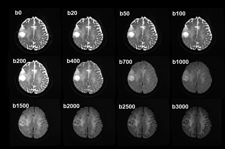
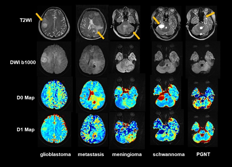
The prospective study was approved by our Institute Review Board. Five patients with brain tumor (1 with glioblastoma, 1 with metastasis, 1 with meningioma, 1 with schwannoma, 1 with papillary glioneuronal tumor (PGNT)) were included in this preliminary study. Precise tumor subtype was determined by post-surgical pathological analysis. All MRI examinations were performed on a 3.0T scanner (uMR790, United Imaging Healthcare, Shanghai, China) before surgery.Routine MRI sequences included: 3D T1-weighted imaging (T1WI), 3D T2-weighted imaging (T2WI), 3D T2 fluid-attenuated inversion-recovery (T2-FLAIR). Axial DWI was performed using 12 b-values (0, 20, 50, 100, 200, 400, 700, 1000, 1500, 2000, 2500, 3000). Other DWI protocol parameters were: TR/TE = 5016/108 ms, FOV 230*220 mm2, Matrix 144*138, 22 slices, slice thickness/gap 5/1.5 mm. The total acquisition time for DWI was 6 min 57 s. The VDC model applied here assumed the diffusion coefficient to obey [4]:
$$D(b) = D_{0}e^{-bD_{1}}$$ where D0 represent the diffusivity at b0, and D1 is associated to the curvature of diffusivity with b-value, accounting for non-Gaussian diffusion behavior. The resultant expression of diffusion-weighted signal to varying b-value should be [4]:
$$S(b) = S_{0}exp[-(D_{0}/D_{1})(1-e^{-bD_{1}}]$$
Signal-to-noise ratios (SNR) for b3000 was evaluated in white and grey matter for all subjects. Rician noise correction was applied for all original diffusion-weighted images. D0 and D1 values were obtained voxel-by-voxel using Levenberg-Marquardt nonlinear fitting algorithm in MATLAB. Region of interests (ROI) was manually decided on DWI b1000 to cover the whole tumor. Conventional ADC was obtained from DWI b0 and b1000.
Results
A typical series of DWI image was shown in Figure 1. SNR for white and grey matter in DWI b3000 was 11.4 and 7.2, separately. D0 and D1 mappings were obtained for each subject, as displayed in Figure 2.The average VDC parameters, as well as ADC, were reported below. Case1: glioblastoma, D0 1.29±0.23 μm2/ms, D1 0.29±0.09 μm2/ms, ADC = 0.89±0.17 μm2/ms. Case 2: metastasis, D0 1.92±0.61 μm2/ms, D1 0.58±0.32 μm2/ms, ADC = 0.95±0.44 μm2/ms. Case 3: meningioma, D0 1.27±0.43 μm2/ms, D1 0.63±0.33 μm2/ms, ADC = 0.77±0.10 μm2/ms. Case 4, schwannoma, D0 2.45±0.68 μm2/ms, D1 0.24±0.30 μm2/ms, ADC = 2.31±0.44 μm2/ms. Case 5, PGNT, D0 1.12±0.54 μm2/ms, D1 0.24±0.34 μm2/ms, ADC = 1.06±0.13 μm2/ms.
Discussion & Conclusion
This study is only a preliminary experience of applying VDC to human brain tumors. Difference distribution in tumor was found between D0 and D1. For cases with large D1 (> 0.5 μm2/ms, metastasis, meningioma), difference between ADC and D0 was quite large (~ 50% ADC). Spatial distribution of D1, which is associated with the signal curve deviation from Guassian diffusion, could help to detect the heterogeneity of complexity in tumor tissues. A previous study has illustrated the ability of VDC to differentiate high-grade pediatric tumors from low-level ones, and reported that a combination of D0 and D1 would increase the diagnosis performance [5].This study illustrates the potential of applying a simple, pure empirical non-Gaussian diffusion model VDC on differentiating tumor subtypes. Our following plan is a prospective study with large cohort to explore the diagnosis effect of VDC in tumor subtype differentiation.
Acknowledgements
No acknowledgement found.References
1. Jensen JH, Helpern JA, Ramani A, Lu H, Kaczynski K. (2005) Diffusional kurtosis imaging: the quantification of non-Gaussian water diffusion by means of magnetic resonance imaging. Magn Reson Med, 53:1432–40.
2. Novikov DS, Fieremans E, Jensen JH, Helpern JA. (2011) Random walks with barriers. Nat Phys, 7(6):508–14
3. Zhou XJ, Gao Q, Abdullah O, Magin RL. (2010) Studies of anomalous diffusion in the human brain using fractional order calculus. Magn Reson Med 63:562–9.
4. Magin RL, Karaman MM, Hall MG, Zhu W, Zhou XJ (2019). Capturing complexity of the diffusion-weighted MR signal decay. Magnetic Resonance Imaging, 56, 110-118.
5. Jay Fu, William Hou, Muge Karaman, and Xiaohong Joe Zhou (2022). Differentiation of Pediatric Brain Tumor Grades Using High b-Value Diffusion MRI with a Varying Diffusion Curvature Model, ISMRM, 4025
Figures