1717
Pathological contrast enhancement in different brain diseases in synthetic T1-weigthed images derived from 3D quantitative transient imaging
Graziella Donatelli1,2, Gianmichele Migaleddu1, Matteo Cencini3, Paolo Cecchi1,2, Luca Peretti3,4, Claudio D'Amelio5, Guido Buonincontri3, Michela Tosetti2,3, Mirco Cosottini5, and Mauro Costagli3,6
1Neuroradiology Unit, Azienda Ospedaliero-Universitaria Pisana, Pisa, Italy, 2IMAGO 7 Research Foundation, Pisa, Italy, 3Laboratory of Medical Physics and Magnetic Resonance, IRCCS Stella Maris, Pisa, Italy, 4University of Pisa, Pisa, Italy, 5Neuroradiology Unit, University of Pisa, Pisa, Italy, 6DINOGMI, University of Genoa, Genoa, Italy
1Neuroradiology Unit, Azienda Ospedaliero-Universitaria Pisana, Pisa, Italy, 2IMAGO 7 Research Foundation, Pisa, Italy, 3Laboratory of Medical Physics and Magnetic Resonance, IRCCS Stella Maris, Pisa, Italy, 4University of Pisa, Pisa, Italy, 5Neuroradiology Unit, University of Pisa, Pisa, Italy, 6DINOGMI, University of Genoa, Genoa, Italy
Synopsis
Keywords: Head & Neck/ENT, Contrast Agent
Contrast enhancement, a marker of blood-brain barrier breakdown and active inflammation, provides crucial information in brain disease. Quantitative Transient Imaging (QTI) enables robust quantitative T1, T2 and PD mapping. 30 patients with brain tumors, multiple sclerosis and limbic encephalitis underwent a 3T-MRI brain exam which included conventional T1-weighted and QTI sequences acquired before and after contrast media administration. Synthetic T1-weighted images were obtained from the QTI maps. At radiological inspection, all pathological contrast enhancements in conventional images were visible in the synthetic T1-weighted images obtained from postcontrast QTI and showed the same patterns of contrast enhancement.Introduction
Contrast enhancement, a marker of blood-brain barrier breakdown and active inflammation, provides crucial information in many brain diseases. Recently, a quantitative MR technique called Magnetic Resonance Fingerprinting (MRF)1 – and, in particular, the implementation called Quantitative Transient Imaging (QTI)2 – has shown highly repeatable and reproducible T1, T2 and PD mapping3. To date, the ability of MRF-derived synthetic images in depicting brain diseases has not yet been assessed thoroughly. Here we assessed whether postcontrast 3D QTI-derived synthetic T1-weighted images are able to capture pathological contrast enhancement in different brain diseases.Methods
This study includes 30 adult patients (aged 54±18 years old, 15 males) who underwent a 3T-MRI exam of the brain with intravenous contrast media administration for clinical purposes, by using an MR750 scanner (GE Healthcare, Chicago, USA).- 12 patients had primary malignant or benign brain tumors;
- 2 had long-term epilepsy-associated tumors;
- 3 had brain metastasis;
- 11 had inflammatory diseases including multiple sclerosis, Baló's concentric sclerosis, Susac syndrome and limbic encephalitis;
- 1 patient had multiple sclerosis and meningioma;
- 1 patient had cavernous angioma.
Results
At radiological inspection of conventional images, 18 patients had contrast enhancing lesions. Eight patients had primary malignant brain tumors, 2 had meningiomas, 3 had brain metastasis (8 enhancing lesions overall), 4 had multiple sclerosis (16 enhancing demyelinating lesions overall) and 1 had limbic encephalitis. All pathological contrast enhancements in conventional images were visible in the synthetic T1-weighted images obtained from postcontrast QTI maps and showed the same patterns of contrast enhancement: homogeneous, non-homogeneous, gyriform or ring-like. Figures 1-5 show representative cases of each disease group with contrast enhancing lesions.Conclusion
Synthetic T1-weighted images obtained from post-contrast QTI maps are able to show pathological contrast enhancement in a wide range of brain diseases.Acknowledgements
This study was funded by the Italian Ministry of Health and co-funded by the Health-Service of Tuscany (grant: GR-2016-02361693).References
- Ma D, Gulani V, Seiberlich N, et al. Magnetic resonance fingerprinting. Nature. 2013;495(7440):187-192. doi:10.1038/NATURE11971
- Gómez PA, Cencini M, Golbabaee M, et al. Rapid three-dimensional multiparametric MRI with quantitative transient-state imaging. Sci Rep. 2020;10(1):1-17. doi:10.1038/s41598-020-70789-2
- Buonincontri G, Kurzawski JW, Kaggie JD, et al. Three dimensional MRF obtains highly repeatable and reproducible multi-parametric estimations in the healthy human brain at 1.5T and 3T. Neuroimage. 2021;226:117573. doi:10.1016/j.neuroimage.2020.117573
- Kurzawski JW, Cencini M, Peretti L, et al. Retrospective rigid motion correction of three-dimensional magnetic resonance fingerprinting of the human brain. Magn Reson Med. 2020;84(5):2606-2615. doi:10.1002/mrm.28301
- Jiang Y, Ma D, Seiberlich N, Gulani V, Griswold MA. MR fingerprinting using fast imaging with steady state precession (FISP) with spiral readout. Magn Reson Med. 2015;74(6):1621-1631. doi:10.1002/mrm.25559
- Peretti L, Cencini M, Cecchi P, Donatelli G, Costagli M, Tosetti M. PySynthMRI: An open-source Python tool for Synthetic MRI. In Proc. Annual Meeting of the Int Soc Magn Reson Med. 2022: program number 2784
Figures
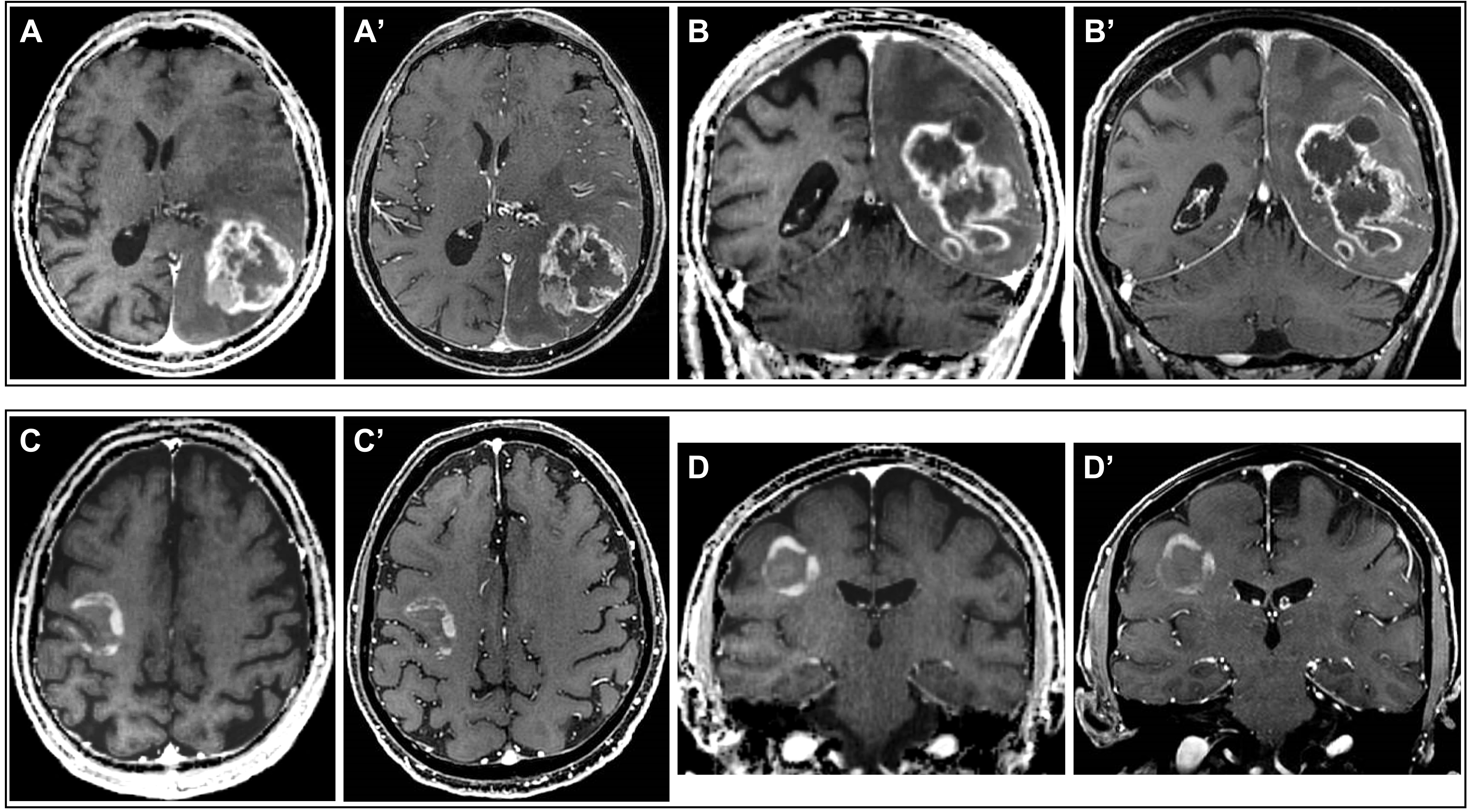
Figure 1. Postcontrast QTI-derived
synthetic (A-D) and conventional FSPGR (A’-D’) T1-weighted images in two patients
with pathology-confirmed
diagnosis of glioblastoma (upper row) and
diffuse astrocytoma (bottom row). In both cases synthetic images documented in
detail the non-homogeneous pattern of contrast enhancement.
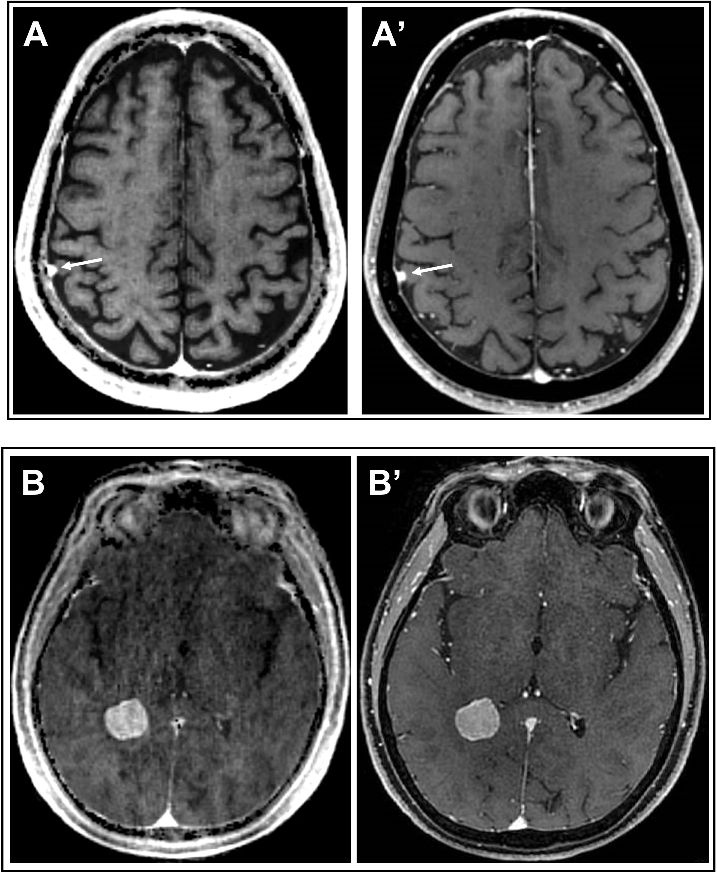
Figure 2. Postcontrast QTI-derived
synthetic (A, B) and conventional FSPGR (A’, B’) T1-weighted images in two
patients with meningioma (small-sized in one patient -upper row-, and intraventricular
medium-sized lesion in the other patient -bottom row-). Both homogeneously enhancing lesions are clearly visible on
synthetic images.
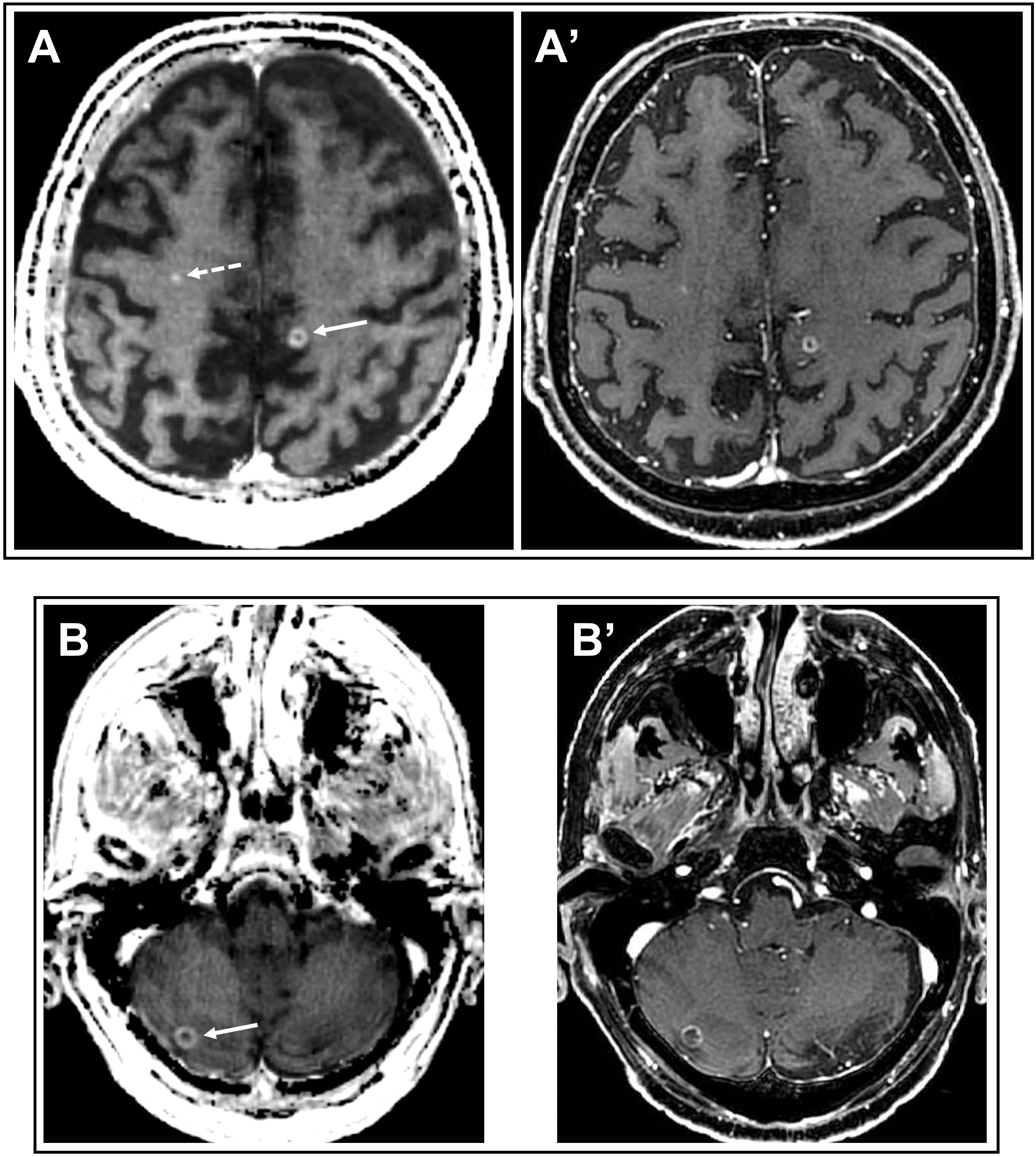
Figure 3. Postcontrast QTI-derived
synthetic (A, B) and conventional FSPGR (A’, B’) T1-weighted images in two
patients with lung cancer brain metastasis. Both ring-enhancing (solid arrows) and
punctiform homogeneously enhancing metastasis (dashed arrow in A) are revealed
by synthetic T1-weighted images.
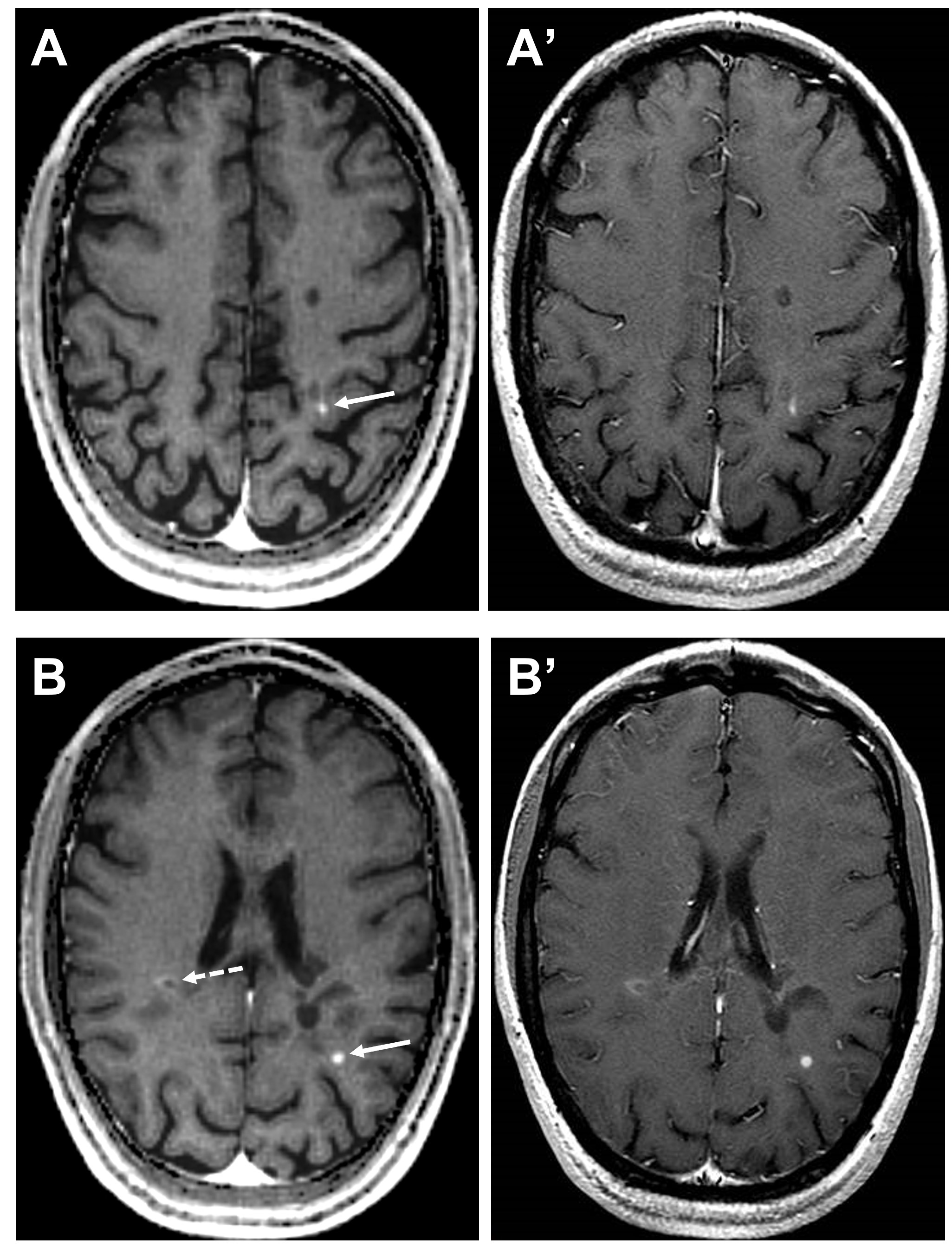
Figure 4. Postcontrast QTI-derived
synthetic (A, B) and conventional SE (A’, B’) T1-weighted images in a patient
with relapsing remitting multiple sclerosis. Small demyelinating lesions with
homogeneous (solid arrows) or ring-enhancement (dashed arrow in B) are clearly
visible in synthetic images.
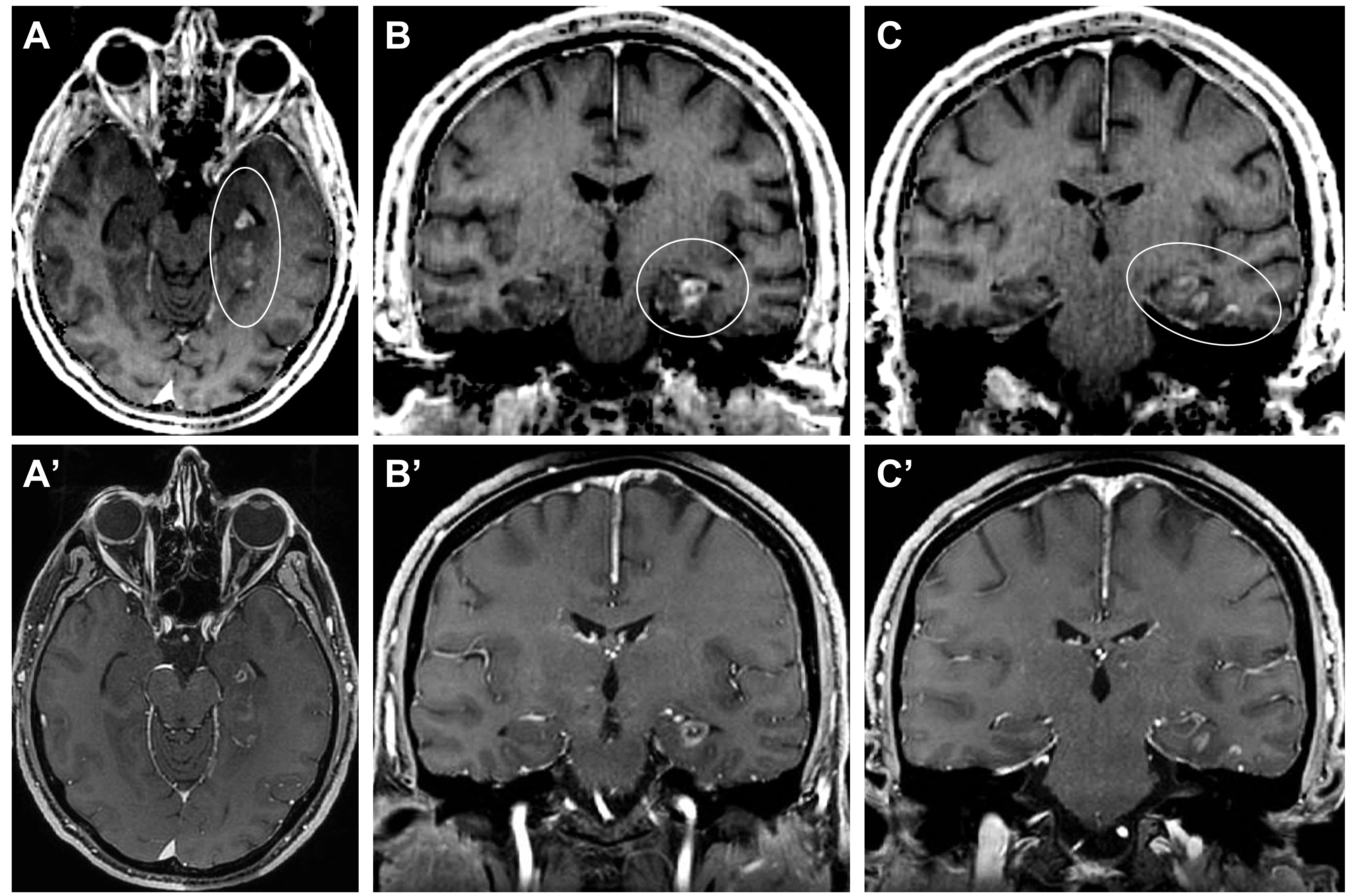
a patient with limbic encephalitis. Multiple
areas of gyral enhancement involve hippocampus, parahippocampal, fusiform and
inferior temporal giri of the left hemisphere.
DOI: https://doi.org/10.58530/2023/1717