1707
Feasibility of diffusion tensor imaging of the brachial plexus using iZOOM1Department of Radiology, Chiba University Hospital, Chiba, Japan, 2Diagnostic Radiology and Radiation Oncology, Graduate School of Medicine, Chiba University, Chiba, Japan, 3Philips Japan, Tokyo, Japan, 4Philips Healthcare (Shanghai) Ltd., Shanghai, China, 5Chiba University Hospital, Chiba, Japan
Synopsis
Keywords: Nerves, Diffusion Tensor Imaging
The brachial plexus is difficult to obtain diffusion tensor image (DTI) with good image quality due to the inhomogeneity of the magnetic field and motion artifacts. Zoom imaging based on 2D RF (iZoom) could both reduce the distortion dramatically and has better fat suppression effects. In this study, we compared DTI using conventional Zoom with that using iZoom for the brachial plexus and diffusion tractography with iZoom created higher mean length and fiber count than the conventional Zoom sequence, suggesting its feasibility as a new method for diffusion tensor imaging for the brachial plexus.Introduction
Diffusion tensor imaging (DTI) is a sequence that uses multiple diffusion-weighted images (DWI) with different directions of motion probing gradient (MPG). Since this sequence can produce anisotropy of water molecules, it can image nerve fibers indirectly. Although it has been used mainly in the brain, it has recently begun to be applied for the evaluation of peripheral nerves such as the sciatic nerve and brachial plexus.1 The brachial plexus is an area that is easily affected by peripheral neuropathy, so it is often the target of DTI. However, it is difficult to obtain DTI with good image quality due to the inhomogeneity of the magnetic field and motion artifacts. The reduced FOV (Zoom) method has been reported to be effective in reducing distortion.2 3 However, this method uses non-coplanar 90 and 180 selection slices with outer volume suppression, which theoretically suffers from crosstalk artifacts from neighboring slices. To avoid such artifacts, Zoom is basically applying interleaved scan packages for odd and even number slices, resulting in doubled scan time if the TR is kept constant. On the other hand, iZoom applies a tilted 2D Echo-Planar RF excitation with only tilting the k-space along the phase-encoding direction. iZoom is not suffering from crosstalk artifacts thanks to tilted 2D RF excitation, separation of scan packages is not needed, resulting in shorten the scan time compared to conventional Zoom. Furthermore, iZoom inherently has better fat suppression effects, is allows to use weaker (lower flip-angle) fat suppression pre-pulse, which reduces the magnetization transfer effects, hence leads to improve the signal-to noise ratio (SNR).456 In this study, we compared DTI using conventional Zoom with that using iZoom for the brachial plexus and investigated the feasibility of iZoom in the peripheral nerve.Methods
Nine volunteers: 2 women (age 26 ± 2.5 years) and 7 men (age 28.7 ± 2.8 years) with electrophysiological studies demonstrating the absence of peripheral neuropathy were examined on 3T MR unit (Ingenia 3T, Philips Healthcare). Scan parameters are shown in Figure 1. Diffusion-weighted axial images were acquired with a 16-channel torso array coil using the conventional Zoom and iZoom methods, respectively.1. Tractography measurement Fiber tracking was performed for nerve roots C6 , C7and spinal cord (using DTI FiberTrack application provided by Philips). The mean length (the average length in mm for all streamlines belonging to a fiber tract) and fiber count (the number of DTI streamlines extracted for a fiber tract) were compared. The settings for fiber tracking were as follows: maximum angle = 20°, minimum FA = 0.2.
2. Visual assessment Two independent blinded readers compared Zoom and iZoom by evaluating four ordinal (1 worst to 5 best) image quality criteria (distortion artifacts, ghosting and motion artifacts, quality of fat suppression, overall image). (Fig. 2)
Tractography measurement and visual assessment between Zoom and iZoom were compared using Wilcoxon signed-rank test.
Results
1. Mean lengths were longer for iZoom than Zoom (P<0.001), and fiber counts were higher for iZoom than Zoom (P<0.005).2. Three image quality criteria (Ghosting/Motion artifacts, quality of fat suppression, and overall image scores were rated significantly higher in iZoom than in Zoom for both readers (P <0.005 for reader1 and P = 0.005 for reader 2) (Fig. 3). The Distortion Artifact evaluation showed no significant difference between both sequences for both readers (reader 1, P = 0.052; reader 2, P = 0.681).
Discussion
The brachial nerve is susceptible to failure for fat suppression due to air distortion and motion artifacts from respiratory motion because it passes near the pulmonary apex. Zoom is applying interleaved scan packages, therefore prone to image misalignment due to motion. However, iZoom is not needed the separation of scan packages. iZoom had better quality of fat suppression than Zoom (Fig4). Therefore, the image misalignment and failure fat suppression may have influenced tractography (Fig5). iZoom can be an effective way to provide DTI with improved image quality, and a potential application for research and clinical diagnosis in diffusion imaging for the brachial plexus.Conclusion
In the brachial plexus, iZoom was feasible as a scan method of DTI in the peripheral plexus.Acknowledgements
No acknowledgement found.References
1. Ryckie G. , Alexander W, Irvin T et al. Diffusion tensor imaging of the roots of the brachial plexus:a systematic review and meta‑analysis of normative values. Clin Transl Imaging. 2020;8(6):419-431
2. Wilm BJ, Svensson J, Henning A, Pruessmann KP, Boesiger P, Kollias SS. Reduced field-of-view MRI using outer volume suppression for spinal cord diffusion imaging. Magn Reson Med. 2007 Mar;57(3):625-30. doi: 10.1002/mrm.21167.
3. Jeong, H., et al. High resolution human diffusion tensor imaging using 2D navigated multi-shot SENSE EPI at 7 Tesla. Magn Reson Med. 2013, 69 (3): 793-802.
4. Wu ZG, Zhang J, Fang WX, Huang F, B1 insensitive zoomed FOV imaging, ISMRM., 2015; 0953.
5. Banerjee S, Nishimura DG, Shankaranarayanan A, Saritas EU. Reduced field-of-view DWI with robust fat suppression and unrestricted slice coverage using tilted 2D RF excitation. Magn Reson Med. 2016 Dec;76(6):1668-1676. doi: 10.1002/mrm.26405.
6. Zhigang Wu et al. iZoom with 2 order flow compensated diffusion for Improving cardiac diffusion imaging: a preliminary study, ISMRM,2022;4137
Figures
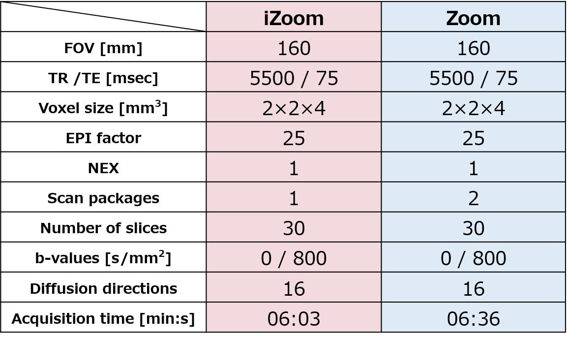
Figure 1. Scan Parameters
Scan parameters were set so that the acquisition time would be the same.
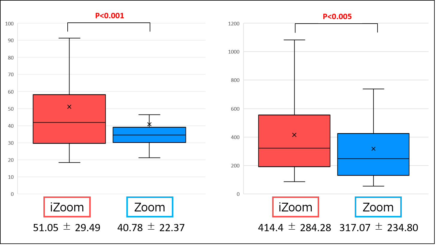
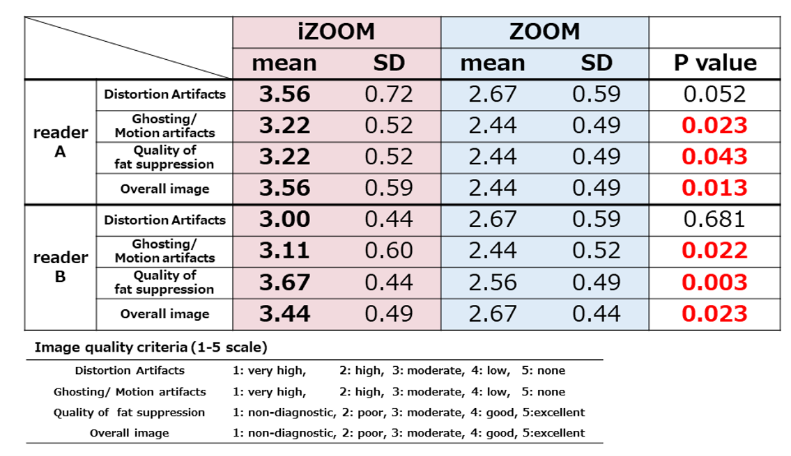
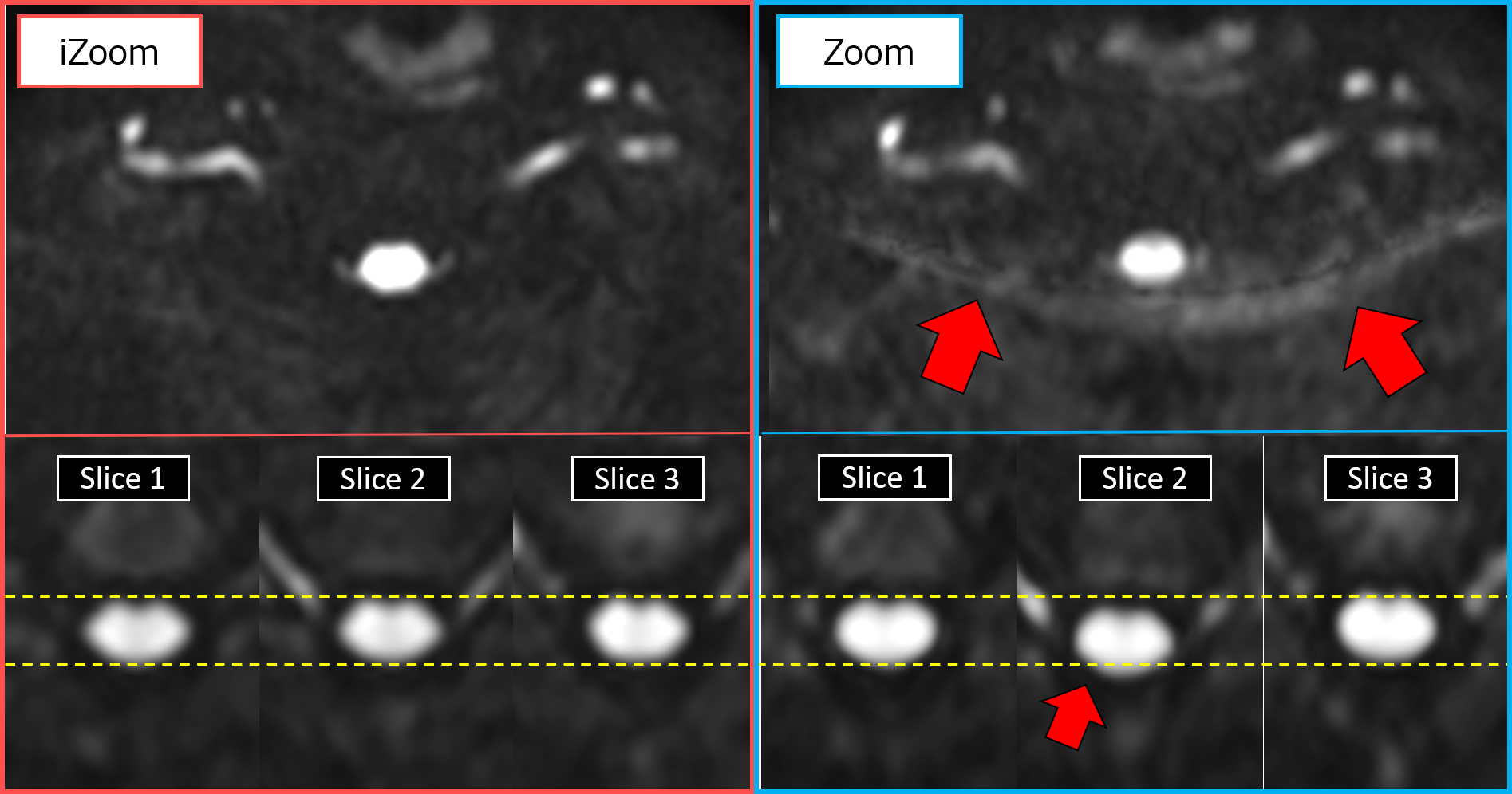
Figure 4. (Upper column) Cases of fat suppression failure. iZoom showed better fat suppression than Zoom. (Lower column) Cases of slices misalignment due to motion. Because Zoom applies interleaved scan packages, misalignment tends to occur from slice to slice.
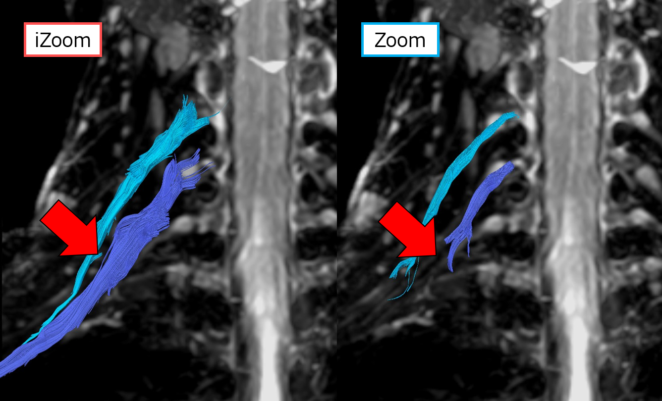
Figure 5. Tractography image of brachial nerve C6. The fiber lengths and counts were better in iZoom than Zoom.