1701
Magnetic resonance imaging of fetal aorta
LIN GANG1
1Xijing Hospital, Air Force Military Medical University, Xi'an, Shaanxi, China
1Xijing Hospital, Air Force Military Medical University, Xi'an, Shaanxi, China
Synopsis
Keywords: Vessels, Cardiovascular
Key words:fetal cardiovascular; coarctation of aortic arch;imaging technology
Prenatal ultrasound diagnosis has always been the first choice of screening for perinatal birth defects. However, due to the influence of many factors, the difficulty of diagnosis is increased, even for experienced experts, so there is an urgent need to find better supplements and alternatives. With the development of the treatment of cardiovascular diseases, many congenital aortic diseases can be alleviated or cured by perinatal surgery or elective surgery, so early diagnosis can reduce perinatal morbidity and mortality.the author has made some innovative explorations in the MRI scanning method of fetal cardiovascular.
Introduce
For abnormal changes in fetal vascular diameter, such as mild coarctation of the aorta, usually the blood flow velocity does not increase, and color Doppler blood flow imaging does not show aliasing. Therefore, the diagnosis of coarctation of the aorta needs to be combined with the long axis section of the aortic arch. Compared with the popularity of routine magnetic resonance examination methods for other parts of the fetus, fetal aortic arch examination has no efficient and accurate scanning method to realize its MRI long-axis profile display due to its unique anatomical structure, environment and inability to use gating and contrast agents.Method
MR examination was performed in 30 pregnant women who were 24 to 39 weeks of gestation and had the need for MR examination or suspected aortic isthmus coarctation by ultrasound. The multi-group concentric sector scanning localization technique, the negative interval scanning, the improved three-point localization technique and film sequence scanning were applied in fetal aortic arch angiography. According to the actual situation of the fetus during the scanning, one or more combinations of the technology above would be used to obtain the long axis profile of the fetal aortic arch.Image quality was evaluated using a 5-point scale by two senior radiologists (double blinding), which4-5 points means good, 3-5 points means meeting the diagnosis, and 1-2 points means not meeting the diagnosis. ICC values of two radiologists were calculated. The diameters of the ascending aorta, descending aorta, arch of the aorta and isthmus of the fetus were measured which was the average value measured by two radiologists.Results
The long axis profile of fetal aortic arch was successfully displayed in 27 of 30 cases, the success rate was 90.0%. The scanning time was 19.6±7.5min. The quality of all images met the diagnostic requirements. The good rate was 92.6% (25/27), with an average score of 4.1±0.5. The ICC value of image quality was 0.8 (95% CI: 0.5-0.9). MRI diagnosis showed that 18 cases of fetal aortic arch were narrowed, 9 cases of fetal aortic arch had no abnormality. The diameters of ascending aorta, descending aorta, arch and isthmus in 18 fetuses with stenosis were 4.1 ± 0.8 mm, 4.2 ± 0.5 mm, 4.3 ± 0.4mm and 2.8 ± 0.3mm, respectively. The diameters of ascending aorta, descending aorta, arch and isthmus in 9 normal fetuses were 4.1 ± 1.0 mm, 3.9 ± 0.7mm, 4.2 ± 0.8 mm and 3.4 ± 0.7mm, respectively. Fetal aortic arch isthmus and inner diameter of MR diagnosis had high consistency of 88.9% (24/27) with those by ultrasound diagnosis. Meanwhile, MR can also show the complete continuous circular bending (similar to "stick" of the long axis of the aortic arch section), which is helpful to observe the whole picture of fetal aorta development at different gestational ages.Discussion
Compared with foreign research, it also solves the problems of fetal cardiovascular scanning time and image quality, such as compressed sensing (CS)[2] technology and new open source image processing technology [3]to generate three-dimensional fine image of fetal heart, etc. We based on the characteristics of the fetus in magnetic resonance (NMR) long axis of the aortic arch section scanning methods, innovation sets the multiple sets of fan with center plane scan and improved positioning method scan, according to real-time situation of the fetus in the examination again, using the two methods or one of two kinds of combination, which can effectively obtain the high quality of the long axis of the fetal aortic arch section. Multi-group fan-shaped scan can obtain the sections of different deflection angles at one time with the same scanning time, which makes the inspection have real-time dynamic effect and can effectively avoid the scanning center deviation caused by fetal movement.Conclusion
The long axis section of fetal aortic arch can be obtained effectively by using the new MR scanning technique, which fills the gap that long axis section of fetal aortic arch is difficult to implement by two-dimensional MR scanning. This technique can make MR to be a good supplement of ultrasound examination and provide more intuitive and comprehensive imaging information for prenatal diagnosis.Acknowledgements
No acknowledgement found.References
[1] LIOYD DF, VAN A,MEROM JF, PUSHPARAJAH K,et al . An exploration of the potential utility of fetal cardiovascular MRI as an adjunct to fetal echocardiography[J]. Prenat Diagn, 2016, 36(10): 916 925. [2] ROY CW, SEED M, KINGDOM JC, MACGOWAN CKet al. Motion compensated cine CMR of the fetal heart using radial under sampling and compressed sensing [J]. J Cardiovasc Magn Reson.2017,19(1):29.[3] LIOYD DFA, PUSHPARAJAH K, et al. Three-dimensional visualisation of the fetal heart using prenatal MRI with motion-corrected slice-volume registration: a prospective, single-centre cohort study[J] .Lancet. 2019, 393(10181):1619-1627.Figures
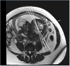
Figure 1. Axial scanning centers of multiple groups of same-center sector surfaces;
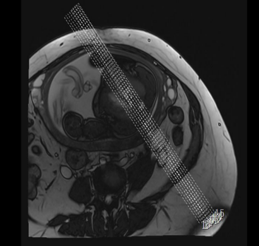
Figure 2 .Scanning directions of multiple groups of same-center sector plane
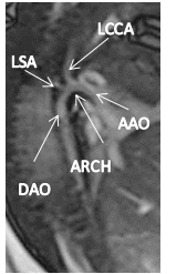
Figure 3 shows the complete long axis section of the aortic arch (crutch + three hairs) DAO: descending aorta;; ARCH: arch of the aorta; LSA: left subclavian artery; LCCA: left common carotid artery; AAO: ascending aorta;
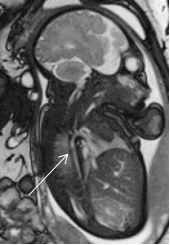
Figure 4. Long axis of aortic arch (bright blood) of fetus at 33 weeks of gestation;
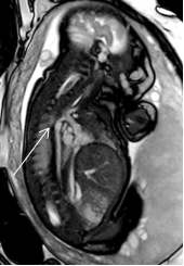
Figure 5. Aortic arch stenosis (bright blood) at 35 W;
DOI: https://doi.org/10.58530/2023/1701