1697
Non-Contrast Carotid Artery Imaging using 3D TOF and Time-SLIP bSSFP with centric ky-kz k-space trajectory.
Vadim Malis1, Won Bae1,2, Yoshimori Kassai3, Marin A Mcdonald1, and Mitsue Miyazaki1
1Radiology, UC San Diego, San Diego, CA, United States, 2VA San Diego Healthcare System, San Diego, CA, United States, 3Canon Medical, Ōtawara-shi, Japan
1Radiology, UC San Diego, San Diego, CA, United States, 2VA San Diego Healthcare System, San Diego, CA, United States, 3Canon Medical, Ōtawara-shi, Japan
Synopsis
Keywords: Vessels, Vessels, Non-Contrast MRA
Carotid artery stenosis (CAS) is one of the most common causes of acute ischemic stroke. MRA techniques are routinely used for the diagnostics of CAS, yet one of their drawbacks remains as a long scanning time. In this study we acquired and compared two sets of images of carotid arteries: 3D time-of-flight (TOF), time spatial labeling inversion pulse (Time-SLIP) balanced steady-state free precession (bSSFP) and their accelerated versions that utilize centric ky-kz acquisition pattern (FAST3D).Introduction
Carotid artery stenosis (CAS) is a frequent disease seen in general practice. It is one of the most common causes of acute ischemic stroke, accounting for around 20% of occurrences. [1] Thus, it is critical to diagnose, prevent, and treat it early. In general, the combination of non-contrast MRA and plaque imaging like black blood is utilized for the diagnosis of CAS. Non-contrast MRA techniques are routinely utilized for CAS screening, [2] yet among the challenges is a long scan time. In this study, we compared the depiction of the carotid artery using: 3D time-of-flight (TOF) and flow-in 3D balanced steady-state free precession (bSSFP) with time spatial labeling inversion pulse (Time-SLIP).[3, 4] Both techniques were scanned using 3D MRA with and without centric ky-kz acquisition pattern (FAST3D) [5].Methods
Four healthy (51 ± 13 years) subjects were scanned on a clinical 3T scanner (Vantage Galan 3T, Canon Medical Systems, Japan) after signing an IRB-approved consent form. Images were acquired using an Atlas Head and Neck SPEEDER coil. The scanning protocol consisted of the following series: (i) 3D TOF and (ii) 3D TOF FAST3D both with 11 slabs covering the aortic arch to middle cerebral arteries without gating; (iii) 3D bSSFP and (iv) 3D bSSFP FAST3D; scanned with PPG gating and Time-SLIP tag pulse (TI=1200ms) applied to the carotid artery region in the coronal orientation. Maximum intensity projection (MIP) images were reconstructed for all the series and further improved using a Hessian-based Frangi filter [6]. For Time-SLIP images spinal cord was manually cut out of the volume prior to filtering. Detailed acquisition parameters are shown in Table 1.Results
Figure 1 shows MIP in axial, coronal, and sagittal orientation of regular and FAST3D TOF series. The arteries are well depicted with FAST3D sequence without compromising the image quality. Note a tremendous scan time reduction in TOF FAST3D for 11-slab acquisition from the neck to middle cerebral arteries in 3:40 minutes in Table 1. Figure 2 shows the vessel structures extracted automatically using the Frangi filter. MIP images in coronal orientation for Time-SLIP with 3D bSSFP and zoomed-in carotid artery regions are shown in Figure 2. Similar image quality between standard and accelerated sequences can be visually appreciated. Again, tremendous scan time reduction (less than 2 minutes) in Time-SLIP 3D bSSFP with FAST3D, as shown in Table 1.Discussion
Acquisition windows of FAST3D are extended due to the filling of ky-kz k-space trajectory, which causes blurring around the aortic arch. We have increased the parallel imaging reduction factor (SPEEDER) to reduce the acquisition window. Both images of 3D TOF and Time-SLIP bSSFP with FAST3D show compatible image quality, as compared to those without FAST3D, yet achieving substantial scan time reductions.Conclusion
Both TOF and Time-SLIP bSSFP with FAST3D provide remarkable scan time reduction with good image quality.Acknowledgements
This work was supported by Canon Medical Systems, Japan (grant 35938).References
[1] Arasu R., et. al., Aust J Gen Pract. 50(11) (2021).[2] Westwood ME., et. al., BMJ. 324(7331):198 (2002).
[3] Miyazaki M., et. al., JMRI 35:1-19 (2012).
[4] Miyazaki M., et. al., EJR 80:9-23 (2011).
[5] Busse RF., et. al., MRM 60:640-649, (2008).
[6] Frangi., et. al., MICCAI. 1(1496) (1998).
Figures
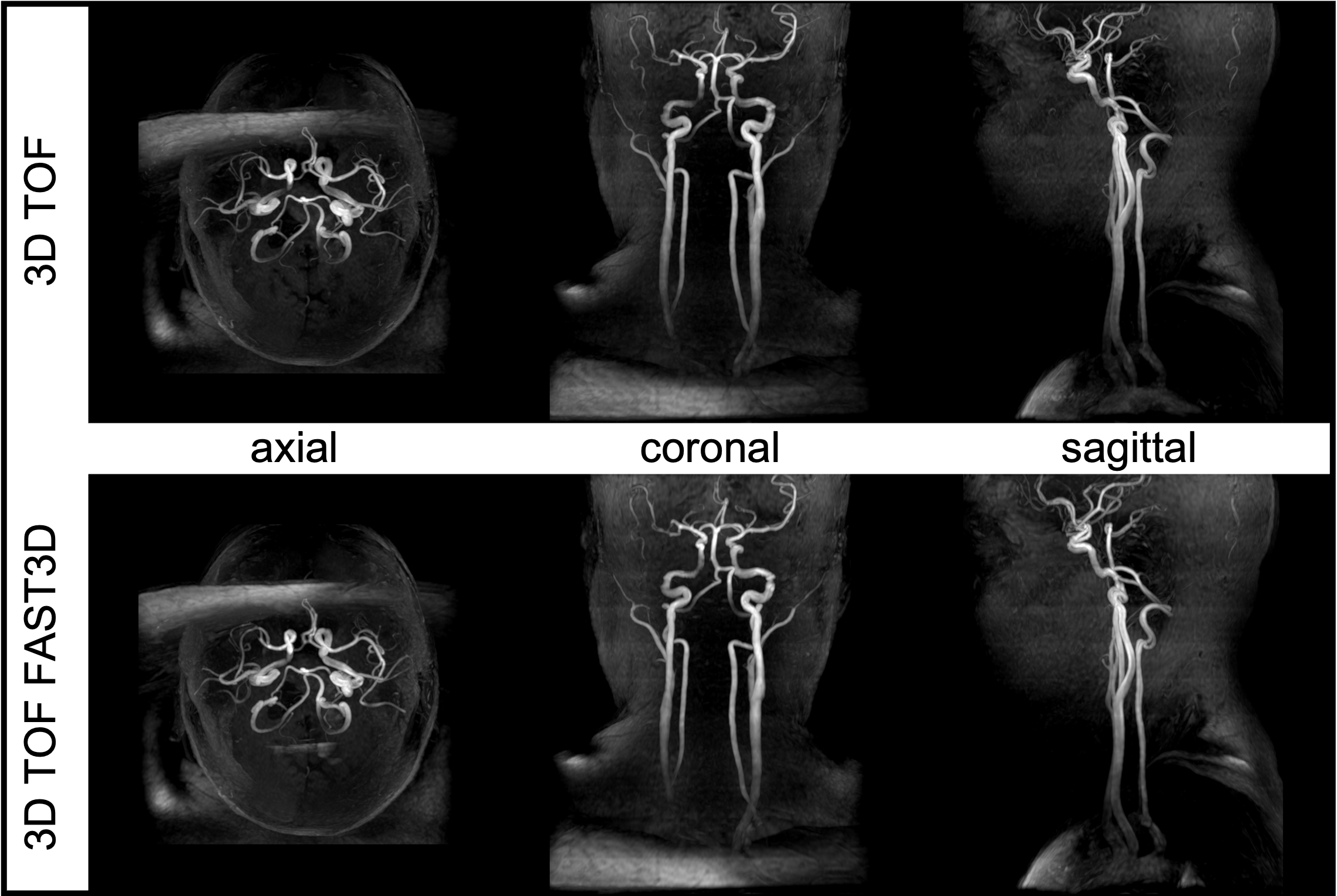
Figure 1: MIP images of axial, coronal, and sagittal orientations for standard (top row) and FAST3D TOF series without eliminating unwanted signals.
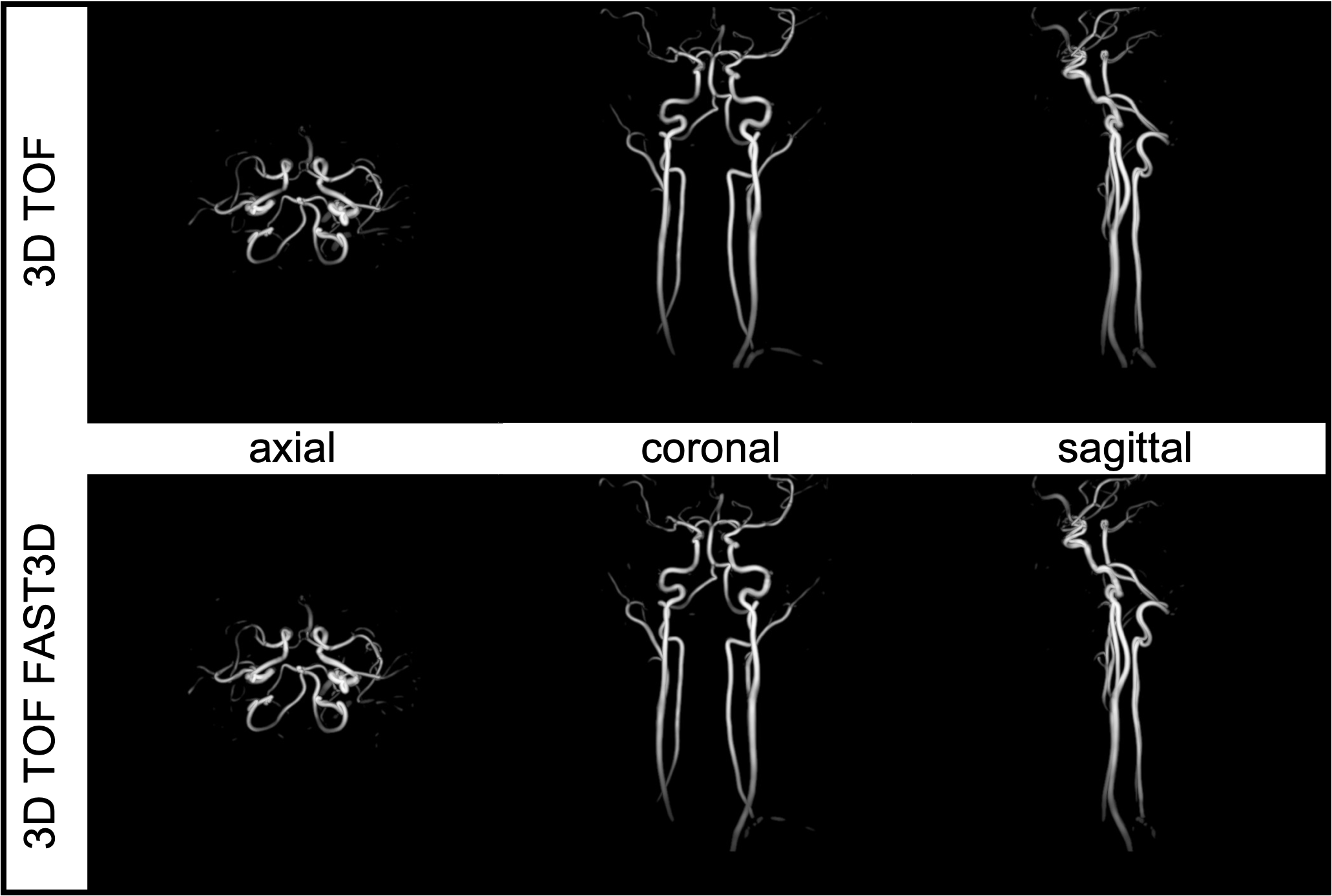
Figure 2: MIP images of carotid arteries and vessel structures extracted using the Frangi filter from the TOF (top) and TOF FAST3D (bottom) image series.
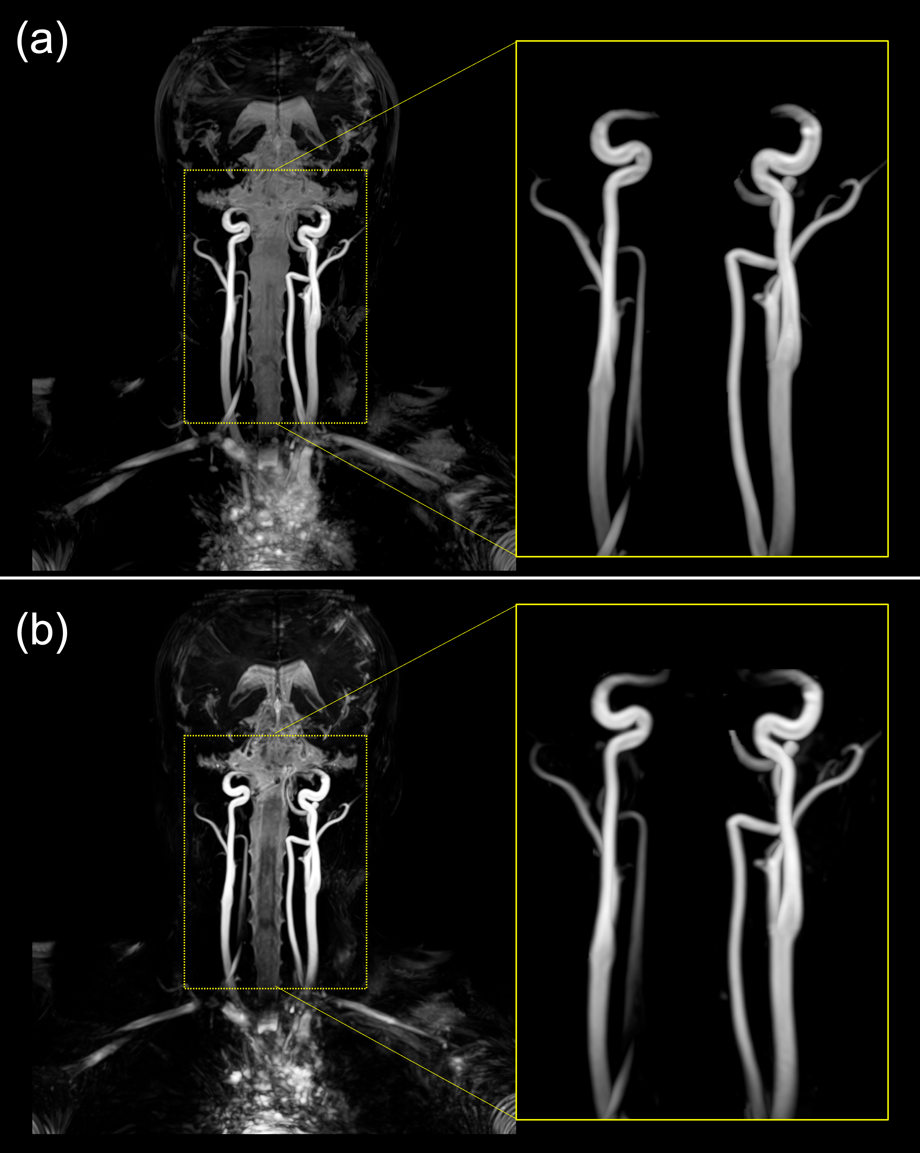
Figure 3: MIP images of the flow-in Time-SLIP 3D bSSFP images acquired with the regular sequence (a) and FAST3D (b) demonstrating similar quality.
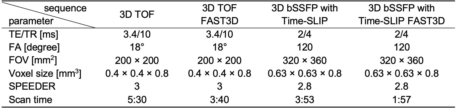
Table 1: Scanning parameters
DOI: https://doi.org/10.58530/2023/1697