1689
Ferumoxytol-Enhanced 5D ROCK-MUSIC with Low-rank Tensor Reconstruction: Initial Feasibility in Pediatric Congenital Heart Disease
Zixuan Zhao1, Hsu-Lei Lee2, Guowen Shao1, Zhengyang Ming1, Fei Han3, Dan Ruan1, Anthony Christodoulou2, J. Paul Finn1, and Kim-Lien Nguyen1,4
1University of California, Los Angeles, Los Angeles, CA, United States, 2Cedars-Sinai Medical Center, Los Angeles, CA, United States, 3Siemens Health Solutions, Los Angeles, CA, United States, 4VA Greater Los Angeles Healthcare System, Los Angeles, CA, United States
1University of California, Los Angeles, Los Angeles, CA, United States, 2Cedars-Sinai Medical Center, Los Angeles, CA, United States, 3Siemens Health Solutions, Los Angeles, CA, United States, 4VA Greater Los Angeles Healthcare System, Los Angeles, CA, United States
Synopsis
Keywords: Heart, Cardiovascular, Congenital Heart Disease, Ferumoxytol
ROCK-MUSIC is a 4D whole heart MRI pulse sequence that has shown promise for fast imaging in pediatric congenital heart disease (CHD) without dependence on the ventilator respiratory gating signal. Recent research in cardiac multitasking with low rank tensor (LRT) reconstruction allows for decomposition of cardiorespiratory motion. In this preliminary work, we showed that LRT reconstruction can be applied to the ROCK-MUSIC acquisition to enable higher temporal resolution with cardiorespiratory resolved imaging. When compared to L1-ESPIRiT, the LRT approach is able to achieve comparable image quality.Introduction
Researchers have proposed an accelerated 4D-MRI technique that leverages incoherent, rotating Cartesian k-space sampling with ferumoxytol enhancement (ROCK-MUSIC), which has shown considerable potential in pediatric congenital heart disease (CHD).1 As originally developed, ROCK-MUSIC employs a combined compressed sensing and parallel imaging reconstruction (ESPIRiT) for multiphase, whole-heart imaging.2 However, respiratory motion and high temporal resolution remain challenging. Cardiac multitasking for multidimensional imaging conceptualizes cardiac and respiratory phases as different time dimensions by using low rank tensor (LRT).3 We hypothesize that LRT reconstruction can be used with ROCK-MUSIC acquisition to enable cardiorespiratory motion resolved 5D-imaging of pediatric CHD patients.Methods
ROCK-MUSIC MRI was implemented based on a 3D gradient-recalled-echo (GRE) sequence to generate the rotating Cartesian spiral-in pattern. The retrospective data was obtained from eight pediatric patients (age range, 5 days to 3.1 years old) with known CHD. The patients were scanned on a clinical 3.0T magnet (Magnetom TIM Trio®, Siemens Medical) ([TE/TR] = 1.2ms/2.9ms, 0.8-1.1mm3 isotropic resolution, TA = 4.35-6.12min, flip angle=19°) during the steady-state intravascular distribution of ferumoxytol. All the data were reconstructed using ESPIRiT approach. For LRT reconstruction, the acquired self-gating (SG) lines in the ROCK pattern were used for respiratory and cardiac binning where the number of respiratory and cardiac bins are 6 and 20 respectively. We evaluated the image quality between the two reconstruction techniques using a Likert score of 1-4, with 4 denoting best image quality (cardiac chamber boundaries strongly delineated with homogenous signal in chamber lumen on all phases, fine detail visible on most or all phases) and little or no image artifacts. We used a stack of 2D short axis (SA) slices to quantify the contrast to noise ratio (CNR). The slope of the blood-myocardium edge at the interventricular septum was quantified to assess the sharpness of the images. A larger slope equates to a sharper image. Left ventricular end-diastolic volume (EDV), end-systolic volume (ESV), and left ventricular ejection fraction (LVEF) were derived from each strategy and compared using Bland-Altman analysis. CNR was computed in the septum and ventricular blood pool.Results
For each study, 6 respiratory x 20 cardiac = 120 cardiorespiratory phases were reconstructed using the LRT approach and 9 cardiac phases were generated from ESPIRiT approach. Fig. 1 provides an illustration of 5D ROCK-MUSIC reconstructed using LRT where the systolic and diastolic views in 6 respiratory phases are presented. Fig. 2 shows a comparison of cine image quality across several phases using both reconstruction methods for two patients. Relative to ESPIRiT reconstructions, LRT showed comparable diaphragmatic and cardiac boundary sharpness. Fig. 3 presents the edge-sharpness function (ESF) and the modulation transfer function (MTF) analyses for a 2-month-old female patient. The quantitative ESF and MTF confirmed the comparable qualitative depiction of image quality. The mean Likert score of the ESPIRiT and LRT are 3.6 and 3.3 (p=0.77), respectively. One LRT reconstruction failure was considered an outlier and may have been caused by complex motion patterns reflecting the motion binning. The mean sharpness slope values at the septum were 0.54 ±0.09 and 0.59±0.17 for ESPIRiT and LRT methods (p=0.60), respectively. Based on Bland-Altman analysis of ventricular cardiac function, the mean bias values between ESPIRiT and LRT were minimal (LVEDV 0.6%, LVESV 0.1%, and LVEF 0.1%). LRT reconstructions showed a significant improvement in CNR over ESPIRiT reconstructions by an average of 14.9±0.4%.Discussion and Conclusion
In this preliminary study with a small cohort, we showed early feasibility for using LRT reconstruction with ROCK-MUSIC MRI acquisition to enable cardiac and respiratory phase-resolved images in pediatric CHD. Although ROCK-MUSIC acquisitions were designed to depict cardiac phase-resolved images using the ESPIRiT reconstruction method, the flexibility of the ROCK-MUSIC acquisition allows for LRT reconstruction. LRT enabled higher temporal resolution with 120 cardiorespiratory phases while maintaining comparable image quality to ESPIRiT. Further confirmation in a larger cohort will be needed to fully assess the reliability of LRT for multidimensional image reconstruction.Acknowledgements
No acknowledgement found.References
[1] Han, F.; Rapacchi, S.; Khan, S.; Ayad, I. et al. Four-dimensional, multiphase, steady-state imaging with contrast enhancement (MUSIC) in the heart: a feasibility study in children. Magn Reson Med, 74, n. 4, p. 1042-1049, Oct 2015.[2] Uecker, M.; Lai, P.; Murphy, M. J.; Virtue, P. et al. ESPIRiT--an eigenvalue approach to autocalibrating parallel MRI: where SENSE meets GRAPPA. Magn Reson Med, 71, n. 3, p. 990-1001, Mar 2014.[3] Christodoulou, A. G.; Shaw, J. L.; Nguyen, C.; Yang, Q. et al. Magnetic resonance multitasking for motion-resolved quantitative cardiovascular imaging. Nat Biomed Eng, 2, n. 4, p. 215-226, Apr 2018.Figures
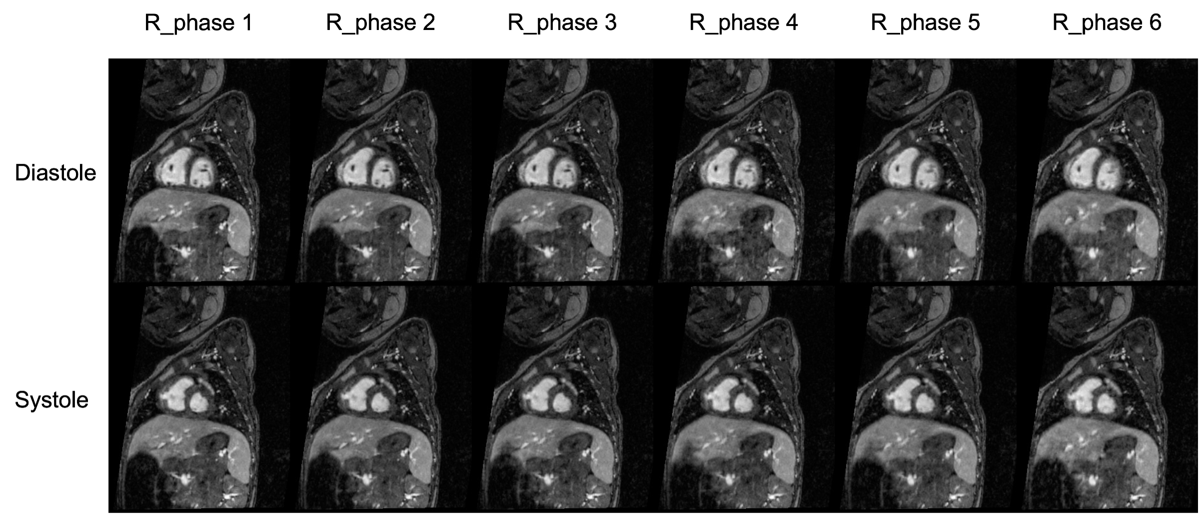
5D ROCK-MUSIC images using low-rank tensor reconstruction. Six respiratory phases are reconstructed where each respiratory phase contains 20 cardiac phases. Systolic and diastolic images for six respiratory phases are shown for an 11-month-old female patient (weight 6.67 kg) with complex congenital heart disease.
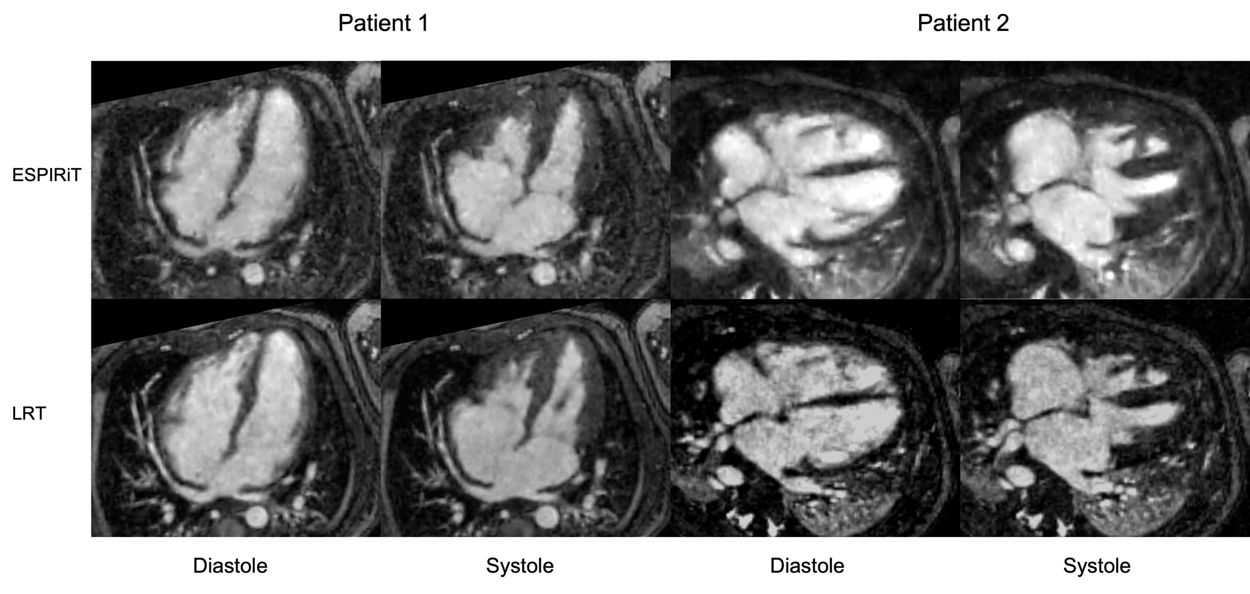
Comparison of ESPIRiT and low-rank tensor (LRT) reconstructued cine images for two patients in 4-chamber view at end-systole, and end-diastole.
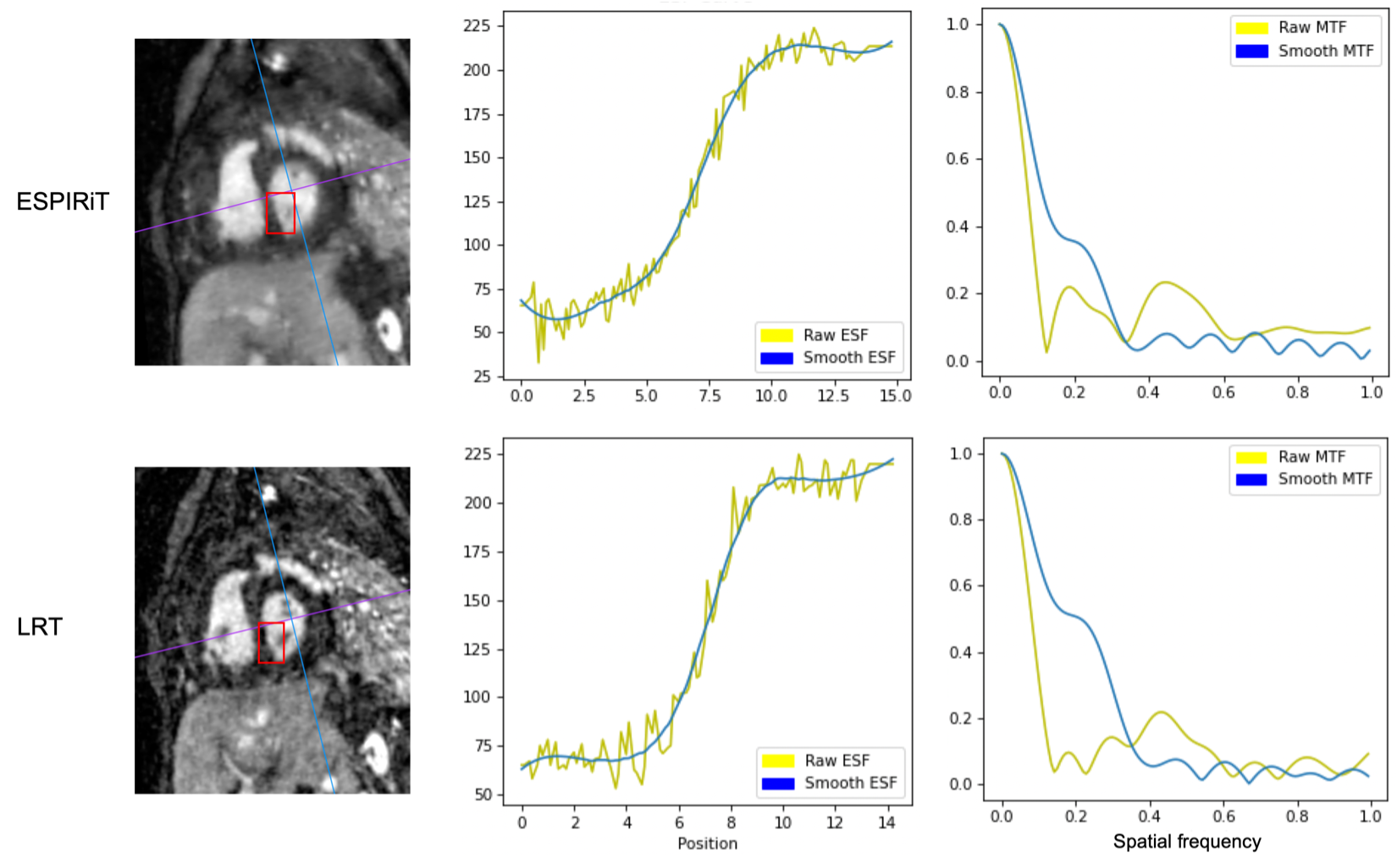
Comparison of the edge-spread function (ESF) and the modulation transfer function (MTF) between ESPIRiT and low-rank tensor (LRT) reconstruction. A short-axis mid-diastolic view from a female patient (2-month old, 3.77 kg) was used for ESF and MTF analysis, which support the sharpness and resolution consistency of the two reconstruction methods.
DOI: https://doi.org/10.58530/2023/1689