1672
Application of fractional order calculus diffusion model in predicting the differentiation in esophageal squamous cell carcinoma
Keke Zhao1, Shaoyu Wang2, Hongkai Zhang1, Jinrong Qu1, and Hailiang Li1
1Henan Tumor Hosptial, Zhengzhou, China, 2MR Scientific Marketing, Siemens Healthineers, Shanghai, China
1Henan Tumor Hosptial, Zhengzhou, China, 2MR Scientific Marketing, Siemens Healthineers, Shanghai, China
Synopsis
Keywords: Cancer, Cancer, esophageal squamous cell carcinoma
A total of 50 patients with locally advanced ESCC were prospectively enrolled in this study. Diffusion Weighted Imaging (DWI) with multiple high b-values was performed before surgery, and a new set of parameters (D, β and m) from a fractional order calculus (FROC) diffusion model were acquired. The grade of differentiation about ESCC was assessed. We found that β and D value exhibited non-significant difference, and m value showed a significant difference between different grades ESCC (P =0.029).Introduction
To explore the value of a new set of parameters (D, β and m) from a fractional order calculus (FROC) diffusion model in predicting the grade of differentiation in esophageal squamous cell carcinoma (ESCC)[1, 2].Methods
This study was approved by the Hospital Ethics Committee, and the written informed consent form was waived. A total of 50 patients with ESCC were prospectively enrolled from October 2020 to September 2021, and the grad of differentiation was confirmed by postoperative pathology. MR scanning was performed within 1 week before surgery on a 3T MR scanner(MAGNETOM Skyra, Siemens Healthineers, Erlangen, Germany), DWI sequence was performed with 12 b-values(b, 0, 50, 100, 150, 200, 400, 500, 600, 800, 1000, 1500, 2000s/mm2). DWI parameters map from fractional order calculus (FROC) diffusion model were reconstructed calculated using a unified in-house developed software called BoDiLab, which is based on Python 3.7. The parameters included apparent diffusion coefficient (D), fractional order parameter b (which correlates with tissue heterogeneity), and a microstructural quantity m [3]. The degree of differentiation of low-, middle- and high-grade were assessed, and these values were compared using the one-way ANOVA between the low, middle and high-grade tumor groups, respectively.Results
Twenty-two patients were identified as low grade, 23 as middle grade, and 5 as high grade by pathology. D, β and m of different grade groups were shown in Table 1, and only m showed significant difference between different grade groups (P =0.029). Patients were stratified into high group and middle & low group, ROCs of D, β and m for high group and middle & low group were shown in Table 2. m (0.756) had a higher AUC than both D (0.627) and β (0.533).Conclusion
Among the new set of parameters in high b-value DWI, m may be used to predict the grade of differentiation in ESCC patients.Discussion
Our study found a new set of parameters from a fractional order calculus (FROC) diffusion model may be used as an effective functional imaging technique to predict the grade of differentiation in ESCC patients, and the m value showed a significant difference between different grades ESCC (P=0.029). The m value has been suggested as a measure of diffusion mean free length, but its biologic interpretation remains intriguing[3].Acknowledgements
No acknowledgement found.References
1. Song, T., et al., The value of intravoxel incoherent motion diffusion-weighted imaging in predicting the pathologic response to neoadjuvant chemotherapy in locally advanced esophageal squamous cell carcinoma. Eur Radiol, 2021. 31(3): p. 1391-1400.
2.Lu, Y., et al., The value of GRASP on DCE-MRI for assessing response to neoadjuvant chemotherapy in patients with esophageal cancer. BMC Cancer, 2019. 19(1): p. 999.
3. Sui, Y., et al., Differentiation of Low- and High-Grade Pediatric Brain Tumors with High b-Value Diffusion-weighted MR Imaging and a Fractional Order Calculus Model. Radiology, 2015. 277(2): p. 489-96.
Figures
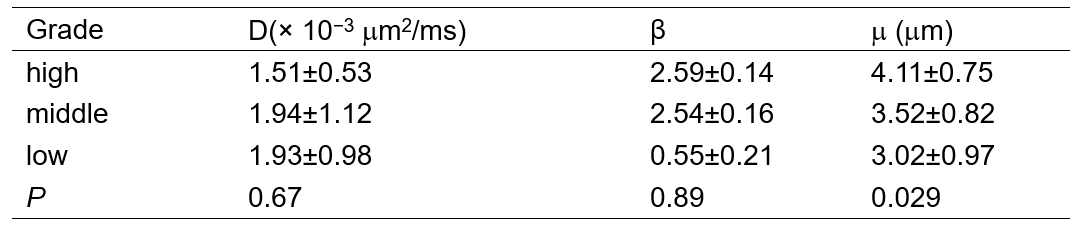
Table 1. D, β and m of different grade groups
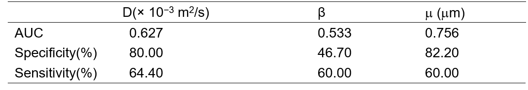
Table 2. ROCs of D, β and m for high group and middle
& low group
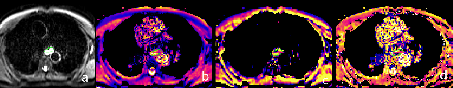
Figure1: 57y, male, ESCC. a: b0 map; b: FROC_D; c: FROC_β;d:FROC_μ
DOI: https://doi.org/10.58530/2023/1672