1666
Standardized volume and IVIM parameters of spleen in patients with acute leukemia: the value of evaluating tumor burden and prognosis1The Second Hospital of Shanxi Medical University, Taiyuan, China
Synopsis
Keywords: Cancer, Diffusion/other diffusion imaging techniques, spleen
This study investigated the values of standardized volume and intravoxel incoherent motion (IVIM) parameters of spleen in evaluating tumor burden and prognosis in newly diagnosed acute leukemia (AL). Eighty-five AL patients and seventy-four healthy volunteers were recruited and underwent IVIM in the abdomen on a 3.0T MRI system. The results showed standardized volume and IVIM parameters of spleen were associated with tumor burden and treatment response in AL, and 218.1cm3 was the threshold of standardized spleen volume for predicting treatment response, which indicated that they can be potentially useful in tailoring the individualized treatment plan for each patient.
INTRODUCTION
Acute leukemia (AL) is considered as metastatic disease, which remains challenging to treat1. The spleen is a frequent organ of leukemia metastasis (up to 80% of patients on autopsy), and the common clinical manifestation is splenomegaly 2. Splenomegaly, raised serum lactate dehydrogenase (LDH) and high peripheral white blood cell (WBC) counts are clinical markers of high tumor burden, which are related to poor prognosis in AL3,4.At present, spleen palpation is a simple and brief clinical evaluating test for the volume of spleen. However, the spleen can be palpable only when it is two to three times its normal size. Some imaging techniques (ultrasound, computed tomography) used to assess spleen morphology, but spleen volume (SV) depend on individual factors. Although splenomegaly is a useful qualitative indicator for evaluating the tumor burden of AL, it is necessary to study the value of quantitatively standardized SV in assessing tumor burden and further determine a standardized volume threshold in regards to prognosis for guiding clinical practice precisely.
Functional tumor burden based on imaging can assess the function and activity of tumor, which has been regarded as an effective supplement to tumor volume burden in recent years5,6. Diffusion-weighted imaging (DWI) has been applied in assessing the spleen in hematologic malignancies, such as multiple myeloma and lymphomas7-9. Intravoxel incoherent motion (IVIM) is a DWI-based method with multiple b values and assumes a bi-exponential relationship between the signal intensity and the b value, which would enable quantitative parameters separately reflect tissue water diffusion related to tissue cellularity (pure diffusion coefficient [D]) and tissue microcapillary perfusion (pseudo-diffusion coefficient [D*], represented the rate of microcapillary blood flow; pseudo-perfusion fraction [f], represented the fraction of vascular volume)10. We hypothesis IVIM parameters could be used to reflect pathophysiologic changes of spleen based on variation of cellularity and angiogenesis in patients with AL.
In this study, we sought to study whether the standardized volume and IVIM parameters of spleen were associated with tumor burden and their values of evaluating treatment response in newly diagnosed AL.
METHODS
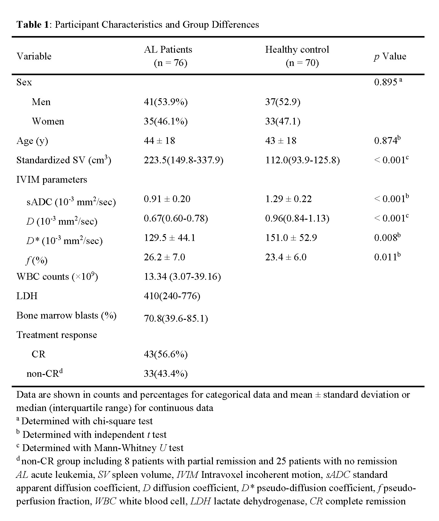
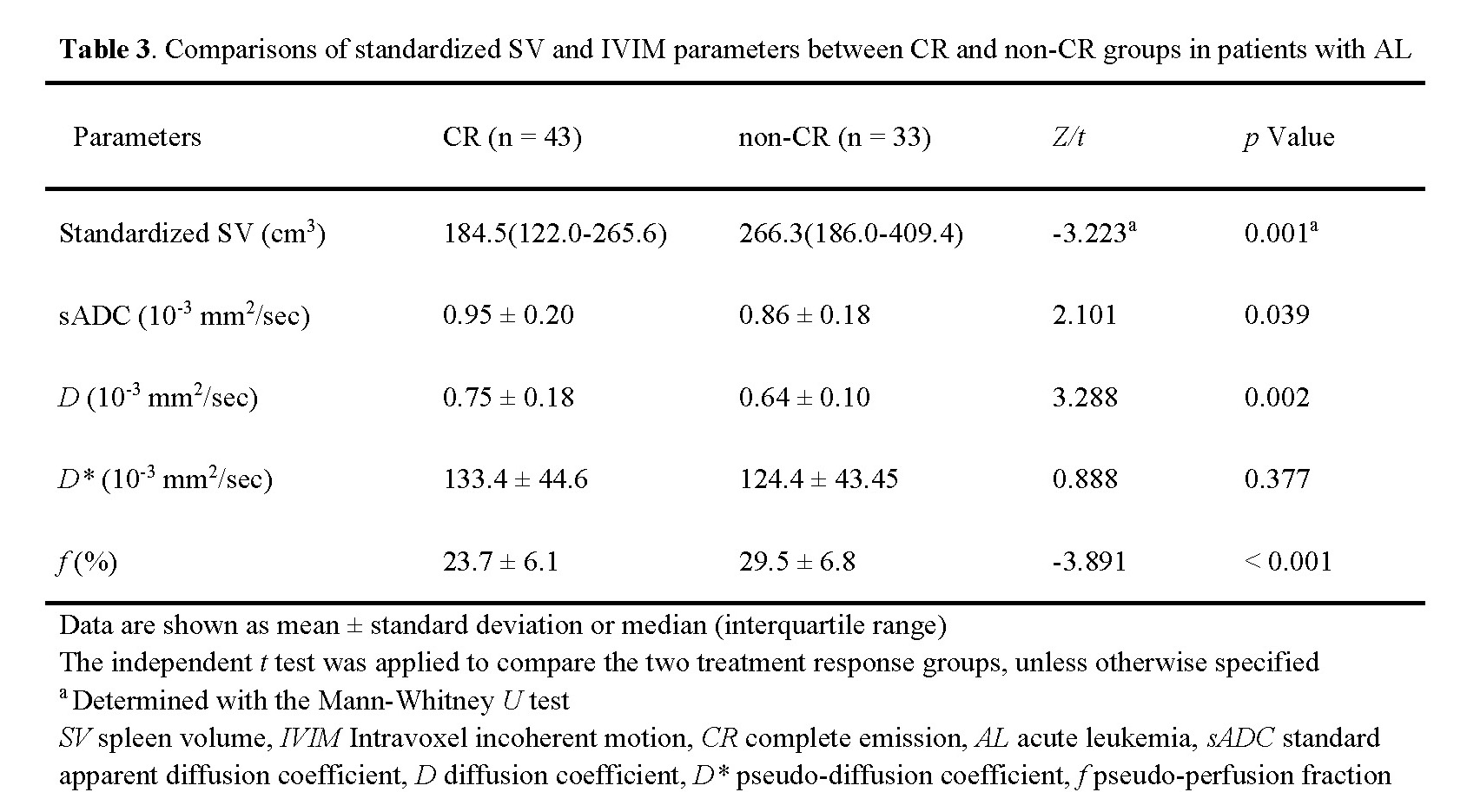
This prospective study enrolled patients with newly diagnosed AL and healthy volunteers. All participants underwent IVIM DWI of the abdomen. The standardized volume and IVIM parameters (standard apparent diffusion coefficient [sADC]; pure diffusion coefficient [D]; pseudo-diffusion coefficient [D*]; and pseudo-perfusion fraction [f]) of spleen were measured by two radiologists. Patients’ clinical biomarkers of tumor burden including WBC counts, LDH and bone marrow blasts were collected. AL patients were divided into complete remission (CR) and non-CR group according to the response after the first induction chemotherapy. Statistical evaluations were performed by using independent t test, Mann-Whitney U test, Spearman correlation, multivariate logistic regression and ROC curve.RESULTS
76 AL patients (CR: n=43; non-CR: n=33) and 52 healthy volunteers were evaluated. Standardized spleen volume (SV), sADC, D and f were significantly different between AL patients and healthy volunteers, which were correlated with the clinical tumor burden, and significantly different between CR and non-CR groups (all p<0.05). 218.1cm3 was the threshold of standardized SV for predicting treatment response. D and f were the independent predictors (hazard ratio=0.004, p=0.011; hazard ratio=1.141, p=0.002, respectively), and their combination achieved an AUC of 0.805 in predicting treatment response.DISCUSSION
Compared with qualitative morphological assessment of spleen size (whether the spleen is enlarged), the standardized SV excluding the effect of individual difference can be used to accurately quantify the severity of splenomegaly. Our results demonstrated larger standardized SV was correlated with higher WBC counts and LDH. Simultaneously, our study found that sADC, D and f values showed significant correlation with clinical biomarkers of tumor burden. Thus, standardized SV and IVIM parameters can probably be used to assess tumor burden quantitatively. Prior studies have reported the probable reason of splenomegaly include: (1) Increased cellularity with more blasts which enter the spleen through large and fenestrated sinusoidal vessels in the red pulp1. (2) Pathological angiogenesis of leukemic spleen, which can promote the rapid expansion of malignant blasts in the spleen and the progression of the disease11. In our study, larger standardized SV was correlated with lower sADC, lower D and higher f values, furtherly indicating sADC, D (cellularity) and f (angiogenesis) may reflect the changes of pathophysiology in splenomegaly.IVIM parameters showed significant differences between AL patients and healthy volunteers in our study. In addition, the lower sADC and D values indicated an unfavorable treatment response, which may reflect the diffusion disorder caused by hypercellularity. The f value from IVIM estimates perfusion by using flowing blood as an internal marker, which is more applicable for clinical practice [12]. Our study suggested that higher f value tended to show an unfavorable treatment response after the first induction chemotherapy in AL. Furthermore, the results showed that f and D values of spleen were independent predictors of treatment response.
CONCLUSION
Standardized volume and IVIM parameters of spleen may be viable imaging markers of tumor burden and can be used to evaluate prognosis in newly diagnosed AL.Acknowledgements
No acknowledgement found.References
1. Whiteley AE, Price TT, Cantelli G, et al. Leukaemia: a model metastatic disease. Nat Rev Cancer. 2021;21(7):461-475.
2. Viadana E, Bross ID, Pickren JW. An autopsy study of the metastatic patterns of human leukemias. Oncology. 1978;35(2):87-96.
3. Ma S, Shi Y, Pang Y et al. Notch1-induced T cell leukemia can be potentiated by microenvironmental cues in the spleen. J Hematol Oncol. 2014;7:71.
4. Smith ML, Hills RK, Grimwade D. Independent prognostic variables in acute myeloid leukaemia. Blood Rev. 2011;25(1):39-51.
5. Lee EYP, An H, Perucho JAU, et al. Functional tumour burden of peritoneal carcinomatosis derived from DWI could predict incomplete tumour debulking in advanced ovarian carcinoma. Eur Radiol. 2020;30(10):5551-5559.
6. Dall'Olio FG, Marabelle A, Caramella C et al. Tumour burden and efficacy of immune-checkpoint inhibitors. Nat Rev Clin Oncol. 2022;19(2):75-90.
7. Rasche L, Kumar M, Gershner G, et al. Lack of Spleen Signal on Diffusion Weighted MRI is associated with High Tumor Burden and Poor Prognosis in Multiple Myeloma: A Link to Extramedullary Hematopoiesis? Theranostics. 2019;9(16):4756-4763.
8. Littooij AS, Kwee TC, Barber I, et al. Accuracy of whole-body MRI in the assessment of splenic involvement in lymphoma. Acta Radiol. 2016;57(2):142-151.
9. Kharuzhyk S, Zhavrid E, Dziuban A, et al. Comparison of whole-body MRI with diffusion-weighted imaging and PET/CT in lymphoma staging. Eur Radiol. 2020;30(7):3915-3923.
10. Le Bihan D, Breton E, Lallemand D, et al. Separation of diffusion and perfusion in intravoxel incoherent motion MR imaging. Radiology. 1988;168(2):497-505.
11. Shaked Y, Cervi D, Neuman M, et al. The splenic microenvironment is a source of proangiogenesis/inflammatory mediators accelerating the expansion of murine erythroleukemic cells. Blood. 2005;105(11):4500-4507.
12. Li J, Li W, Niu J, et al. Intravoxel Incoherent Motion Diffusion-weighted MRI of Infiltrated Marrow for Predicting Overall Survival in Newly Diagnosed Acute Myeloid Leukemia. Radiology. 2020;295(1):155-161.
Figures
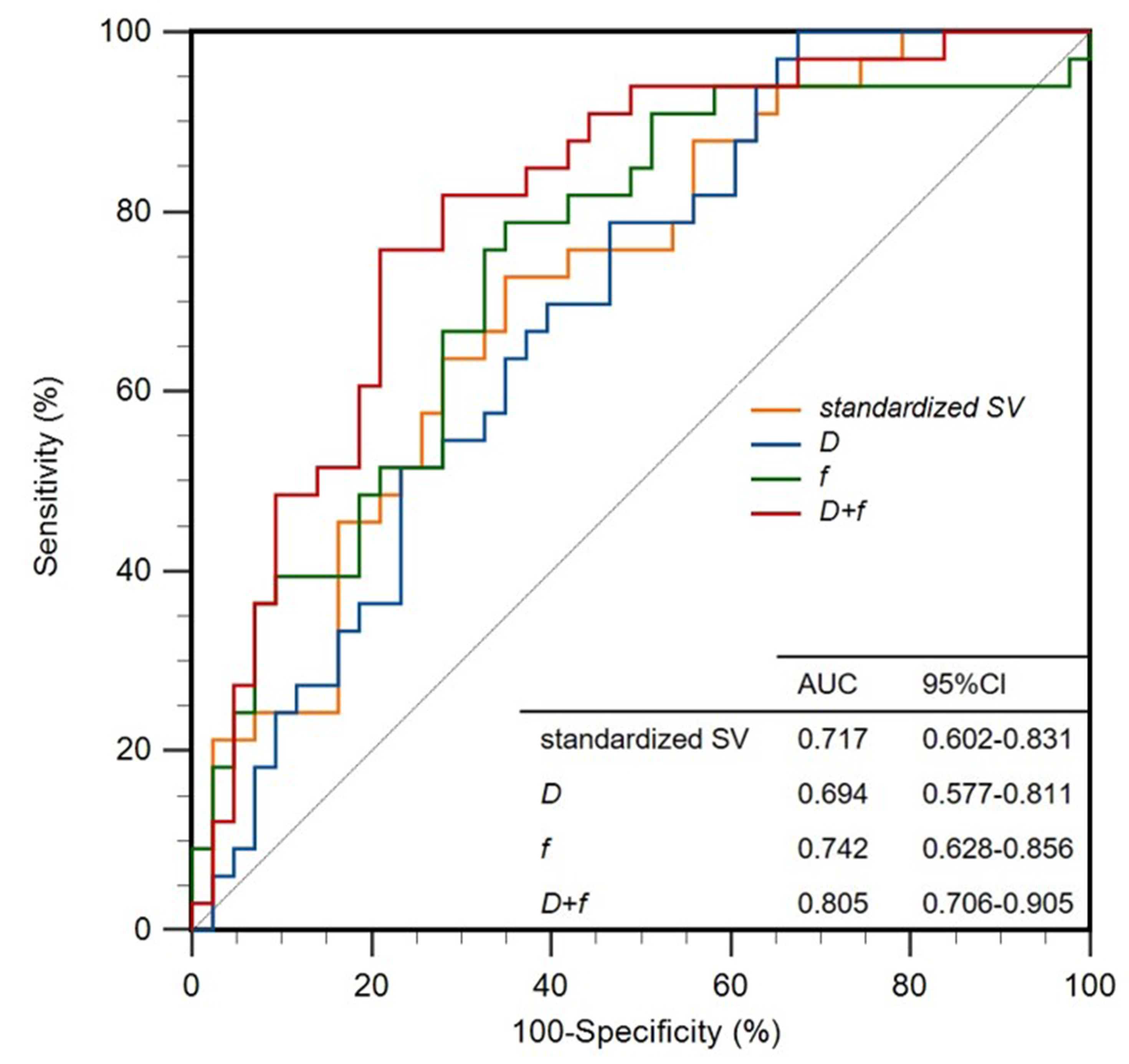
Figure 2. Receiver operating characteristic curves of standardized SV (AUC = 0.717), D (AUC = 0.694), f (AUC = 0.742) and D combined with f (AUC=0.805) for predicting treatment response in patients with newly diagnosed AL.