1649
Deep Learning Based Reconstruction for Multi-shot DWI of the Breast: A Preliminary Study1Department of Medical Imaging, National Taiwan University Cancer Center and National Taiwan University College of Medicine, Taipei, Taiwan, 2Department of Medical Imaging, National Taiwan University Hospital and National Taiwan University College of Medicine, Taipei, Taiwan, 3GE Healthcare, Taipei, Taiwan, 4GE Healthcare, Menlo Park, CA, United States, 5GE Healthcare, Houston, TX, United States, 6GE Healthcare, Boston, MA, United States
Synopsis
Keywords: Breast, Machine Learning/Artificial Intelligence, Deep learning reconstruction, Multi-shot DWI
Diffusion-weighted imaging (DWI) in the breast is limited by image distortion, which can be improved with multi-shot DWI (MUSE). We conducted a pilot study to investigate the impact of deep-learning reconstruction (DLRecon) on MUSE image quality. Compared with the non-DL MUSE images, the MUSE DLRecon showed higher SNR without altering the mean ADC value. Moreover, the higher shots MUSE DL with reduced NEX could provide less-distortion DWI with comparable SNR and scan time to 2-shot MUSE imaging, which is commonly used in the clinical setting. Preliminary results indicate the feasibility of MUSE-DWI in the breast with higher number of shots.INTRODUCTION
Diffusion-weighted imaging (DWI) is increasingly used as a non-contrast MRI technique for breast tumor detection and characterization. Compared with the single-shot EPI, multiplexed sensitivity encoding (MUSE), a multi-shot segmented technique, expands on existing sensitivity-encoding techniques by acquiring k-space with an interleaved trajectory with the aim of achieving better spatial resolution and reduced geometric distortion [1-3]. However, it is very challenging to apply higher number of shots in MUSE under clinically feasible scan time. Reducing the number of signal averages can proportionally reduce scan time of MUSE with increased shots, but at the cost of reduced SNR. In this preliminary study, we evaluated the deep learning-based (DL) noise reduction strategy [4] to: (1) achieve improved SNR of MUSE and (2) achieve higher shots MUSE with reduced distortion and comparable SNR under similar scan time for breast MUSE-DWI.METHODS
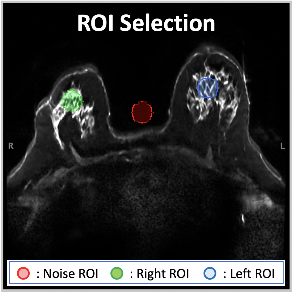
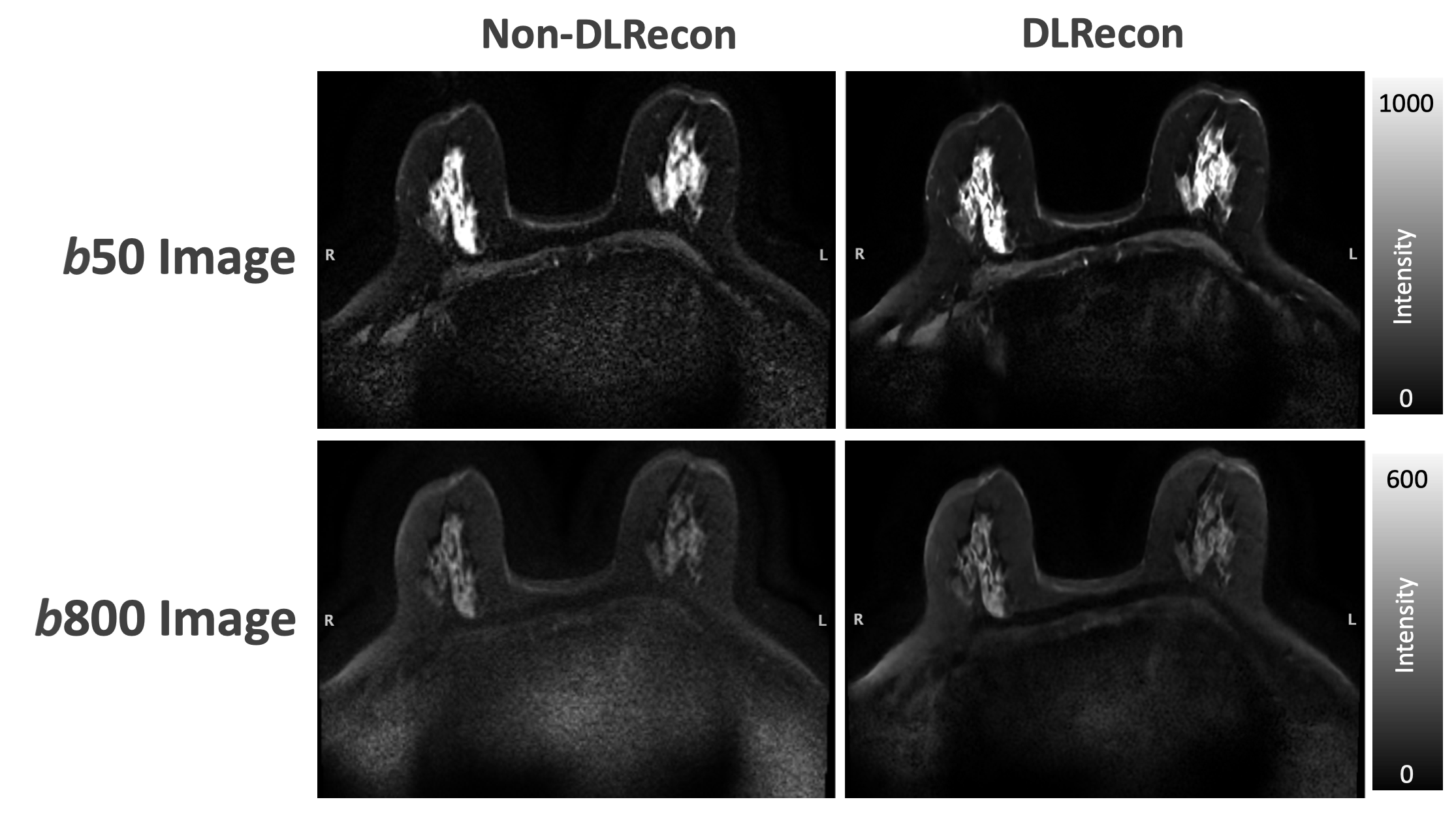
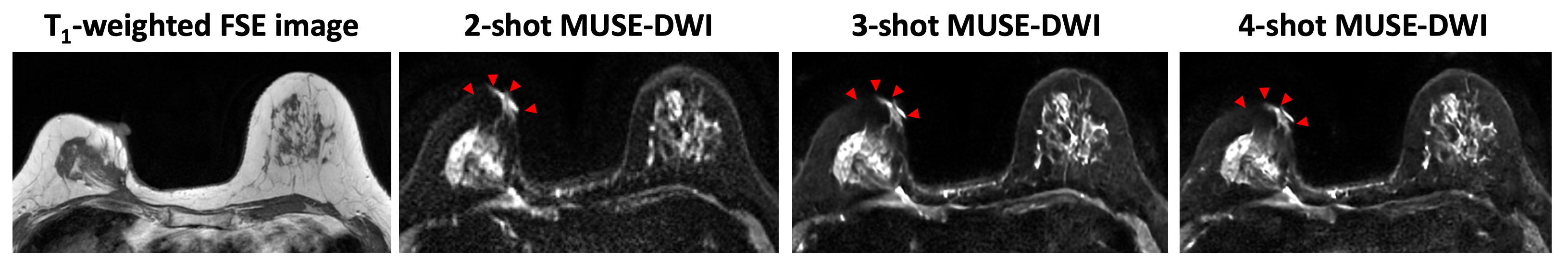
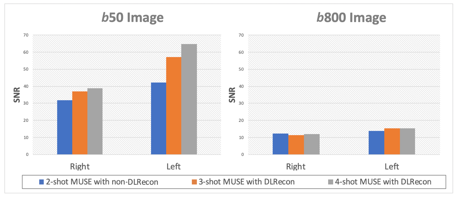
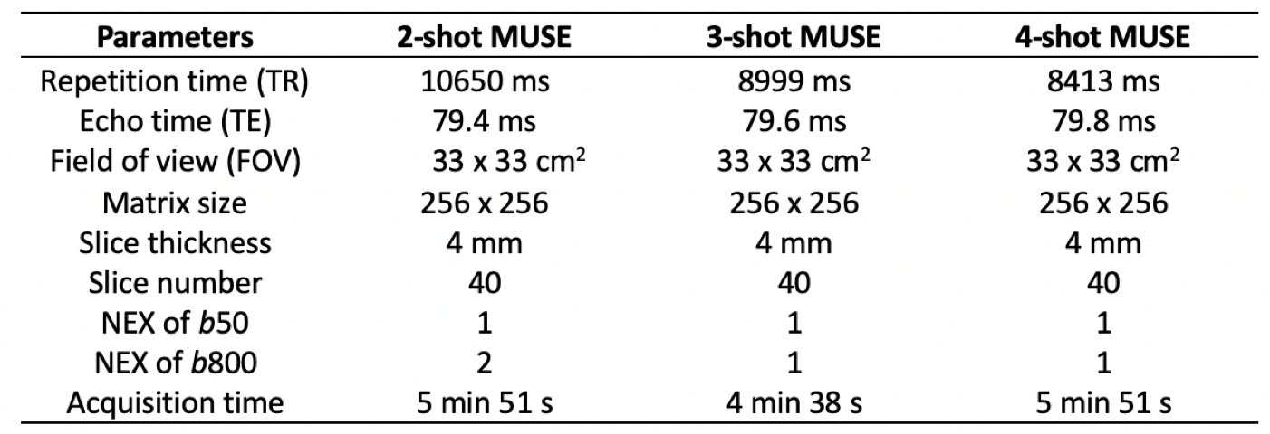
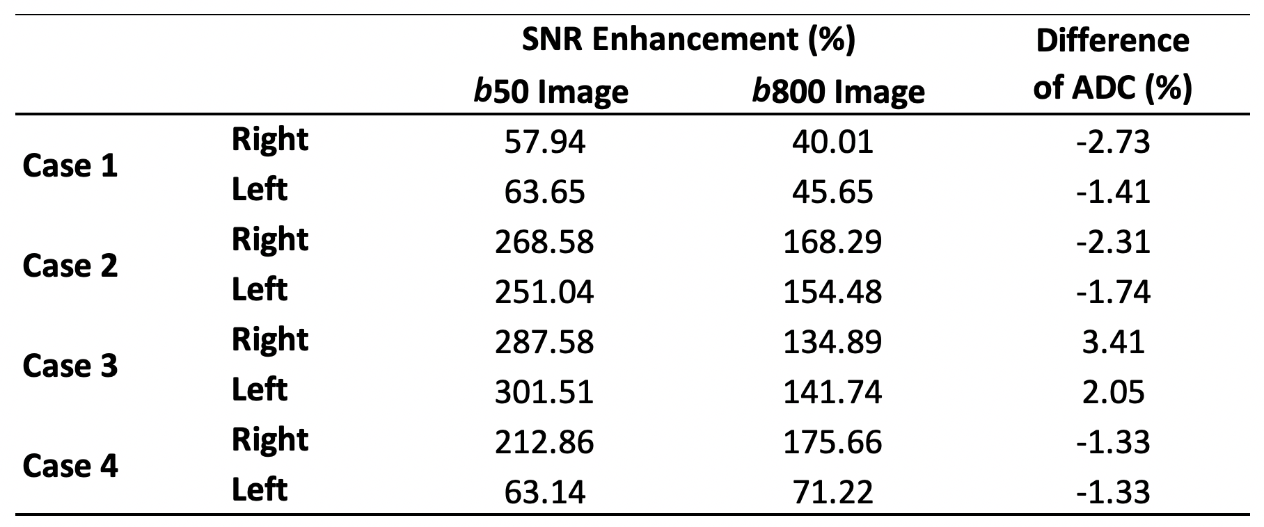
We acquired 2 sets of data to investigate the improvement of image quality and the feasibility of deep learning reconstruction (DLRecon) in MUSE-DWI. In the dataset 1, breast images were collected from 4 female participants, and the parameters were listed as follows: TR, 10650 ms; TE, 79.4 ms; FOV, 33 x 33cm2; matrix size, 256 x 256; slice thickness, 4 mm; numbers of b-value, 2 (50 and 800); NEX, 2. The SNR was calculated by 3 ROIs (background, right, and left breast regions; Figure 1), and the quantitative ADC maps with DLRecon and non-DLRecon were also compared. In the dataset 2, we acquired MUSE images with 2-, 3-, and 4-shot for one female participant to investigate the feasibility of higher shots MUSE- DWI DLRecon (The parameters were listed in the Table 1). All the data were performed in 3T SIGNA Architect MRI.RESULTS and DISCUSSION
The MUSE DL images showed less noise in the background, compared with MUSE non-DLRecon (Figure 2). In the quantitative analysis, the MUSE DL images showed improvements from 57% to 301% and 40% to 175% in SNR on b50 and b800 images, respectively (Table 2). The difference of ADC between MUSE DL and non-DLRecon ranged from -2.73% to +3.41%. In the Figure 3, there was obviously less distortion on the 3- and 4-shot MUSE images than the 2-shot MUSE images, and the SNR and scan time of 3- and 4-shot MUSE DL were comparable to the 2-shot MUSE non-DLRecon (Table1, Figure 4).CONCLUSION
Our results showed the apparent SNR improvement in MUSE DL with no significant change in quantitative ADC values. Moreover, higher shots MUSE DL demonstrated less-distortion DWI with comparable SNR and scan time to 2-shot MUSE. Our preliminary result showed that the MUSE DL could be beneficial for the region susceptible to distortion and high density of diffusion direction needed in the complex diffusion modelling with feasible scan time in the breast.Acknowledgements
No acknowledgement found.References
1. Baxter GC, Patterson AJ, Woitek R, Allajbeu I, Graves MJ, Gilbert F. Improving the image quality of DWI in breast cancer: comparison of multi-shot DWI using multiplexed sensitivity encoding to conventional single-shot echo-planar imaging DWI. Br J Radiol. 2021 Mar 1;94(1119):20200427.
2. Hu Y, Ikeda DM, Pittman SM, Samarawickrama D, Guidon A, Rosenberg J, Chen ST, Okamoto S, Daniel BL, Hargreaves BA, Moran CJ. Multishot Diffusion-Weighted MRI of the Breast With Multiplexed Sensitivity Encoding (MUSE) and Shot Locally Low-Rank (Shot-LLR) Reconstructions. J Magn Reson Imaging. 2021 Mar;53(3):807-817.
3. Daimiel Naranjo I, Lo Gullo R, Morris EA, Larowin T, Fung MM, Guidon A, Pinker K, Thakur SB. High-Spatial-Resolution Multishot Multiplexed Sensitivity-encoding Diffusion-weighted Imaging for Improved Quality of Breast Images and Differentiation of Breast Lesions: A Feasibility Study. Radiol Imaging Cancer. 2020 May 29;2(3):e190076.
4. Matthew J. Middione, Alimohammad S. Moalem, Cheng William Hong, Arnaud Guidon, Daniel B. Ennis, and Ryan L. Brunsing (2022). Multishot EPI and a Deep Learning-Based Noise Reduction Strategy for High Resolution Pancreatic DWI. International Symposium of Magnetic Resonance in Medicine 2022 Conference.
Figures