1595
Point-of-Care Knee MRI at 0.064T: Expanded Evaluation and Initial Comparison with Clinical Knee MRI at 3T
Jennifer M Watchmaker1, Ding Xia2, Idoia Corcuera-Solano1, Etan Dayan1, Justin Ngeow1, Fang Liu3, Zahi Fayad2, Mingqian Huang1, and Li Feng2
1Diagnostic, Molecular and Interventional Radiology, Icahn School of Medicine at Mount Sinai, New York, NY, United States, 2Biomedical Engineering and Imaging Institute, Icahn School of Medicine at Mount Sinai, New York, NY, United States, 3Athinoula A. Martinos Center for Biomedical Imaging, Harvard Medical School, Boston, MA, United States
1Diagnostic, Molecular and Interventional Radiology, Icahn School of Medicine at Mount Sinai, New York, NY, United States, 2Biomedical Engineering and Imaging Institute, Icahn School of Medicine at Mount Sinai, New York, NY, United States, 3Athinoula A. Martinos Center for Biomedical Imaging, Harvard Medical School, Boston, MA, United States
Synopsis
Keywords: Tendon/Ligament, Low-Field MRI, knee, hardware
At last year’s ISMRM, a pioneering work was presented to demonstrate the initial feasibility and performance of point-of-care knee MRI at 0.064T using a Hyperfine Swoop portable MRI scanner. This work extended that study for further evaluation in an expanded cohort of healthy subjects and subjects with knee pathology, and compare low-field imaging with clinical 3 Tesla imaging in a subset of volunteers.Introduction
MRI of the knee joint at 1.5-3 Tesla has been the gold-standard for evaluating both acute and chronic pathology including ligamentous injury, bony edema, and fracture1,2. While high-field imaging provides impeccable detail of the joint, cartilage, and surrounding structures, obtaining these studies can be challenging due to limited accessibility, high cost, and long-wait times. At last year’s ISMRM, a pioneering work was presented to demonstrate the initial feasibility and performance of point-of-care knee MRI at 0.064T using a Hyperfine Swoop portable MRI scanner. It has been shown that low-field imaging is sufficient to identify the six tendons and ligaments of the knee joint in volunteers3. This work extended that study for further evaluation in an expanded cohort of twenty-nine subjects, including both volunteer and patients with knee pathologies. In addition, we also show the initial comparison of paired knee images acquired at both 0.064T and clinical 3T.Methods
Twenty-nine subjects (13 male, age range 26-73 years) were recruited for this prospective study. Three of whom had underlying knee pathology (Table 1). Low-field imaging was performed on a prototype portable Swoop MR system (Hyperfine, Guilford, CT) equipped with a flexible coil for the lower extremity. The Swoop MRI scanner has a field strength of 0.064 T with gradient strengths of 26 mT/m (z-axis) and 25 mT/m (x- and y-axis). The imaging protocol consisted of two steady-state GRE (PSIF) acquisitions and a fluid-sensitive Short Tau Inversion recovery (STIR) acquisition. Total scan time was approximately 30 minutes. Resolutions were 1.5 x 1.5 x 3.0 mm3 for all the acquisitions. Imaging parameters for the PSIF imaging were: TR=11-12ms, TE=8-9ms, and 8 averages. Imaging parameters for the STIR imaging were: TR/TE=1300/4.25ms. Five subjects also underwent subsequent 3T MRI of the same knee for comparison. High-field imaging was performed at 3 Tesla following a routine clinical protocol implemented at our hospital. All low-field images were pooled for blind assessment of diagnostic quality for identifying six major tendons and ligaments of the knee (quadriceps tendon, patellar tendon, posterior cruciate ligament (PCL), anterior cruciate ligament (ACL), iliotibial band, lateral collateral ligament, and medial collateral ligament) an ordinal scale (Table 2).Results
Exams were successfully completed in all the subjects. Figure 1A shows a representative sagittal PSIF image identifying the ACL origin and insertion, with clear ability to trace fibers throughout entire course (Score = 4); Figure 1B shows a representative sagittal PSIF image identifying the PCL (Score = 4); and Figure 1C shows a representative coronal PSIF image identifying the medial collateral ligament. The maximum score was assigned for the quadriceps tendon, patellar tendon and PCL and patellar tendon for all subjects (scores of 2,3,4, respectively). In 26/29 subjects the IT band received the maximum score. The average score for identification of the ACL, LCL, and MCL was 3.5, 2.6, and 3.8, respectively. Figure 2 shows sagittal PSIF images of the PCL and ACL at 0.064T (A and B) and 3T (C and D), respectively. Figure 3 shows case examples of each participant with knee pathology; Figure 3A is a patient who underwent ACL reconstruction with tibial tunnel creation, the integrity of which can be assessed at low-field. Figure 3B shows a patient with total knee arthroplasty with minimal metal artifact from the implant. Figure 3C shows a patient with a clinical diagnosis of patellofemoral pain syndrome, and edema in Hoffa's fat pad can be appreciated on STIR imaging.Conclusion
In this work, we have demonstrated that low-field MRI is sensitive in the detection of the ligaments and tendons of the knee joint in an expanded cohort, and that at both low and high-field the structural integrity of commonly injured ligaments can be evaluated. In addition, we show that low-field imaging is suitable for use in post-operative patients and patients with metal implants. Incorporation of low-field imaging may serve to provide timely diagnoses of tendinous/ligamentous tear, to evaluate edema which often portends fracture occult on plain radiographs, and may prove to be a facile modality to ensure integrity of structural repair post-operatively.Acknowledgements
Authors would like to acknowledge the funding support from Hyperfine Inc.References
1. ACR Appropriateness Criteria®Acute Trauma to the Knee. In:2019:https://acsearch.acr.org/docs/69419/Narrative.
2. ACR Appropriateness Criteria® Chronic Knee Pain. In:2018:https://acsearch.acr.org/docs/69432/Narrative.
3. Watchmaker J. et. al. Portable, Low-Field MRI for Evaluation of the Knee Joint. In. International Society for Magnetic Resonance in Medicine 31st Annual Meeting.2022:https://archive.ismrm.org/2022/1689.html.
Figures
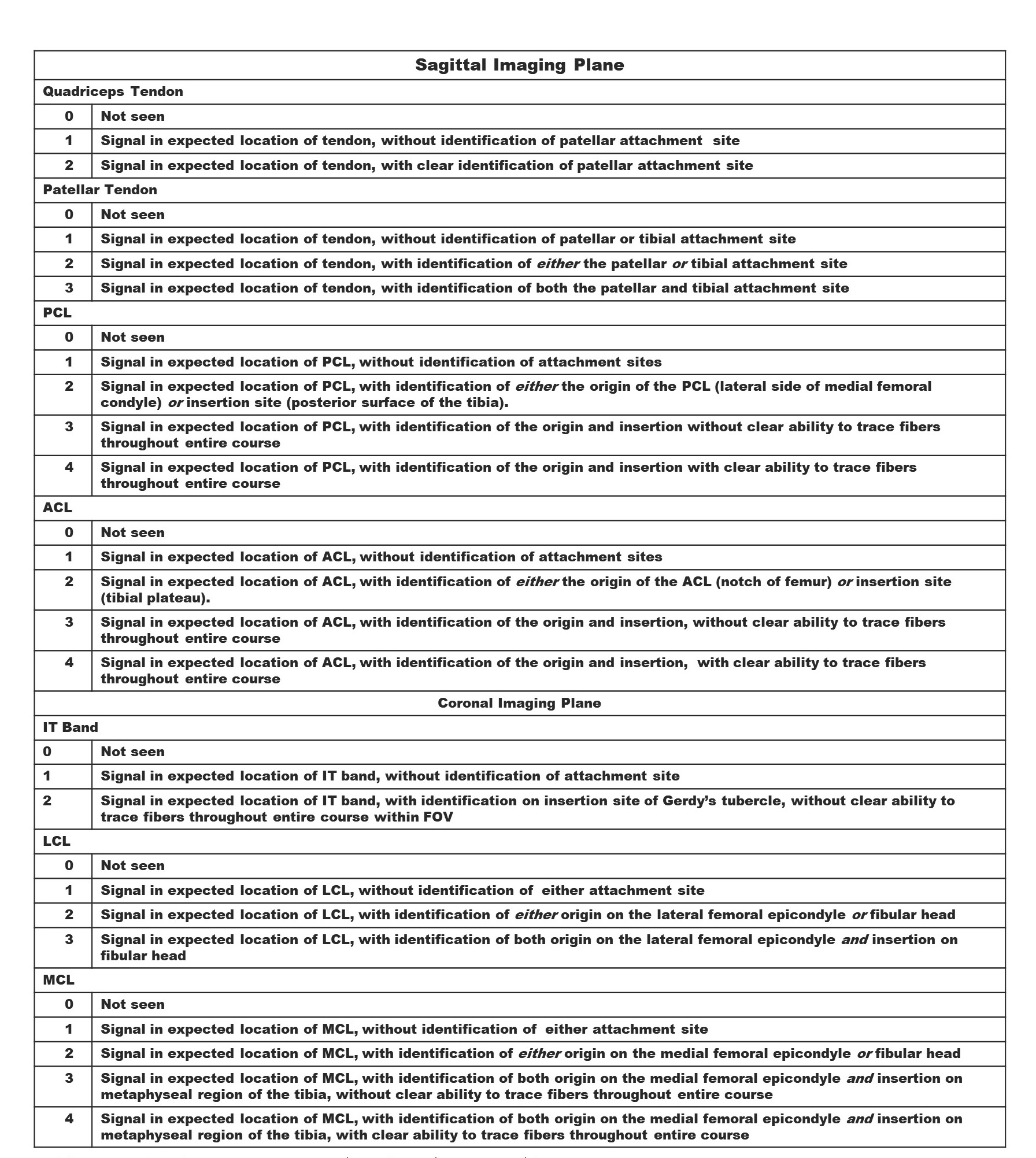
Table 2. Ordinal scoring system for identification of knee structures.
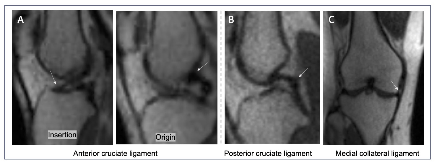
Figure 1. Signal in expected location of ACL, with identification of the origin and insertion (a). Signal in the expected location of the IT band with identification of the insertion (b). Signal in expected location of PCL with ability to trace fibers throughout (c). Signal in expected location of the medial collateral ligament with ability to trace fibers throughout (d).
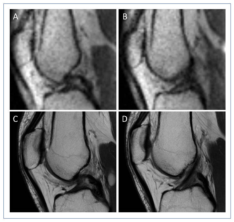
Figure 2. Sagittal 0.064 T and 3T proton-density weighted images of knee joint in a healthy subject. Intact posterior cruciate ligament (PCL) and anterior cruciate ligament (ACL) is appreciated at low field sagittal PSIF imaging (a,b). In comparison, high-field imaging confirms structural integrity of the PCL (c) and ACL (d).
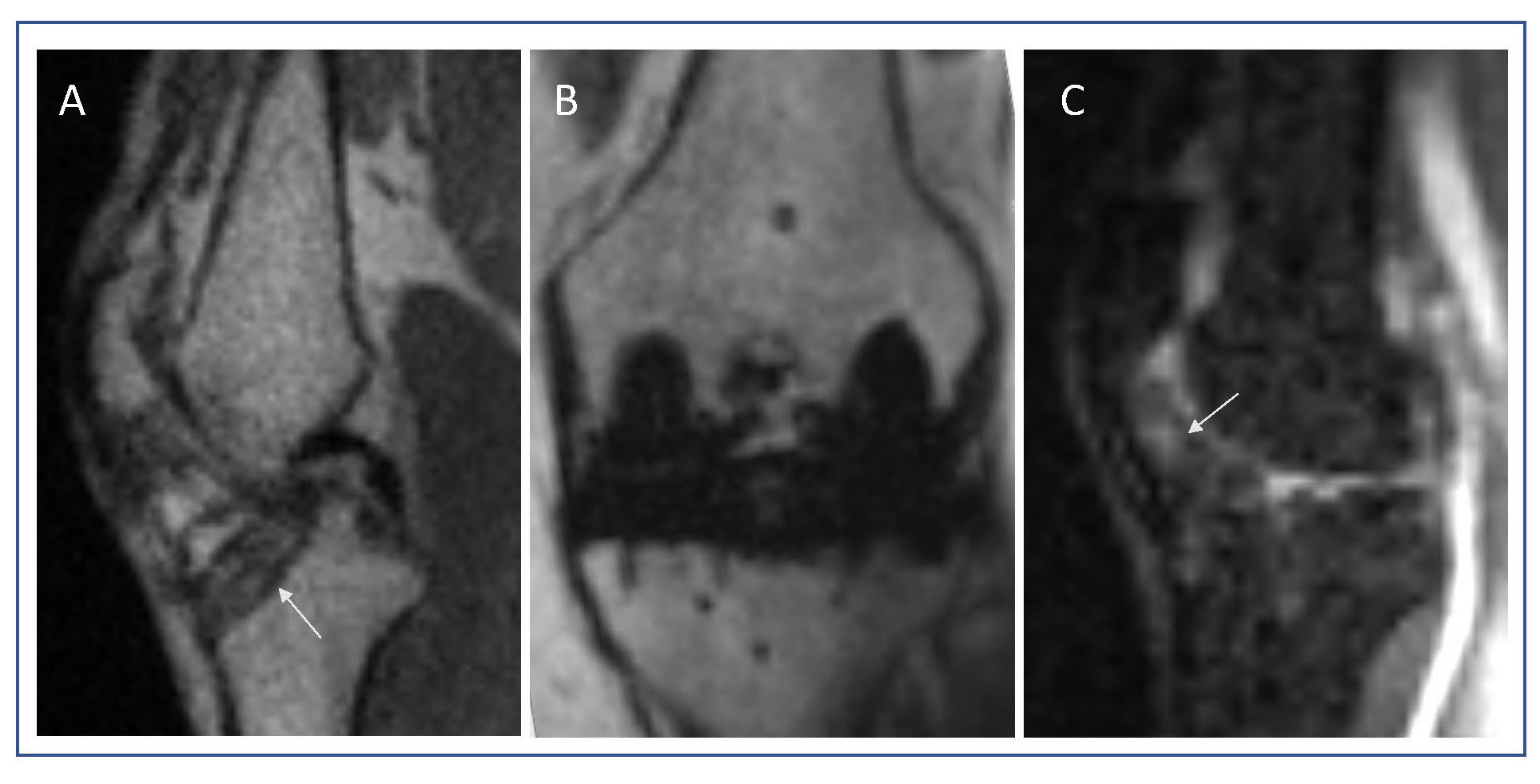
Figure 3. 0.064 T image of knee joint in subjects with knee pathology. Arrow denotes an intact tibial tunnel, an area of potential post-operative complication on sagittal PSIF image (a). Metal implant with minimal artifact seen in patient who underwent prior total knee arthroplasty (b). Scarring of Hoffa’s fat pad is well appreciated on sagittal STIR imaging in patient with clinical diagnosis of patellofemoral pain syndrome (c).
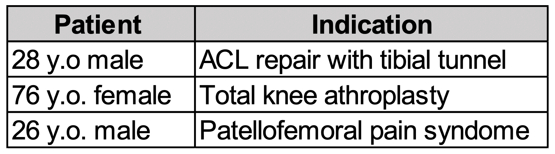
Table 1. Patient demographics.
DOI: https://doi.org/10.58530/2023/1595