1593
Accelerated imaging of resected lymph nodes at high spatial resolution using a portable low-field MRI scanner
Arthur de Lange1, Lejla Alić1, Bennie ten Haken1, and Frank F.J. Simonis1
1TechMed Centre, University of Twente, Enschede, Netherlands
1TechMed Centre, University of Twente, Enschede, Netherlands
Synopsis
Keywords: Low-Field MRI, New Devices
After sentinel lymph nodes are detected using SPIONs and excised, their characterization is important to detect possible metastases. In this research a low-field (0.5T) tabletop MRI scanner was tested for this purpose using 4x accelerated high resolution 3D acquisition. Both simulations and experiments on excised pig lymph nodes showed promising results, with the accelerated scans showing similar image quality with respect to fully sampled datasets. This protocol shows lymph nodes can be imaged at 0.25 mm isotropic resolution within reasonable scan times. Clinical usage should be proven by scanning true metastatic lymph nodes.Introduction
Lymph node (LN) metastasis is an important prognostic factor in a variety of cancer types. A sentinel lymph node biopsy (SLNB) aims to remove the first draining LN to exclude further metastases. The SLN is found by locating peritumorally administered tracer. Superparamagnetic iron oxide (SPIO) was recently suggested as such a tracer enabling diagnostic imaging and perioperative detection1,2. This study evaluates the feasibility of analyzing resected LNs directly using a portable low-field MRI scanner.Methods
Resected porcine LNs were injected with 5 µl SPIO tracer (Magtrace, Endomagnetics Limited, UK) and subsequently placed in a sealed test tube containing formaldehyde (Figure 1-left). A magspec benchtop system (Pure Devices, Rimpar, Germany) with an increased bore of 15mm diameter at a field strength of 0.5T (Figure 1-right) was used for imaging. The magnet was connected to an external gradient amplifier (DC-600), an RF amplifier (RF-100) and a Low noise amplifier (LNA).T1 and T2 weighted 3D volumes of 14×14×14 mm³ were acquired using Cartesian sampling with a matrix size of 32×32×32 resulting in an isotropic resolution of 0.438 mm, see Table 1 for further imaging parameters. Since both scans took unpractically long, scan times were accelerated by a factor of 4 by simulating an a variable density 2D Poisson disc acquisition pattern3 followed by Compressed Sense (CS) reconstruction4. Subsequently, the undersampling pattern was actually implemented on the portable scanner with the same FOV and a 56×56×56 matrix to obtain T1-weighted images at 0.25 mm isotropic resolution. All resulting images were inspected visually and the structural similarity index (SSIM)5 was calculated with respect to the fully sampled dataset.Results
Scans of the injected LNs can be seen in Figure 2, in which the SPIO tracer is clearly visible as a black spot. The images reconstructed after simulated undersampling show similar results as the fully sampled data with almost no visible differences. The SSIM between the original and undersampled images is 0.9 (T1w) and 0.75 (T2w). Actually acquired T1w accelerated scans in Figure 3 show only slight blurring and a SSIM of 0.88.Discussion
All images show a clear distinction between fat, nodal tissue and SPIO tracer proving the feasibility of imaging LNs using a portable MRI scanner and imaging time can be effectively reduced using undersampling combined with CS reconstruction. Clinical input is required to decide the best balance in spatial resolution and imaging time. Additionally, the protocol needs to be tested on human LNs with metastases to see with which contrast those can be most accurately detected.Conclusion
A portable MRI scanner can be used for on-site high resolution imaging of excised lymph nodes with accelerated acquisition times. Clinical relevance needs to be proven by comparison of resected nodes with pathology results.Acknowledgements
No acknowledgement found.References
1. R Mahieu et al, New Developments in Imaging for Sentinel Lymph Node Biopsy in Early-Stage Oral Cavity Squamous Cell Carcinoma, 2020 Cancers 12.10 3055, 2. S Waanders et al, A handheld SPIO-based sentinel lymph node mapping device using differential magnetometry, 2016 Phys. Med. Biol. 61 8120, 3. E Levine et al, “3d Cartesian MRI with compressed sensing and variable view sharing using complementary poissondisc sampling, MRM 2017, 77.5, 1774–1785. 4. M Lustig et al, Sparse MRI: The application of compressed sensing for rapid MR imaging, 2007 MRM 58.6 1182, 5. Z Wang et al, Image quality assessment: from error visibility to structural similarity, 2004 IEEE transactions on image processing 13.4 600Figures
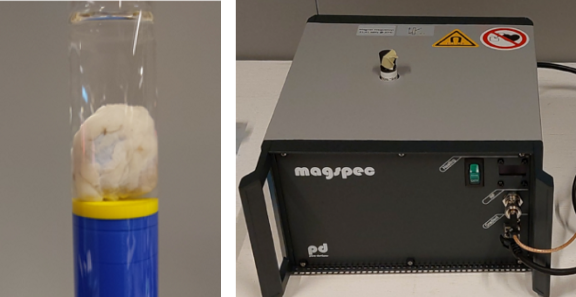
Figure 1: A
resected porcine lymph node in a test tube (left) and the portable magnet
(right)
Table 1: Scan parameters
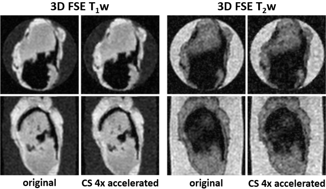
Figure 2: High resolution isotropic scans of a porcine lymph node with SPIO tracer (in black) showing a simulated 4x undersampling of data
hardly affects image quality
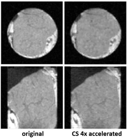
Figure 3: T1w
scans of a porcine lymph node without tracer showing the implemented 4x undersampling gives similar results to the fully sampled dataset
DOI: https://doi.org/10.58530/2023/1593