1570
Whole-Tumour Histogram Analysis of Multiple Diffusion Metrics for Meningiomas Grade1Department of Radiology, First Affiliated Hospital of Fujian Medical University, Fuzhou, China, 2Department of Radiology, National Regional Medical Center, Binhai Campus of the First Affiliated Hospital, Fujian Medical University, Fuzhou, China, 3Key Laboratory of Radiation Biology of Fujian higher education institutions, First Affiliated Hospital, Fujian Medical University, Fuzhou, China, 4Philips Healthcare, Shanghai, China, 5Department of Radiology, Fujian Key Laboratory of Precision Medicine for Cancer, First Affiliated Hospital of Fujian Medical University, Fuzhou, China
Synopsis
Keywords: Tumors, Diffusion/other diffusion imaging techniques, Meningiomas; Diffusion-weighted MRI
An accurate assessment of the World Health Organization grade is vital in meningiomas. While many studies have investigated the usefulness of conventional DWI and DTI for noninvasive grading intracranial meningiomas, there are neither any studies comparing three advanced diffusion model including DKI, MAP and NODDI with DTI for predicting meningioma grade. Thus, it is vital to evaluate whether these advanced models derived from diffusion spectrum imaging can also be beneficial in grading meningiomas. Our results suggested that whole tumour histogram analyses of the diffusion metrics from multiple diffusion models are promising methods in grading meningiomas.Introduction
Meningiomas are the most frequent primary intracranial tumour (38.3%) and are classified into 3 grades according to the World Health Organization Classification (WHO)1, 2. The grading of meningiomas has clinical significance for determining a treatment strategy and assessing prognosis. Conventional diffusion tensor imaging (DTI) are currently the most used techniques in clinic and presume a Gaussian diffusion contribution of water molecular within tumours. More advanced diffusion models with high b values, such as Diffusional kurtosis imaging (DKI), have been shown that outperform than conventional DWI and DTI in grading meningiomas3, 4. Recently, mean apparent propagator (MAP) 5.and neurite orientation dispersion and density imaging (NODDI) 6, may reflect the diffusion behavior of water molecules within brain tumours more accurately. Nevertheless, to our knowledge, there are neither any studies on the role of NODDI or MAP in meningioma grading nor any reports comparing DTI, DKI, MAP, and NODDI for predicting meningioma grade. Furthermore, these advanced diffusion models can be simultaneously derived from diffusion spectrum imaging (DSI), which is a method freely reconstructed by using multiple gradient directions and high b values of entire q-space to quantitatively estimate diffusion behaviors of water molecules. The purpose of this study was to evaluate and compare the diagnostic accuracy of DTI, DKI, MAP, and NODDI-based diffusion parameters combined with histogram analysis in grading meningiomas, and to assess the correlations between the diffusion metrics and Ki-67 index.Methods
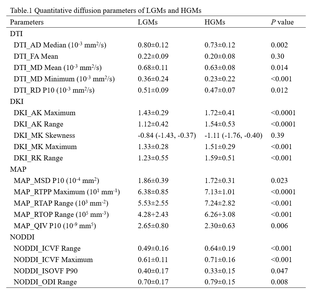
In this study, 122 consecutive patients with histologically proved meningiomas were enrolled. MRI examinations were performed with a 3.0 T MRI unit (Philips Ingenia, Best, Netherlands). The histogram features of multiple diffusion metrics obtained from diffusion tensor imaging (DTI), diffusion kurtosis imaging (DKI), mean apparent propagator (MAP), and neurite orientation dispersion and density imaging (NODDI) in the solid component of tumour were analyzed. All values were compared between high-grade meningiomas (HGMs) and low-grade meningiomas (LGMs) with Man-Whitney U test. Logistic regression analysis was applied to predict the grade. The correlation between diffusion metrics and Ki-67 index was analyzed.Results
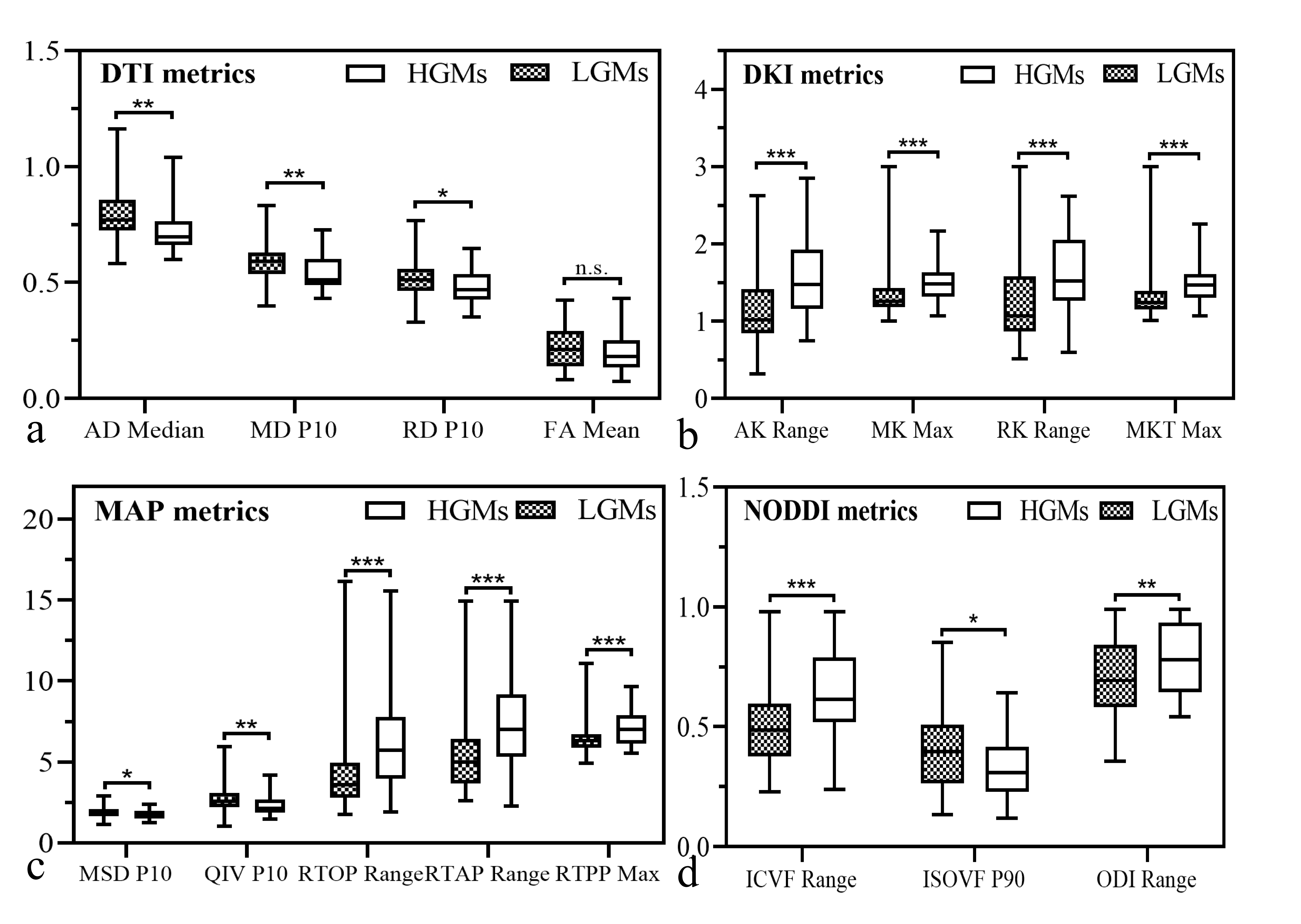
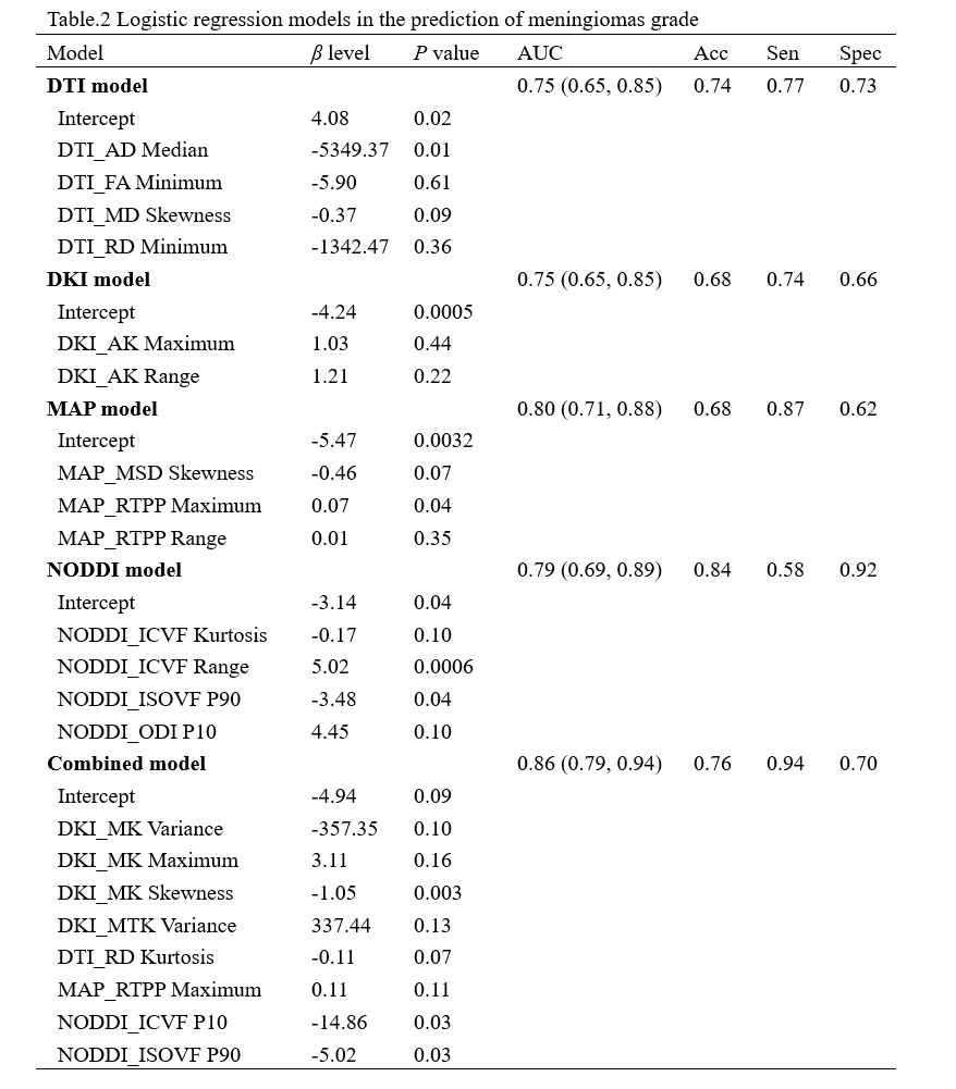
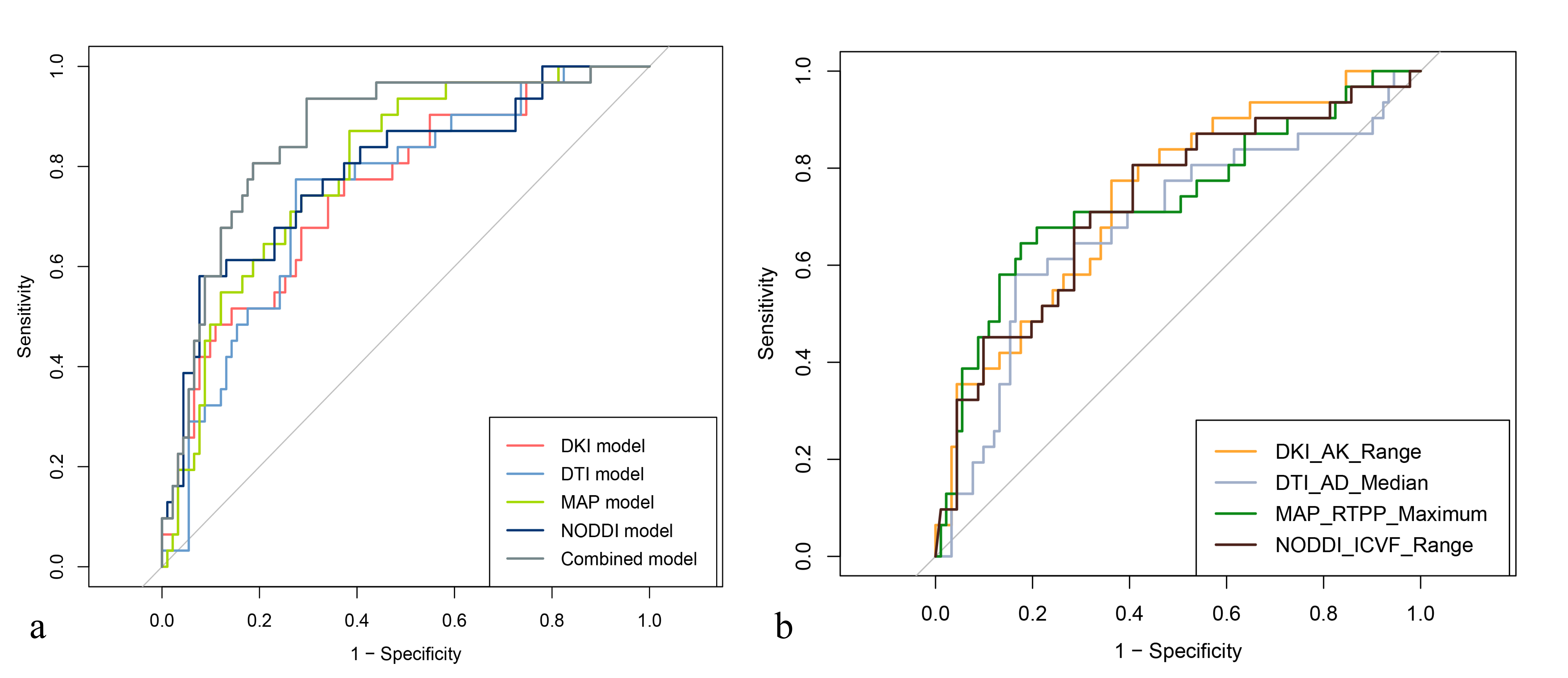
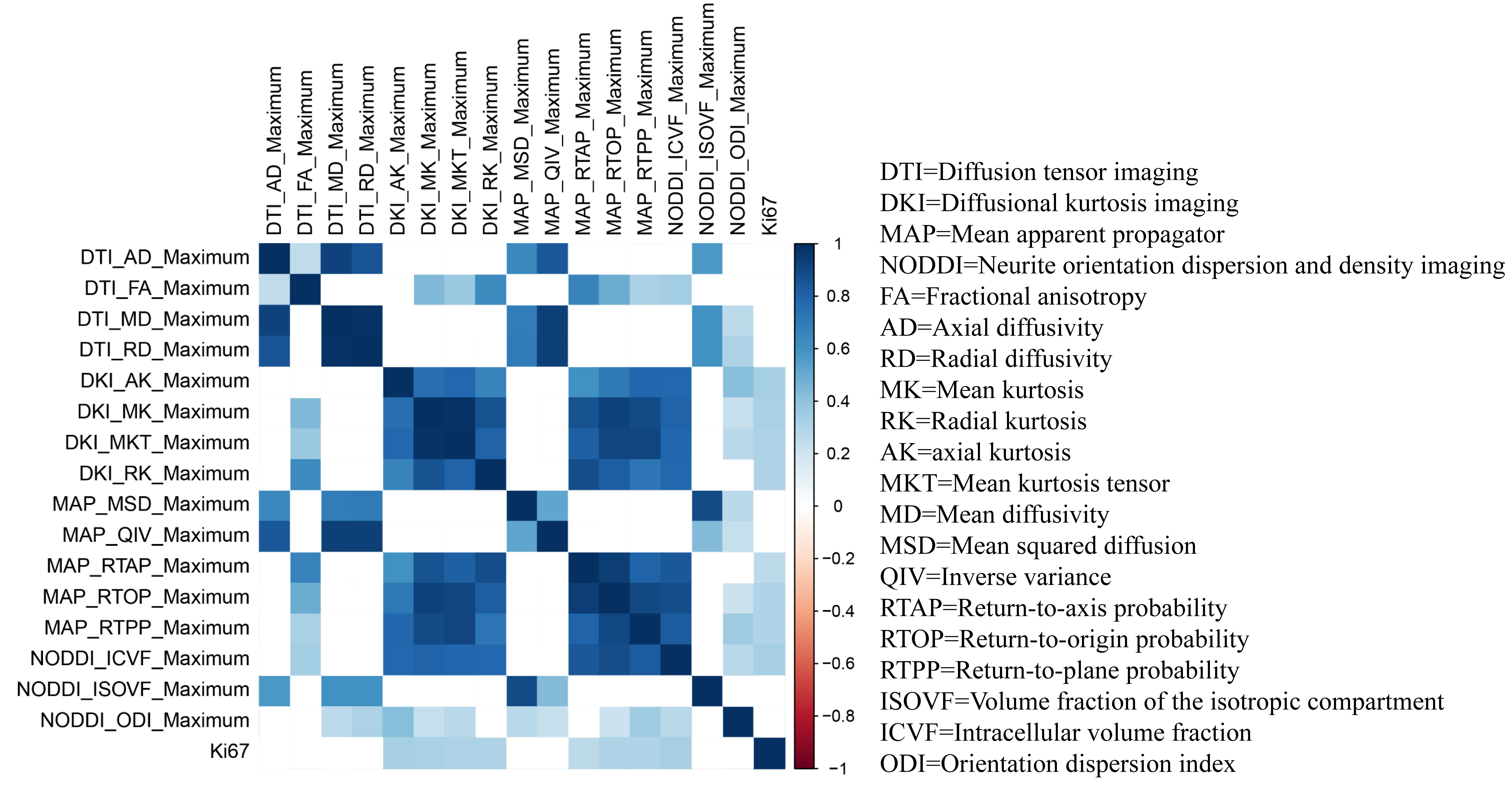
The DKI_AK maximum, DKI_AK range, MAP_RTPP maximum, MAP_RTPP range, NODDI_ICVF range, and NODDI_ICVF maximum values were lower (P<0.0001), while the DTI_MD minimum values were higher in the LGMs than those in the HGMs (P<0.001) (Table 1 and Fig 1). Among the DTI, DKI, MAP, NODDI and combined diffusion models, no significant differences were found in the areas under the receiver operating characteristic curve (AUCs) for grading meningiomas (AUCs, 0.75, 0.75, 0.80, 0.79, and 0.86, respectively; all corrected P>0.05) (Table 2 and Fig 2). Significant positive correlations were found between the Ki-67 index and the DKI, MAP and NODDI metrics (r= 0.26-0.34, all P<0.05) (Fig 3).Discussion
An accurate and noninvasive evaluation of the WHO grade is particularly important for patients with meningiomas. In this study, with a relatively large sample size, our results demonstrated that the whole-tumour histogram features of any diffusion models (DTI, DKI, MAP, NODDI, and the combined diffusion models) were helpful in grading intracranial meningiomas (AUC, 0.75, 0.75, 0.8, 0.79, and 0.86, respectively). Our results also showed that the increased benefit of upgrading the imaging protocol for advanced diffusion model was small, meaning that DTI had similar diagnostic performance compared with the advanced diffusion methods in the prediction of meningiomas grade. In addition, weak correlations were revealed between Ki-67 index and almost diffusion metric of DKI, MAP and NODDI metrics.Conclusions
Whole tumour histogram analyses of the multiple diffusion metrics from four diffusion models are promising methods in grading meningiomas. The DTI model has similar diagnostic performance compared with advanced diffusion models.Acknowledgements
No acknowledgement found.References
1. Ostrom QT, Patil N, Cioffi G, Waite K, Kruchko C, Barnholtz-Sloan JS. CBTRUS Statistical Report: Primary Brain and Other Central Nervous System Tumors Diagnosed in the United States in 2013-2017. Neuro Oncol 2020; 22:iv1-iv96
2. Louis DN, Perry A, Wesseling P, et al. The 2021 WHO Classification of Tumors of the Central Nervous System: a summary. Neuro Oncol 2021; 23:1231-1251
3. Lin L, Bhawana R, Xue Y, et al. Comparative Analysis of Diffusional Kurtosis Imaging, Diffusion Tensor Imaging, and Diffusion-Weighted Imaging in Grading and Assessing Cellular Proliferation of Meningiomas. AJNR American journal of neuroradiology 2018; 39:1032-1038
4. Chen XD, Lin L, Wu J, et al. Histogram analysis in predicting the grade and histological subtype of meningiomas based on diffusion kurtosis imaging. Acta radiologica 2020; 61:1228-1239
5. Avram AV, Sarlls JE, Barnett AS, et al. Clinical feasibility of using mean apparent propagator (MAP) MRI to characterize brain tissue microstructure. NeuroImage 2016; 127:422-434
6. Zhang H, Schneider T, Wheeler-Kingshott CA, Alexander DC. NODDI: practical in vivo neurite orientation dispersion and density imaging of the human brain. NeuroImage 2012; 61:1000-1016
Figures