1559
Radiomic Analyses of the Instrumented Spinal Cord After Treatment for Degenerative Myelopathy and Acute Cord Injuries
Azadeh Sharafi1, Andrew Klein1, and Kevin M Koch1
1Radiology, Medical College of Wisconsin, Milwaukee, WI, United States
1Radiology, Medical College of Wisconsin, Milwaukee, WI, United States
Synopsis
Keywords: Spinal Cord, Radiomics
Quantitative MRI of the spinal cord can provide insightful information about injured or degenerated cord tissue. However, the presence of instrumentation used treat such conditions can compromise the ability to perform such analyses. In this preliminary study, first and second-order radiomics features were computed across axial segmented spinal cord in subjects with 1) degenerative cervical myelopathy and 2) spinal cord injury patients treated with spinal fusion. Radiomics were computed using T1 and T2 weighted metal-artifact suppressed 3D-MSI and analyzed and successfully utilized to model local fusion status within the clinical cohorts and also against non-instrumented control subjects.Introduction
Radiomics is a quantitative technique that extracts multiple features from medical images for tissue classification [1]. In this preliminary study, we aim to investigate using radiomics analysis to differentiate between different disease states and local zones of the instrumented spinal cord. The study seeks to develop potential quantitative metrics that build upon conventional qualitative radiological practices that examine spinal cord textures and intensity patterns on conventional MRI in regions of suspected cord pathology.Methods
MRI acquisition was performed at a 3T scanner using a 21-channel vendor-provided head-neck receiver coil. T1 and T2-weighted metal-artifact suppressed 3D-MSI [2] were acquired with 1.2mm isotropic resolution, using 2x2 auto-calibrated parallel imaging, TE of 8/60ms, and repetition times of 750/2100 ms for T1/T2-weighted image acquisitions, respectively. 8 spectral bins were acquired in both scans to provide sufficient coverage within the spinal cord near titanium fusion hardware at 3T. The study was approved by the local IRB and all subjects provided written consent into the study. Data were collected on 15 healthy controls, 15 patients treated for severe degenerative cervical myelopathy (CSM), and 12 treated for spinal cord injuries (SCI). CSM and SCI subjects were treated using either anterior (A), posterior (P), or both A and P titanium hardware fusions. The spinal cord was segmented in axial images using the semi-automatic technique presented in [3]. Radiomic analysis was performed on each slice using the Python pyradiomics package to extract 96 features, including first order, 2D shape, as well as the Grey Level Co-occurrence (GLCM), Size Zone, run length, dependence, and neighboring gray tone difference matrices. Principal Component Analysis (PCA) analysis was then performed to reduce the dimensionality of the dataset from 96 to 23. The number of components was selected to capture at least 95% of the total variance. Afterward, the logistic regression with 10-fold cross-validation was performed, and the area under the receiver operator characteristic curve (AUC-ROC) was calculated to assess the ability to use radiomic features inside the spinal cord for distinguishing between different cohorts and zones. All analyses utilized methods contained within the scikit-learn python package.Results
Figure 1 shows sample T1 and T2 weighted MRI images and the segmented cord for slices at instrumented (fused) and adjacent segments. Figure 2 depicts a sample slice in a non-instrumented (or adjacent) segment of the cord for SCI, CSM, and control cohorts. Figure 3 displays the AUC-ROC scores distinguishing each cohort and cord segment class when either T1 or T2 or both T1 and T2 extracted features were used. The multinomial logistic regression analysis showed an average of 0.75 accuracy, 0.72 precision, 0.73 recall, and F1-score of 0.72 in classifying the data into Controls, CSM, and SCI groupsDiscussion and Conclusion
The AUC-ROC scores observed in this preliminary study suggest the promising potential for radiomics measures derived from T1 and/or T2-weighted images to distinguish injured, degenerated, and healthy spinal cord tissues. Comparing the AUC-ROC scores measured for either or both T1 and T2 extracted features also suggests that using only T2-weighted extracted features may be sufficient to distinguish between different groups. Future work will expand upon these preliminary analyses using larger cohorts and incorporating clinical symptom scores for the degenerative and injured cohorts.Acknowledgements
This work was supported by the Department of Defense Congressionally Directed MedicalResearch Program, Spinal Cord Injury Research Program, award number W81XWH1910273. Opinions, interpretations, conclusions, and recommendations are those of the authors and are not necessarily endorsed by the Department of DefenseReferences
- van Timmeren, J., Cester, D., Tanadini-Lang, S. et al. Radiomics in medical imaging— “how-to” guide and critical reflection. Insights Imaging 11, 91 (2020)
- Koch K. M., Brau A. C., Chen W., et al. Imaging near metal with a MAVRIC-SEMAC hybrid Magnetic Resonance in Medicine 65:71–82 (2011)
- Koch, K.M. and Nencka A. S., Morphological Assessment of the Instrumented Spinal Cord using Isotropic 3D-MSI MRI. Proc. Intl. Soc. Mag. Reson. Med. 29 (2021)
Figures
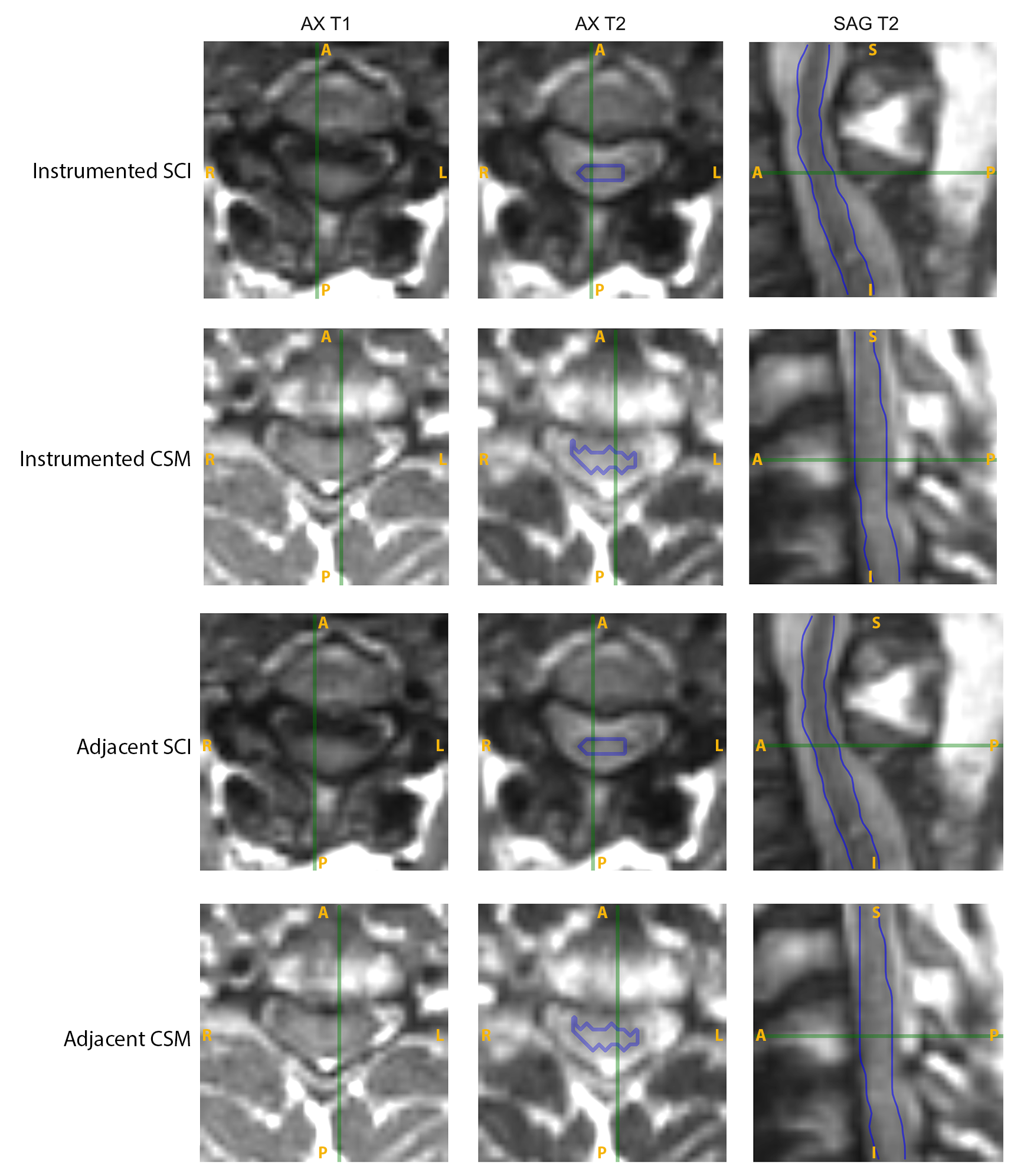
Figure 1 T1- and T2-weighted 3D-MSI MRI of the damaged spinal cord at instrumented (fused) and adjacent segments of SCI and CSM study participants. The segmented spinal cord is shown in blue on the T2-weighted axial and coronal reformat images.
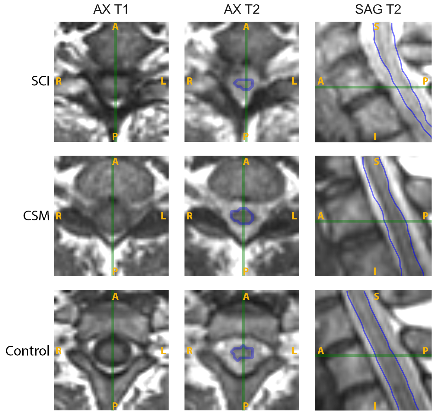
Figure 2. T1- and T2-weighted 3D-MSI MRI of non-instrumented segments in SCI, CSM, and asymptomatic control study participants.
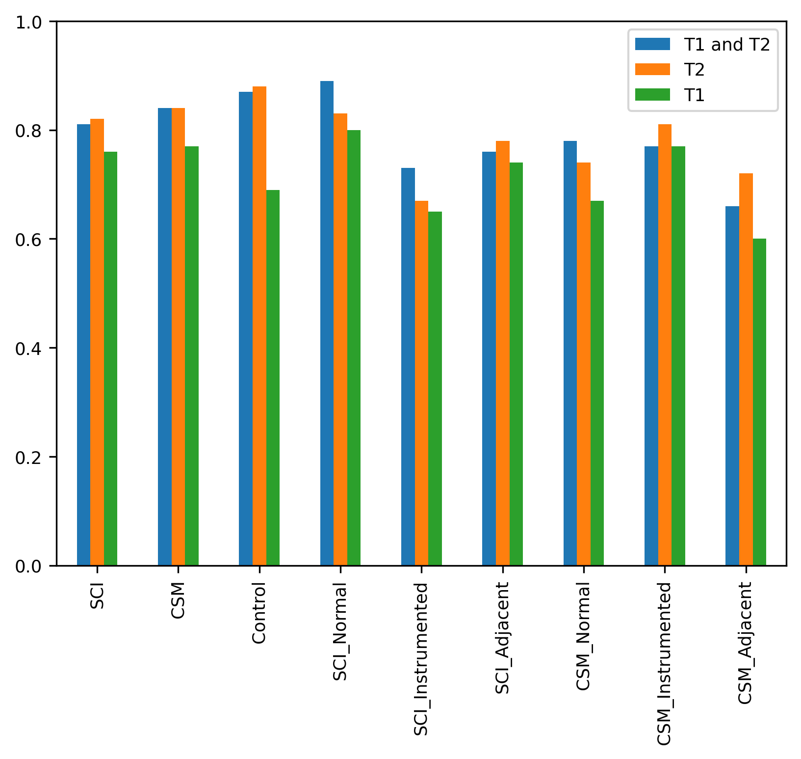
Figure 3. AUC-ROC scores from 10-fold cross-validated logistic-regression in different groups for using T1-weighted, T2-weighted or both sets of extracted radiomic features
DOI: https://doi.org/10.58530/2023/1559