1552
Does spinal CSF perfuse out of the spine?
Diana Vucevic1, Vadim Malis1, Won Bae1, and Mitsue Miyazaki1
1Radiology, UCSD, San Diego, CA, United States
1Radiology, UCSD, San Diego, CA, United States
Synopsis
Keywords: Spinal Cord, Visualization
Cerebrospinal fluid (CSF) perfusion in the spinal canal is one of the greatest mysteries in the scientific community. CSF can be imaged without the use of gadolinium-based contrast agents (GBCA) using our novel spin-labelling MRI technique at 3 Tesla. Perfusion of CSF in the spine can be seen in the thoraco-lumber region from the nerve roots. This CSF outflow may be absorbed by the venous plexus along the dorsal nerve roots. The second pathway It may be from the nerve roots, recirculated to lymphatic tissue and into the thoracic duct.Introduction
Cerebrospinal fluid (CSF) clearance pathways are one of the greatest mysteries in the scientific world. CSF is produced by the choroid plexuses of the brain and travels within the ventricles and subarachnoid space (SAS) to provide buoyancy and maintain homeostasis of the brain and spinal cord. Within recent years, the introduction of a “glymphatic system” proposes that CSF enters the brain along the perivascular space and flushes through the brain parenchyma [1-4]. Contrast MRI studies have shown the parasagittal dura (PSD) as a brain clearance pathway. Non-contrast, spin-labelling MRI of endogenously tagged CSF, shows there is a distinct CSF egress route of the dura mater to the superior sagittal sinus by PSD [5]. However, in comparison to the brain, the mechanism of CSF perfusion of the spine remains unclear. Two mechanisms are hypothesized – through venous pathways or the lymphatic pathways. The purpose of this research work is to investigate the location of CSF spinal perfusion using novel MRI non-contrast, spin-labeling of endogenous CSF.Methods
All MR imaging data was conducted using a clinical 3 Tesla MR imager (Vantage Galan 3T, Canon Medical Systems, Japan). Two healthy subjects (1 female, age 25, and one male, age 30) took part in this preliminary study and were scanned after informed consent. T2- weighted 2D fluid-attenuated inversion recovery (FLAIR) along with T2-weighted 3D centric single-shot fast spin echo (3D cSSFSE) were optimized and used for localization of the thoracic and lumber regions of the spinal canal.Results
Our preliminary work using a non-contrast MRI technique, shows CSF perfuses out of the spine from the nerve roots predominantly around the thoracolumbar region, favored by the right side. Figure 1 A and Figure 2 A show the Time-SLIP tag pulse location. Figure 1B and Figure 2B show the subtracted color maps at each of the inversion time, demonstrating CSF perfusion out from the nerve roots. Figure 1C and Figure 2C show the evolution of tagged CSF signal with their corresponding fits.Discussion
Without the use of gadolinium-based contrast agents (GBCA), our preliminary results using non-contrast MRI show that there is presence of CSF perfusion out of the spine from the nerve roots of the thoracolumbar region, where flow is much slower compared to the upper spinal canal. The absorption of CSF is one to be investigated next. If the CSF is perfusing out of the nerve roots, it is possible it is being absorbed by the venous plexus as SAS is predominant along the dorsal nerve roots. It may also be that CSF perfusion from the nerve roots is recirculated to lymphatic tissue and into the thoracic duct.Conclusion
Our work demonstrates that CSF does perfuse out of the spine along the nerve roots without the administration of GBCA. The perfusion is predominant along the thoracolumbar region. The absorption of this outflow is the next puzzle piece to solve.Acknowledgements
This work was supported by an NIH grant RF1AG076692 (M.M.) and a grant by Canon Medical Systems, Japan (35938).References
[1] Nedergaard M. Science 2013; 340: 1529- 1530.
[2] Iliff JJ, Wang M, Zeppenfield DM, et al. Journal of Neuroscience 2013;33:18190-18199.
[3] Absinta M, Ha SK, Nair G, et al. eLife 2017;6:e29738.
[4] Ringstad G, and Eide PK. Nature Communications 2020;11;354.
[5] Malis V, Bae W, Yamamoto A, McEvoy KL, McDonald MA, Miyazaki M. MRMS in press.
Figures
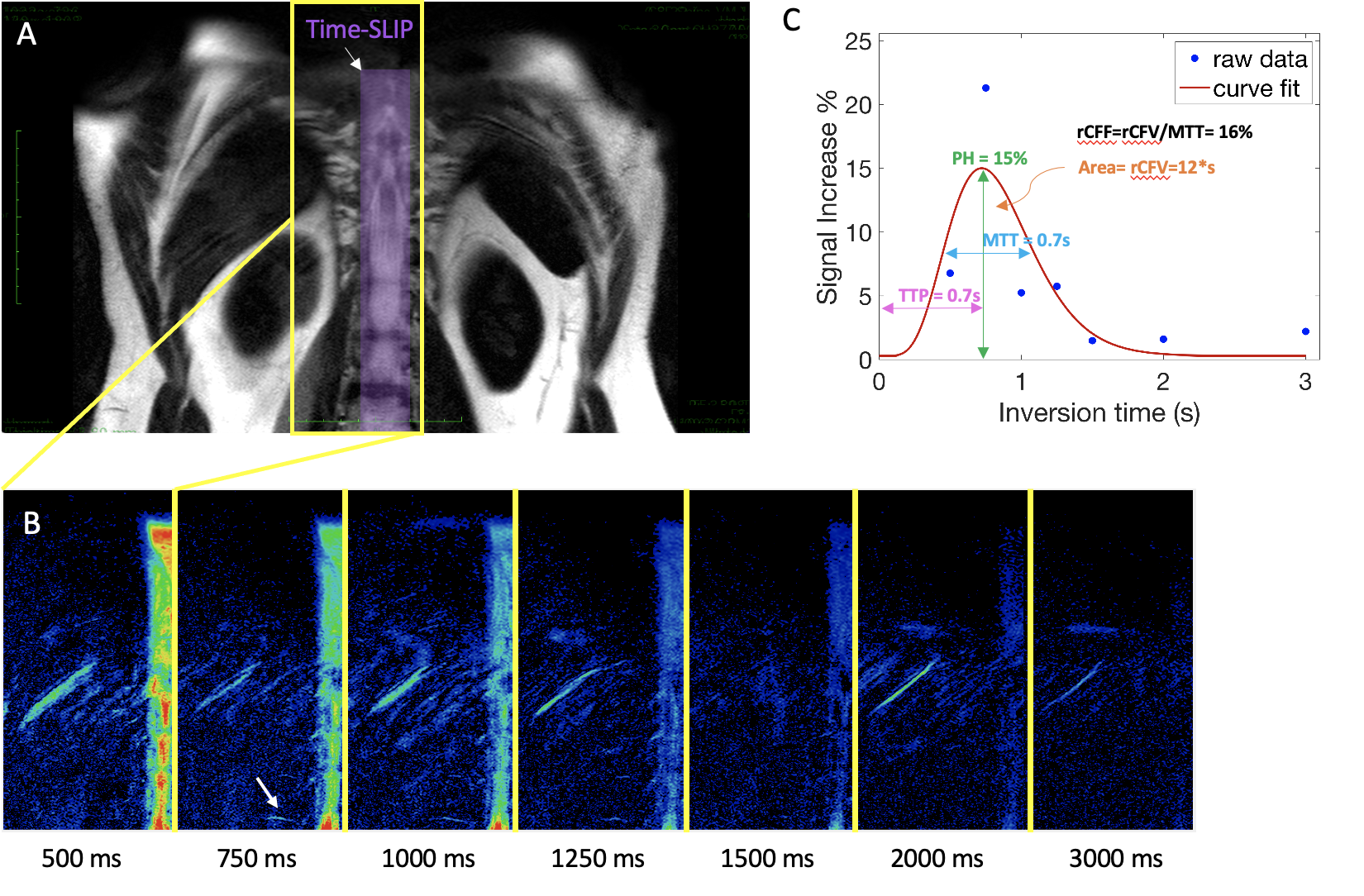
Figure 1. Male participant. A Coronal 3D cSSFSE image with spin labelling tag pulse placed sagittal highlighted in purple. The Time-SLIP tag pulse is 20mm in width down the spinal canal. B Subtracted color map for set of TIs. White arrow showing perfusion at 750ms. C Tagged CSF perfusion signal at the nerve root (white arrow). Peak height (PH) is 15%, mean transition time (MTT) is 0.7s, time to peak (TTP) is 0.7s, relative CSF volume (rCFV) is 12 %*sec under the curve, and relative CSF flow (rCFF) is 16%.
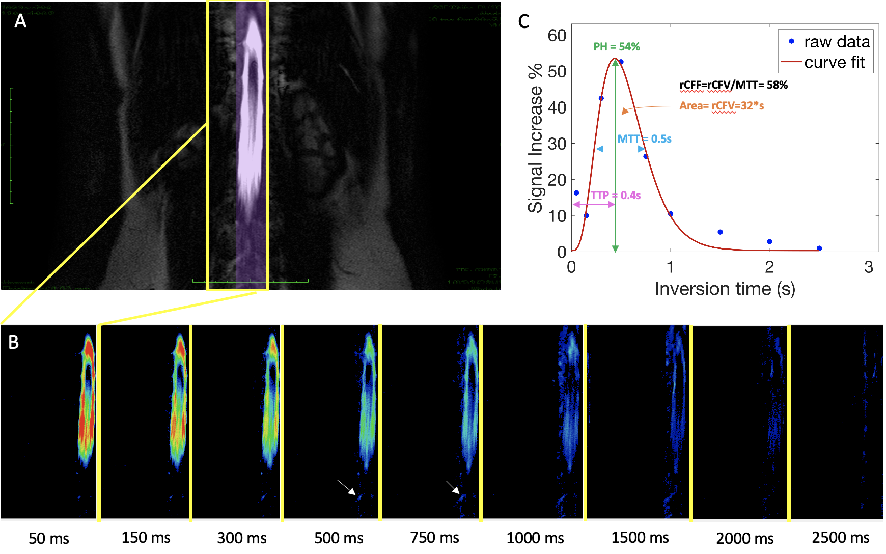
Figure 2. Female participant. A Coronal 3D cSSFSE image with spin labelling tag pulse placed sagittal highlighted in purple. The Time-SLIP tag pulse is 20mm in width down the spinal canal. B Subtracted color map for set of Tis. White arrow showing perfusion at 500ms and 750ms. C Tagged CSF perfusion signal at the nerve root (white arrow). Peak height (PH) is 54%, mean transition time (MTT) is 0.5s, time to peak (TTP) is 0.4s, relative CSF volume (rCFV) is 32%*sec under the curve, and relative CSF flow (rCFF) is 58%.
DOI: https://doi.org/10.58530/2023/1552