1546
In vivo MR vessel size imaging of brain vascular plasticity after experimental spinal cord injury11.Institute of Health and Medical Technology, Hefei Institutes of Physical Science, Hefei Cancer Hospital, Chinese Academy of Sciences, Hefei 230031, P. R. China, Hefei, China, 2Institute of Health and Medical Technology, Hefei Institutes of Physical Science, Hefei Cancer Hospital, Chinese Academy of Sciences, Hefei, China, 31.Institute of Health and Medical Technology, Hefei Institutes of Physical Science, Hefei Cancer Hospital, Chinese Academy of Sciences, Hefei 230031, P. R. China, Hefei,China, China
Synopsis
Keywords: Blood vessels, Blood vessels
Spinal cord injury (SCI) leads to neuronal cell death, axonal damage and demyelination. Brain undergo anatomical changes following SCI. Recently MR vessel size imaging has shown promising application in visualizing neovascularization. In this study we explored the possibility to vascular morphology changes and angiogenesis in the regions along the cranial corticospinal tract (CST) in SCI using vessel size imaging. The results showed increased microvascular density (Density), mean vessel diameter (mVD) and vessel size index (VSI) values in contralateral pyramids four weeks post-injury compared to pre-injury levels. Thus, vessel size imaging could provide valuable information of neovascularization in brain after SCI.
INTRODUCTION
Spinal cord injury (SCI) leads to neuronal cell death, axonal damage and demyelination. Both spinal cord 1 and brain 2,3 undergo anatomical changes following SCI. Recently MR vessel size imaging has shown promising application in visualizing angiogenesis in the brain 4-6. Therefore, in this study we explored the possibility to investigate vascular morphology changes and angiogenesis in the regions along cranial corticospinal tract (CST) in SCI using MR vessel size imaging. We also correlated the MRI findings to immunohistochemistry.METHODS
These studies were performed on six Sprague-Dawley rats weighing from 200–250 g. A T9 spinal cord hemisection was performed as described previously 5. MR scans were carried out on 4 weeks post-injury. All MR studies were performed on a Bruker 7T spectrometer. For in vivo MR studies, rats were anesthetized with a mixture of 2% isoflurane, 30% oxygen and air. T2* weighted imaging was obtained from 2D gradient echo sequence with TR/TE = 800/3.5 ms, flip angle = 50o, matrix size 256 × 256, FOV = 25 mm × 25 mm, 24 slices with slice thickness of 1 mm, a spectral width of 10 kHz, 2 averages. T2 weighted imaging was obtained from the spin-echo (SE) MRI sequence, and the parameters were listed as follows: TR/TE = 4500/35 ms, FOV = 25 mm × 25 mm, matrix size 256 × 256, slice thickness = 1 mm (24 slices, gap = 0); 2 averages; and spectral width = 50 kHz. Diffusions images were acquired using a multi-shot spin-echo echo planar imaging (EPI) sequence with TR/TE = 3000/25 ms, 8 shots, matrix size 128 × 128, FOV = 25 mm × 25 mm, 24 slices with slice thickness = 1 mm, 2 averages. A total of thirty diffusion encoding directions were used at a b-value of 800 s/mm2. Repeated gradient-echo and spin-echo MRI sequence scans were acquired after the injection of ultra-small superparamagnetic iron oxide (USPIO) contrast agent (Shanghai So-Fe Biomedical Co., Ltd.) via the tail vein over approximately 5 min. A dose of 0.02 mL per g of body weight was injected at a speed of 0.02 mL/s. Vessel size images were obtained as described previously 4,5. To quantify CST changes, each MR dataset was analyzed using ImageJ. Statistical comparisons made between the contralateral and ipsilateral levels were carried out by two-tailed Student’s paired t tests.RESULTS
Fig.1 shows that the vascular angiogenesis and morphology changed mainly in contralateral pyramid four weeks post-injury. Significant increases of 134%, 65% and 84% in contralateral pyramid (p < 0.05) 4 weeks post-injury compared to ipsilateral levels when quantified microvascular density (Density), mean vessel diameter in the voxel (mVD), and vessel size index (VSI) values, respectively.DISCUSSION
The increases of microvascular density, mVD, and VSI were observed in the contralateral pyramids in this study, suggestive of angiogenesis or vascular activation in the CST white matter. These MRI findings were confirmed by immunohistological results, i.e. obvious increase in CD31 staining after four weeks after injury, which may be associated with activated astrocytes according to the increased expression of GFAP in those regions.CONCLUSION
MR vessel size imaging is a potential endogenous biomarker for vascular morphology changes and angiogenesis in the brain after spinal cord injury.Acknowledgements
No acknowledgement found.References
1. Cohen-Adad J, et al. Neuroimage. 2011;55(3):1024-33; 2. Wrigley PJ, et al. Cereb Cortex. 2009;19(1):224-32; 3. He G, et. al. Microsc Res Tech. 2017;1–9; 4. Lelacqua GD, et al. Front. Aging Neurosci. 2016;7:241; 5. Lemasson B, et al. Magnetic Resonance in Medicine. 2013;69:18–26; 6. Xu X, et al. Aging (Albany NY) 2020;12(17):17224-17234.
Figures
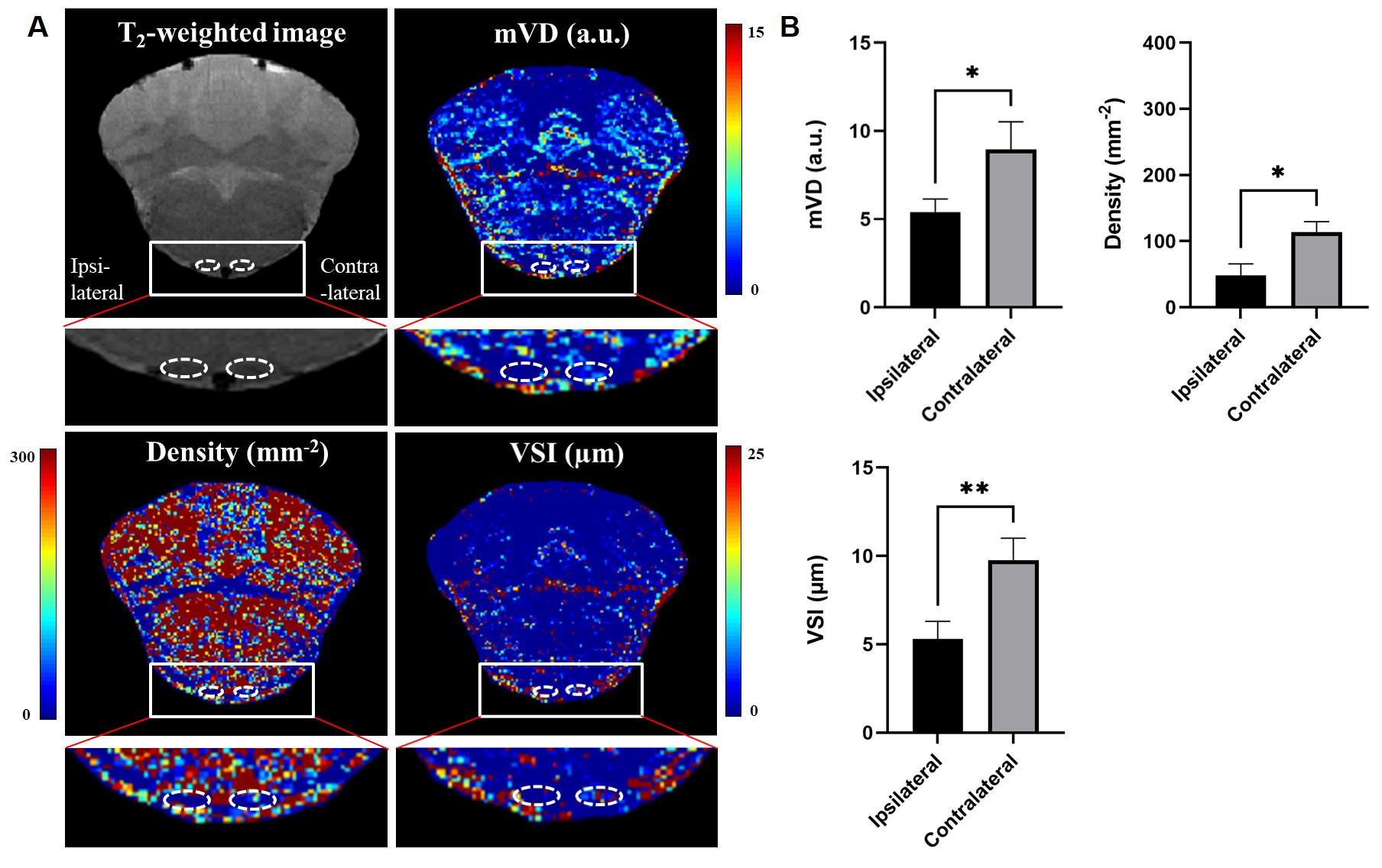
Fig 1. Typical MR vessel size images (A) of a rat brain 4 weeks after spinal cord hemisection injury and (B) quantitative results of microvascular density (Density), mean vessel diameter in the voxel (mVD), and vessel size index (VSI) in ROIs within the ipsilateral and contralateral brain pyramids of animals. “*” indicates significance at P=0.05.