1544
Detecting longitudinal alterations of cerebral white matter associated with breast cancer and chemotherapy using GQI1Department of Medical Imaging and Radiological Sciences, and Department of Artificial Intelligence, Chang Gung University, Taoyuan, Taiwan, 2School of Medicine, Chang Gung University, Taoyuan, Taiwan, 3Department of Psychiatry, Chang Gung Memorial Hospital, Chiayi, Taiwan, 4Department of Diagnostic Radiology, Chang Gung Memorial Hospital, Chiayi, Taiwan, 5Medical Imaging Research Center, Institute for Radiological Research, Chang Gung University and Chang Gung Memorial Hospital at Linkou, Taoyuan, Taiwan
Synopsis
Keywords: Brain Connectivity, Diffusion/other diffusion imaging techniques
In 2020, breast cancer is the most prevalent type of cancer and has the highest incident rate in women worldwide. The extensively used adjuvant chemotherapy might have detrimental effect on human brain and results in chemotherapy-related cognitive impairment (CICI) in breast cancer patients. The present study performed longitudinal design, aiming to investigate the microstructural and macroscale white matter alterations by generalized q-sampling imaging (GQI). Our results suggested that the patients had changes in local white matter integrity and network performance in DAN before treatment and frontal lobe connection after treatment.Introduction
In 2020, breast cancer is the most prevalent type of cancer and has the highest incident rate in women worldwide. The extensively used adjuvant chemotherapy might have detrimental effect on human brain and results in chemotherapy-related cognitive impairment (CICI) in breast cancer patients. Furthermore, patients prior to chemotherapy also reported cancer-related cognitive impairment (CRCI) which might be due to physiological factor or mood symptoms. Previous noninvasive imaging studies showed replicated results of functional and structural alterations in breast cancer patients. The aim of this study was to use generalized q-sampling imaging (GQI), regarded as a technique that can deal with the problem of complex fibers that DTI cannot do, to explore longitudinal changes of white matter integrity and network connections in breast cancer survivors from pre- to post- chemotherapeutic regimens. We hypothesized that mood symptoms and neurotoxicity would affect white matter tracts, especially in the dorsal attention network (DAN) and default mode network (DMN), which are involved in cognitive processing.Methods
All participants were recruited from Chiayi Chang Gung Memorial Hospital. Subjects were eligible if they met the following criteria: female, over the age of 20. These patients were further categorized into pre-treatment (BB, n=65) if they were diagnosed with primary breast cancer and age-matched no-cancer controls (BH, n=71). Some participants came back to have follow-up assessment. In longitudinal study, 28 matched pairs of BB/BBF and 28 matched pairs of BH/BHF were included. At time point one (TP1), baseline data were collected after surgery but before the start of chemotherapy. At time point two (TP2), follow-up data took place at 7.5 months in averaged after completing chemotherapy. The assessment included completion of neuropsychological tests, patient-reported outcomes, and diffusion MRI.Diffusion MRI data were acquired by using 3T MRI (Verio, Siemens). A single-shot spin echo echo-planar imaging (SE-EPI) pulse sequence was performed to obtain diffusion-weighted images in total 193 diffusion-sensitive gradient with b-values 0, 1000, 1500, 2000 s/mm2 along 1, 64, 64, 64 directions, respectively. The eddy current correction was performed by using the diffusion toolbox of the FMRIB software library (FSL, Analysis Group, Oxford, UK) to ensure image quality [1]. After that, Statistical parametric Mapping (SPM, The Wellcome Centre for Human Neuroimaging, London) was applied for normalization and affine non-linear registration into MNI space [2]. Lastly, normalized DWI data were reconstructed with the GQI method. The GQI indices, including GFA maps and NQA maps, and connectivity matrices were obtained from DSI studio (NTU, Taipei, Taiwan) to further analysis [3]. For statistical approaches, voxel-based analysis (VBA) and graph theoretical analysis (GTA) were both performed. Voxel-based two-way repeated measurement ANOVA model was applied to GFA and NQA maps, with time as within-subject factor and group as between-group factor, to investigate group-by-time interaction. Paired t-test were used for assessing the statistically significant changes in the GQI indices from TP1 (BB and BH) to TP2 (BBF and BHF). The network measurement, including topological properties [4] and network-based comparison [5], were also performed in longitudinal assessment.
Results
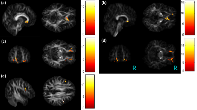
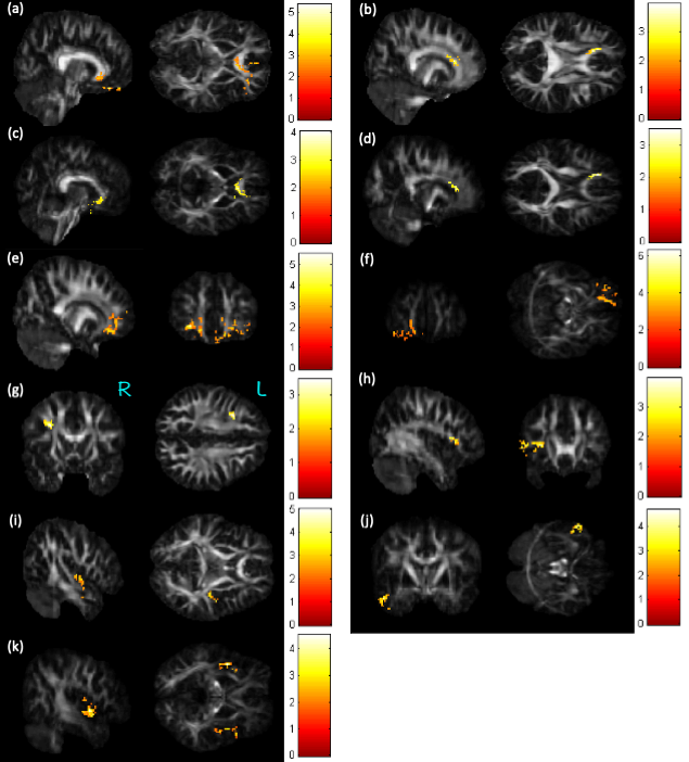
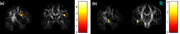
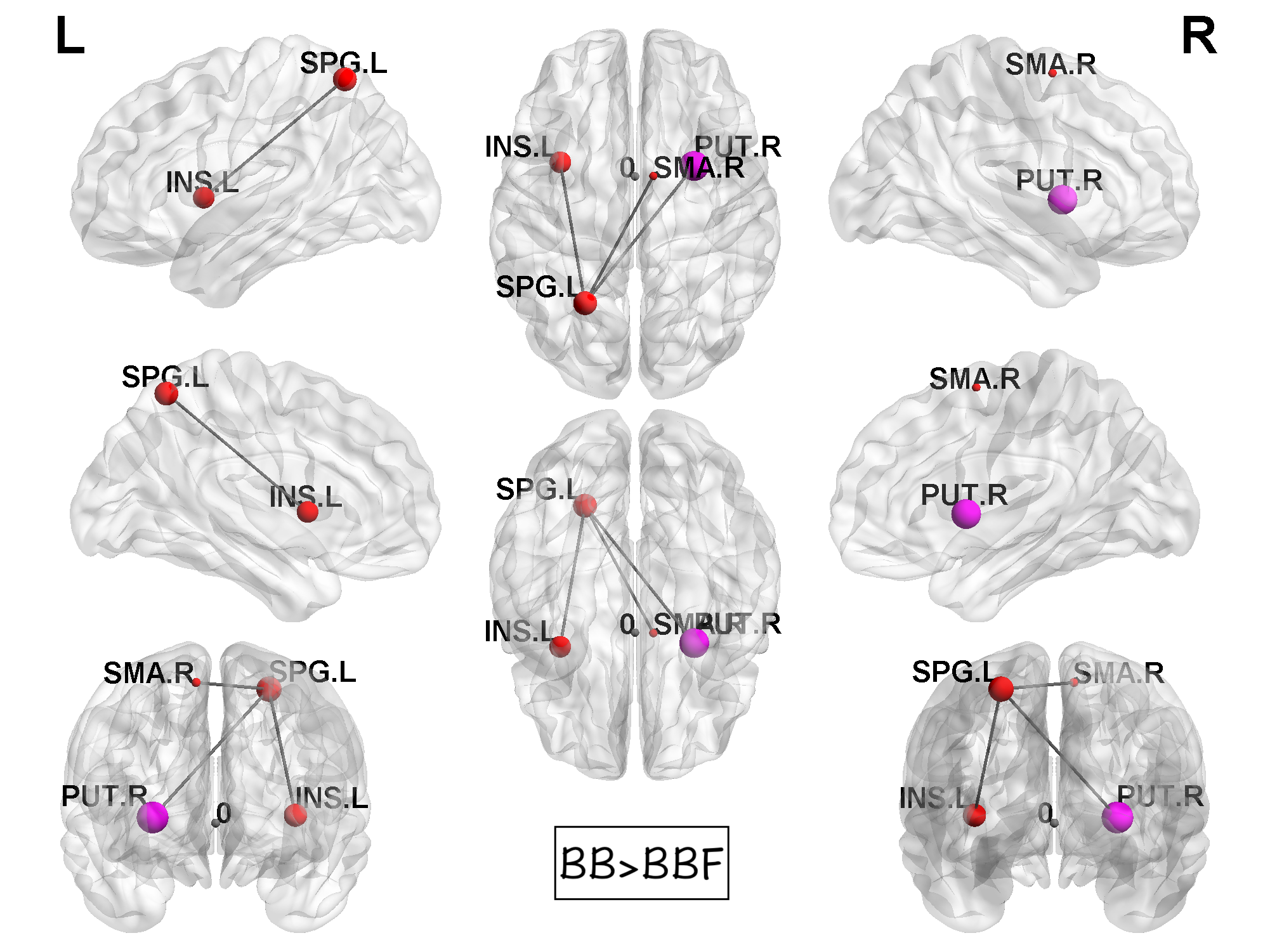
The repeated measurement ANOVA explored significant group by time interactions in the regions of corpus callosum (CC), bilateral orbital part of middle frontal gyrus (ORBmidF), and bilateral middle frontal gyrus (MFG) (Figure 1). Paired t-test detected significant decreased GQI indices in the regions of corpus callosum (CC), left orbital part of middle frontal gyrus (ORBmidF), left middle frontal gyrus (MFG), left inferior frontal gyrus (IFG), right superior temporal gyrus (STG), left middle temporal gyrus (MTG) as well as bilateral insula in patient group (Figure 2) and notable lower GFA values in the regions of right IFG as well as left STG in control group (Figure 3) from TP1 to TP2.The topological data analysis revealed that the AUC differed significantly in some topological properties between BB and BBF, but not between BH and BHF. The results showed significantly lower gamma, lower characteristic path length, higher transitivity, and nearly significantly lower modularity in post-treatment group versus pre-treatment group. Additionally, the NBS detected significantly weaker structural connections among the supplementary motor area, parietal lobe and, subcortical regions in chemotherapy-treated patients compared to baseline (Figure 4).
Discussion
Previous studies have reported that whether breast cancer patients received chemotherapy or not, they suffered from cognitive function impairment. The presence of cancer, stress, and various therapeutic strategies might have an impact on brain. These adverse effects could affect memory, attention, executive function, and processing speed [6]. In this study, we found several white matter tracts disruptions associated with dorsal attention network (DAN) in BC patients prior to chemotherapy. And reductions in white matter integrity associated with default mode network (DMN) in breast cancer patients after receiving chemotherapy.Conclusion
Injuries to the myelin, whether caused by a traumatic stressor or chemotoxicity, can adversely affect cognition. The present study investigated cerebral white matter microstructure and macroscale alterations in breast cancer survivors with and without chemotherapy. Our results suggested that the patients had changes in local white matter integrity and network performance in DAN before treatment and frontal lobe connection after treatment. The results provided evidence of white matter alterations in breast cancer patients, and they may serve as potential imaging markers of cognitive changes.Acknowledgements
This study was supported by research grants MOST107-2221-E-182-054-MY3 and NSTC111-2221-E-182-021 from the National Science and Technology Council, Taipei, Taiwan, respectively. This study was also supported by grantsNMRPD1H0101~3 from Chang Gung University, Taoyuan, Taiwan and CORPG6G0101~3 and CORPG6G0121~3 from Chang Gung Memorial Hospital, Chiayi, Taiwan.References
1. Graham, M.S., et al., Realistic simulation of artefacts in diffusion MRI for validating post-processing correction techniques. NeuroImage, 2016, 125: 1079-1094.
2. Penny, W., et al., Statistical Parametric Mapping: The Analysis of Functional Brain Images. 2007.
3. Yeh, F.C., et al., Generalized q-sampling imaging. IEEE Trans Med Imaging, 2010, 29(9): 1626-35.
4. Hosseini, S.M.H., et al., GAT: A Graph-Theoretical Analysis Toolbox for Analyzing Between-Group Differences in Large-Scale Structural and Functional Brain Networks. PLOS ONE, 2012, 7(7): e40709.
5. Zalesky, A., et al., Network-based statistic: Identifying differences in brain networks. NeuroImage, 2010, 53(4): 1197-1207.
6. Pendergrass, J.C., Set al., Cognitive Impairment Associated with Cancer: A Brief Review. Innov Clin Neurosci, 2018, 15(1-2): 36-44.
Figures