1542
Cortical thickness alterations in patients with T2DM and its correlation with cerebral small vessel diseases and cognitive function1The First Affiliated Hospital of Dalian Medical University, Dalian, China, 2Philips Healthcare, Beijing, China
Synopsis
Keywords: Neurodegeneration, Diabetes, small vessel diseases (CSVD)、cognition、freesurfer
The objective of this study was to explore the changes of cortical thickness in patients with type 2 diabetes mellitus, and the correlation between the thinning cerebral regions and cerebral small vessel diseases (CSVD) burden. The thickness of the left medial orbitofrontal cortex was negatively correlated with the centrum semi-ovale enlarged perivascular space (CSO-EPVS) score and CSVD total burden score. The thickness of the left lateral occipital cortex was negatively correlated with CSO-EPVS score, basal ganglia enlarged perivascular space (BG-EPVS) score, and CSVD total burden score, and positively correlated with mini-mental state examination (MMSE) delayed memory score.
Introduction
The thickness of the cerebral cortex reflects the number of neurons in the cortical column, and the thinning of the cerebral cortex is associated with the decline of cognitive function. The prevalence of cerebral small vessel diseases (CSVD) in patients with T2DM increased [1], and with the development of the severity of CSVD, cognitive ability decreased [2-3]. The purpose of this study was to explore the changes of cortical thickness in patients with type 2 diabetes mellitus (T2DM) and to analyze the correlation between the thinning cerebral regions and cerebral small vessel diseases (CSVD) quantitative scores and cognitive scores.Materials and Methods
Thirty-one patients with T2DM and 24 volunteers as the control group were prospectively collected. MRI examinations were performed with Philips Ingenia CX 3.0T MRI scanner, with T1WI, T2WI, T2 Flair, 3D T1WI, and susceptibility-weighted imaging (SWI) sequences. The CSVD MRI features of all subjects were quantified and scored, including periventricular hyperintense (PVH) and deep white matter hyperintense (DWMH) grades, basal ganglia enlarged perivascular space (BG-EPVS) and centrum semi-ovale enlarged perivascular space (CSO-EPVS) scores, lacune grade, cerebral microbleed (CMB) grade, and CSVD total burden scores were calculated[4]. Detailed neuropsychological scale assessments were performed on all subjects, including mini-mental state examination (MMSE), Montreal cognitive assessment (MoCA), California verbal learning test (CVLT), and symbol digit modalities test (SDMT). FreeSurfer software (Version 6.0.0,http://surfer.nmr.mgh.harvard.edu/) was used for image post-processing, and the cerebral cortex thickness of all subjects was calculated.The difference in cortical thickness between the T2DM group and control group was compared by the general linear model (GLM), and corrected by the false discovery rate (FDR) method. The cortical thickness of brain regions with significant differences between the two groups was extracted. Considering Age, gender, and years of education as covariables, partial correlation analysis was carried out between cortical thickness values and CSVD quantitative scores and cognitive scores.
Results
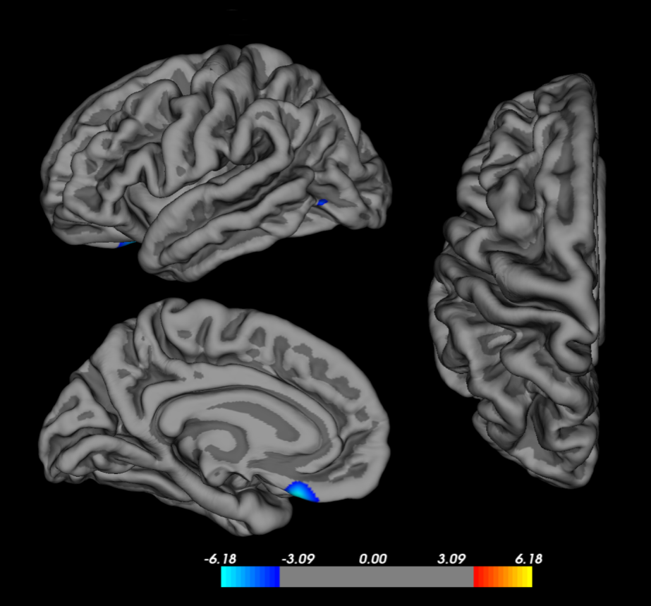
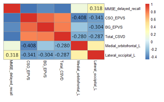
Compared with the control group, the cortical thickness of the left medial orbitofrontal cortex and the left lateral occipital cortex were significantly reduced in the T2DM group (FDR correction, p < 0.05) (Figure 1). Partial correlation analysis showed that the thickness of the left medial orbitofrontal cortex was negatively correlated with the CSO-EPVS score (r=-0.408, p=0.002) and CSVD total burden score (r=-0.280, p=0.038). The thickness of the left lateral occipital cortex was negatively correlated with the CSO-EPVS score (r=-0.341, p=0.011), BG-EPVS score (r=-0.304, p=0.024), and CSVD total burden score (r=-0.287, p=0.034). And positive correlation was observed between the thickness of the left lateral occipital cortex and MMSE delayed memory score (r=0.318, p=0.018) (Figure 2).Discussion
The medial orbitofrontal cortex (mOFC) connects the diencephalon and limbic systems and participates in cognitive functions such as decision-making and execution. In this study, the thickness of mOFC was negatively correlated with the CSO-EPVS score and CSVD total burden score, suggesting that the heavier the burden of CSVD in T2DM patients, the thinner the thickness of mOFC. Therefore, the prevention of cerebrovascular disease may be a valuable way to prevent cognitive decline in patients with T2DM in the future.The lateral occipital gyrus is mainly involved in the visual information transmission pathway, related to object recognition function, and is an important node of a visual transmission pathway, which is from the fusiform gyrus to the lateral occipital gyrus and temporal lobe [5-6]. In this study, the thickness of the left lateral occipital cortex was negatively correlated with CSO-EPVS score, BG-EPVS score, and CSVD total burden score, and positively correlated with MMSE delayed memory score, suggesting that the heavier the burden of CSVD in T2DM patients, the thinner the thickness of the left lateral occipital cortex and the more severe the delayed memory deficit.
Conclusion
This study suggests that the thinning of the cognitive and vision-related cortex in T2DM patients is correlated with increased CSVD burden and cognitive decline.Acknowledgements
No acknowledgement found.References
1. Fang F, Cao R, Luo Q, et al. The silent occurrence of cerebral small vessel disease innonelderly patients with type 2 diabetes mellitus[J]. J Diabetes,2021,13(9): 735-743.
2. Smith EE, Cieslak A, Barber P, et al. Therapeutic strategies and drug development for vascular cognitive impairment[J]. J Am Heart Assoc,2017,6(5): e005568.
3. Lawrence AJ, Brookes RL, Zeestraten EA, et al. Patternand rate of cognitive decline in cerebral small vessel disease: a prospective study[J]. PloS ONE 2015;10(8): e0135523.
4. Staals J , Makin SDJ , Doubal FN , et al. Stroke subtype , vascular risk factors , and total MRI brain small - vessel disease burden [ J ]. Neurology ,2014,83(14):1228-1234.
5. MilnerAD. How do the two visual streams interact with each other[J]? Exp Brain Res, 2017, 235(5): 1297-1308.
6. Goodale MA, Milner AD. Two Visual Pathways-where have they taken us and where will they lead in future[J]? Cortex, 2018, 98: 283-292.
Figures