1522
Three-dimensional Black-blood Thrombus Imaging (BTI) with LIBRE Fat suppression for the diagnosis of Deep Vein Thrombosis1Guangzhou Medical University, Guangzhou, China, 2The First People’s Hospital of Qinzhou, Qinzhou, China, 3Siemens Healthineers, Shanghai, China
Synopsis
Keywords: Vessels, Thrombo-Embolic
Deep vein thrombosis (DVT) can lead to life-threatening pulmonary embolism. Black-blood thrombus imaging (BTI) technique can accurately identify DVT and provide additional information for thrombus staging. However, non-uniform fat suppression of BTI with conventional fat saturation preparation is obvious due to the field inhomogeneities and large field of view (FOV) imaging, which can affect the accuracy of detecting and staging of thrombus. To address this issue, a field inhomogeneity insensitive BTI technique was developed by incorporating LIBRE pulses for fat free and large FOV thrombus imaging.Purpose
Deep vein thrombosis (DVT) is a disease with high morbidity and can lead to life-threatening pulmonary embolism [1]. Recently, a 3D black-blood thrombus imaging (BTI) technique which can effectively suppress venous blood flow to directly visualize and identify thrombosis was developed [2,3]. However, the wide distribution of the thrombus requires large field of view (FOV) for imaging, which results in non-uniform fat suppression due to the inhomogeneity of magnetic fields. Non-uniform fat suppression can affect the accuracy of detecting and staging of thrombus. Fortunately, lipid insensitive binomial off-resonant radio frequency excitation (LIBRE), a novel water excitation module, has been developed and implemented in GRE-based sequences [4]. Previous studies demonstrated that LIBRE is insensitive to magnetic field inhomogeneities and provides better fat suppression in imaging lower extremities [4]. Inspired by these previous studies, we aimed to develop a new BTI technique by incorporating LIBRE for fat free and large FOV thrombus imaging for the diagnosis of DVT.Methods
MR sequence:LIBRE was incorporated into BTI (LIBRE-BTI) to achieve uniform fat suppression. Parameters of LIBRE-BTI were then optimized in numerical simulations, which should meet the following objectives: (i) the effective flip angle of LIBRE should be ~90°; and (ii) the transverse magnetization of fat after LIBRE is close to zero.In vivo study: The optimized LIBRE-BTI sequence was tested on 7 healthy volunteers (5F 2M, age 25.3±4.9 y) and 5 DVT patients (2F 3M, age 52.5±10.5 y). A conventional water excitation (WE) approach (WE-BTI) that uses a 1-2-1 binomial RF pulse was conducted for comparison. The experiments were performed on a 3T MR scanner (MAGNETOM Skyra, Siemens AG, Erlangen, Germany) with an 18-channel body coil and an integrated spine coil. Imaging parameters of LIBRE-BTI and WE-BTI were: TR/TE = 650/12 ms, field of view (FOV) = 413×294 mm2, voxel size = 1.15×1.15×1.20 mm3 (interpolated to 0.57×0.57×0.60 mm3), turbo factor = 45, a centric reordering approach. LIBRE with a sub-pulse of 70° excitation angle, 1.05 ms duration and 380 Hz frequency offset was used in BTI.
Image analysis: All acquired images were loaded to a workstation (Leonardo; Siemens AG, Germany) for image analysis. Signal-to-noise ratio (SNR) and contrast-to-noise ratio (CNR) were calculated to evaluate the fat suppression effect of LIBRE-BTI.
Results
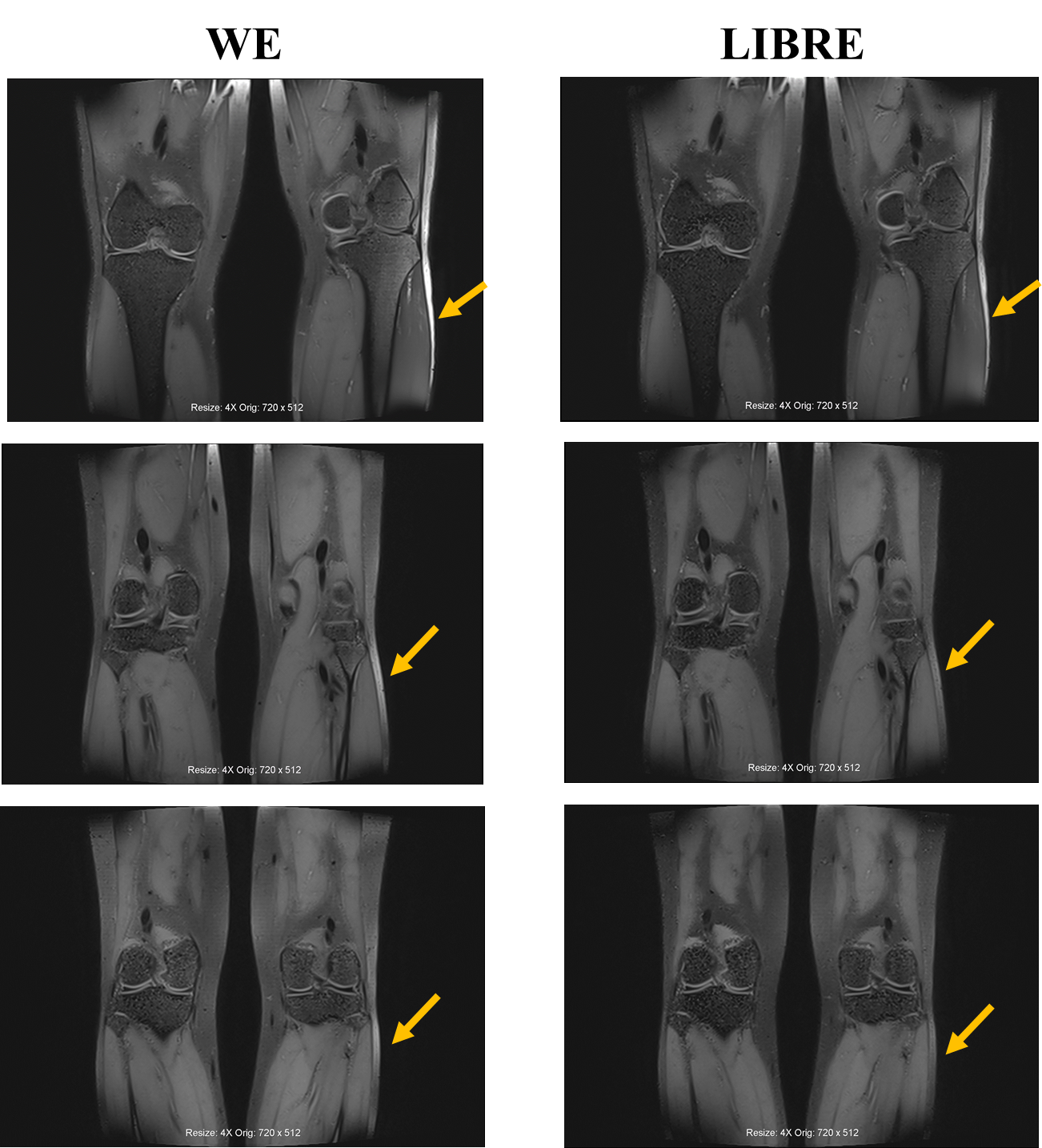
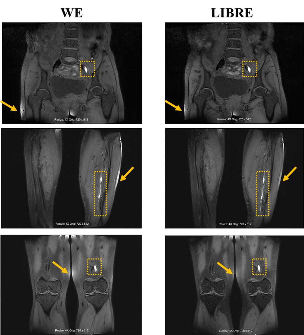
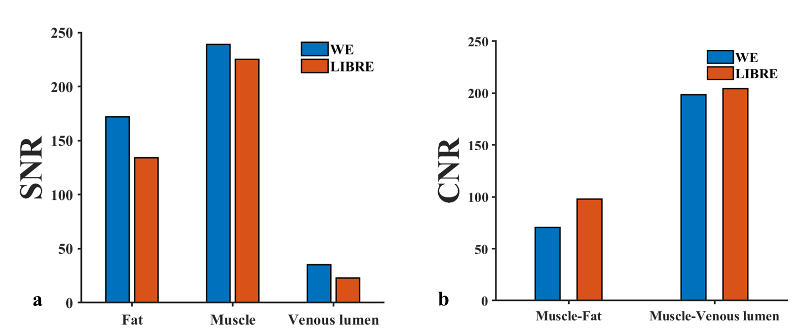
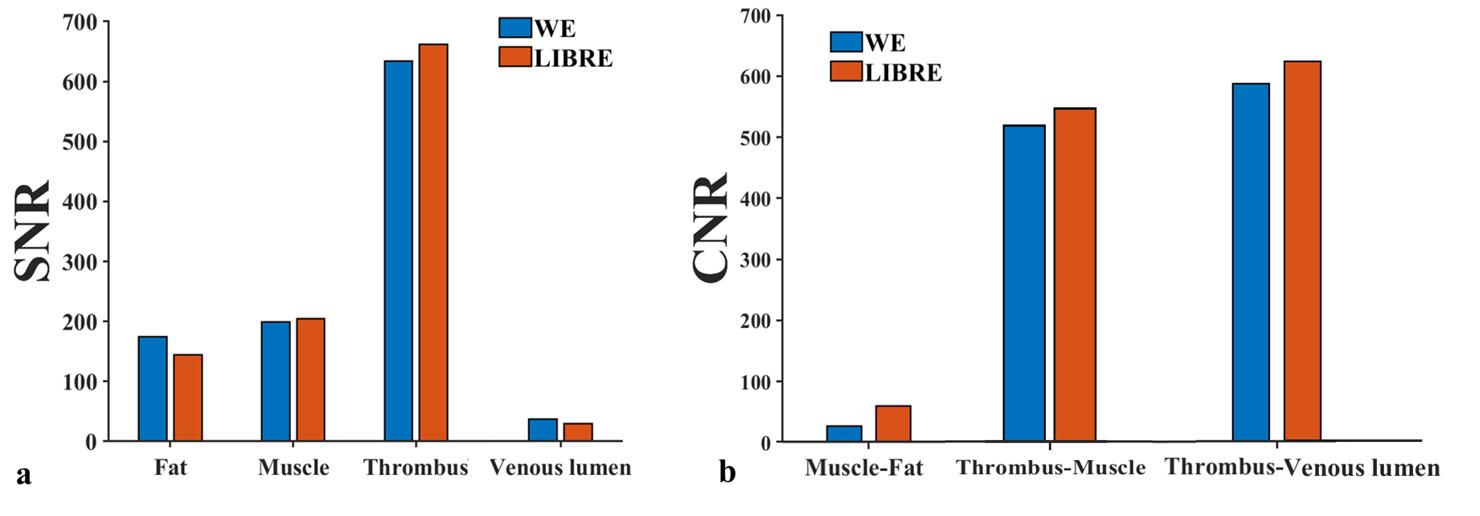
Representative images obtained by LIBRE-BTI and WE-BTI in volunteer and patient experiment were shown in Figure 1&2. LIBRE-BTI showed better fat suppression in the interface between fat and air (yellow arrow in Figure 1&2) and better muscle-to-fat contrast than WE-BTI both in volunteer and patient images. Average SNRs and CNRs of 7 volunteers and 5 patients were shown in Figure 3&4. In volunteer study, LIBRE-BTI got lower average SNRs of fat (133.7 vs. 171.9) and venous lumen (23.2 vs. 34.6) and a higher average CNR of muscle to fat (97.8 vs.70.8) than WE-BTI. In addition, the SNR of muscle (225.3 vs. 238.9) and the CNR of muscle to venous lumen (198.7 vs. 204.2) in LIBRE-BTI and WE-BTI were comparable. In patient study, average fat SNR obtained by LIBRE-BTI was reduced by 17% compared with WE-BTI (143.7 vs. 174.2). SNRs of thrombus and muscle obtained by LIBRE-BTI and WE-BTI were comparable (thrombus: 634.2 vs. 662.5; muscle: 203.7 vs. 199.4). LIBRE-BTI showed 119% improvement in the muscle-to-fat CNR than WE-BTI (60.1 vs. 27.4). Compared with WE-BTI, LIBRE-BTI obtained comparable thrombus to muscle CNR (458.8 vs. 434.7) and comparable thrombus to venous lumen CNR (633.9 vs. 597.3).Discussion
LIBRE provides better fat suppression than conventional WE approach in BTI sequence for the diagnosis of DVT. First, LIBRE-BTI was robust to field inhomogeneities. LIBRE-BTI effectively reduced fat signal at the tissue-air interface. Second, LIBRE-BTI showed an excellent muscle-to-fat contrast with twice average CNR of WE-BTI in patient study. Third, the average SNRs of muscle and thrombus in LIBRE-BTI remained consistent with WE-BTI.Conclusion
LIBRE-BTI can improve fat suppression and thus has great potential for the diagnosis of DVT.Acknowledgements
No acknowledgement found.References
[1] van der Hulle T, Dronkers CEA, Huisman MV, Klok FA: Current standings in diagnostic management of acute venous thromboembolism: Still rough around the edges. Blood Rev 2016, 30(1):21-26.
[2] Xie G, Chen H, He X, Liang J, Deng W, He Z, Ye Y, Yang Q, Bi X, Liu X, Li D, Fan Z: Black-blood thrombus imaging (BTI): a contrast-free cardiovascular magnetic resonance approach for the diagnosis of non-acute deep vein thrombosis. J Cardiovasc Magn Reson 2017, 19(1):4.
[3] Tan M, Mol GC, van Rooden CJ, Klok FA, Westerbeek RE, Iglesias Del Sol A, van de Ree MA, de Roos A, Huisman MV: Magnetic resonance direct thrombus imaging differentiates acute recurrent ipsilateral deep vein thrombosis from residual thrombosis. Blood 2014, 124(4):623-627.
[4] Bastiaansen JAM, Stuber M. Flexible water excitation for fat-free MRI at 3T using lipid insensitive binomial off-resonant RF excitation (LIBRE) pulses. Magn Reson Med. 2018;79(6):3007-3017.
Figures