1511
The risk prediction of ischemic stroke recurrence: a MR vessel wall imaging study
Fei Zhou1, Maoxue Wang1, Ruijing Xin1, Tianshu Yang2, Yongming Dai3, Na Zhang4, Zhanli Hu4, Xin Zhang1, and Bing Zhang1
1Department of Radiology, The Affiliated Drum Tower Hospital of Nanjing University Medical School, Nanjing, China, 2Shenzhen United Imaging Research Institute of Innovative Medical Equipment, Shenzhen, China, 3MR Collaboration, Central Research Institute, United Imaging Healthcare, Shanghai, China, 4Paul C. Lauterbur Research Center for Biomedical Imaging, Shenzhen Institute of Advanced Technology, Chinese Academy of Sciences, Shenzhen, China
1Department of Radiology, The Affiliated Drum Tower Hospital of Nanjing University Medical School, Nanjing, China, 2Shenzhen United Imaging Research Institute of Innovative Medical Equipment, Shenzhen, China, 3MR Collaboration, Central Research Institute, United Imaging Healthcare, Shanghai, China, 4Paul C. Lauterbur Research Center for Biomedical Imaging, Shenzhen Institute of Advanced Technology, Chinese Academy of Sciences, Shenzhen, China
Synopsis
Keywords: Vessel Wall, Vessels
Symptomatic cerebral vascular stenosis diseases include acute ischemic infarction and transient ischemic syndrome. Patients with these diseases often have a high risk of recurrence in the short term, which may have serious adverse consequences. High-resolution MR vessel wall imaging (HR-VWI) technique has provided new possibilities to assess the threat of stroke recurrence due to stenosis caused by intracranial plaque. The present study confirms that plaque morphological parameters obtained by HR-VWI can provide an effective prediction of stroke recurrence risk in patients with intracranial arterial stenosis.Introduction
Intracranial arterial stenosis due to intracranial plaque is a high-risk factor for cerebrovascular diseases including ischemic stroke and transient ischemic attack. Previous studies have shown that these diseases have a high mortality and disability rate and a high risk of recurrence, thus early evaluation and detection are key to prevent adverse outcomes1. High-resolution MR vessel wall imaging is increasingly used in the diagnosis and study of intracranial arterial stenosis due to its ability to qualitatively and quantitatively study intracranial vessel wall lesions2. In this study, we extracted morphological indices of intracranial plaque using high-resolution vessel wall imaging (HR-VWI) and evaluated the effectiveness of this approach in identifying people at risk for ischemic stroke recurrence.Methods
Patients and Questionnaire:The current study was approved by the institutional review board. Written informed consent was obtained from each patient. A total of 126 patients with intracranial stenosis on MRA were recruited, exclusion criteria were: (1) patients who were diagnosed as vasculitis, dissection, vasospasm, Moyamoya disease, or reversible cerebral vasoconstriction syndrome (n = 21); (2) patients with a history of brain surgery (n = 9); (3) patients with incomplete clinicopathological information (n = 7). The detailed information of patients was shown in Table 1. The risk of stroke recurrence for each patient was assessed using the Essen Stroke Risk Score (ESRS), and patients were divided into high-risk group and low-risk group according to their score of ESRS scale (< 3 for low-risk).
Imaging protocol:
MRI Examinations were performed on a 3.0 T MR scanner (uMR 770, United Imaging Healthcare, Shanghai, China). Protocols were: (1) Time-of-flight MRA: repetition time/echo time (TR/TE) = 19.1/3.6 msec, field of view (FOV) = 220×180 $$$mm^{2}$$$, matrix size = 672×438, slice thickness = 0.6 $$$mm$$$, compress sensing-based acceleration factor (uCS) = 3.5, acquisition time = 2 minute 21 seconds; (2) T1-weighted 3D MATRIX (Modulated flip Angle Technique in Refocused Imaging with extended echo train) sequence: TR/TE = 902/13.92 msec, FOV = 192×172 $$$mm^{2}$$$, matrix size = 481×432, slice thickness = 0.4 $$$mm$$$, uCS = 4.9, acquisition time = 6 minute 8 seconds. Post-contrast images were obtained using same T1w MATRIX sequence with identical parameters after intravenous injection of gadolinium contrast (dosage: 0.1 mmol/kg) agent and a 5 minutes interval.
Image analysis:
Preprocessing of HR-VWI image was complicated with a dedicated plaque analysis software (uWS PlaqueTool, United Imaging Healthcare, Shanghai, China). First, a central line extraction algorithm and a vessel wall segmentation algorithm were used to automatically perform curved-planar reconstruction of all intracranial arteries and segmentation of the vessel wall and lumen. Second, the narrow point was manually selected at the most narrowed position of the entire vessel, while the reference points was selected at the site of normal vessel proximal or distal to the lesion site. Then the quantitative parameters including lumen diameter (LD), lumen area (LA), wall thickness (WT), normalized wall index (NWI), stenosis rate and remodeling index (RI) were automatically calculated. Further plaque component analysis was completed at pre-defined narrow point, after manually outlining the plaque and plaque components, the software automatically outputs the percentage of intraplaque hemorrhage (IPH).
Statistical analysis:
Mann-Whitney U test was used for parameters comparison, combined parameter was generated by logistic regression and the discriminatory performance of single and combined parameters was further assessed by receiver operating characteristic (ROC) analysis and DeLong’s test.
Results
Eighty-nine subjects with evidence of intracranial atherosclerosis on clinical imaging were recruited (Table 1). The results demonstrated that RI of high recurrence risk patients was significantly lower than low-risk patients (P = 0.02). Figure 2 showed that RI was the only single parameter which was able to significantly identify high-risk patients (AUC=0.649, P = 0.02). The combined parameter generated by RI and percentage of IPH, could slightly improve the discriminatory performance (AUC=0.693, P < 0.01).Discussion and Conclusion
Our results suggest that imaging indices obtained by HR-VWI can effectively identify people with high risk for stroke recurrence, thus contributing to the early identification and screening accuracy of such high-risk patients.Acknowledgements
No acknowledgement found.References
1. Zhang X, Chen L, Li S, et al. Enhancement Characteristics of Middle Cerebral Arterial Atherosclerotic Plaques Over Time and Their Correlation With Stroke Recurrence. J Magn Reson Imaging. Mar 2021;53(3):953-962. doi:10.1002/jmri.27351
2. Mazzacane F, Mazzoleni V, Scola E, et al. Vessel Wall Magnetic Resonance Imaging in Cerebrovascular Diseases. Diagnostics (Basel). Jan 20 2022;12(2)doi:10.3390/diagnostics12020258
Figures
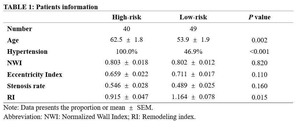
Table 1: Descriptive Statistics of Patients
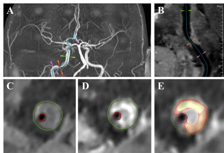
Figure 1: A: TOF
image, orange arrow indicated the narrow point, green and pink arrows are
represent sites; B: A narrow point in the internal carotid artery (ICA) on CPR
image; C: T1WI image at narrow point; d: CE-T1WI image at narrow point; E: The plaque
configuration at the narrow point.
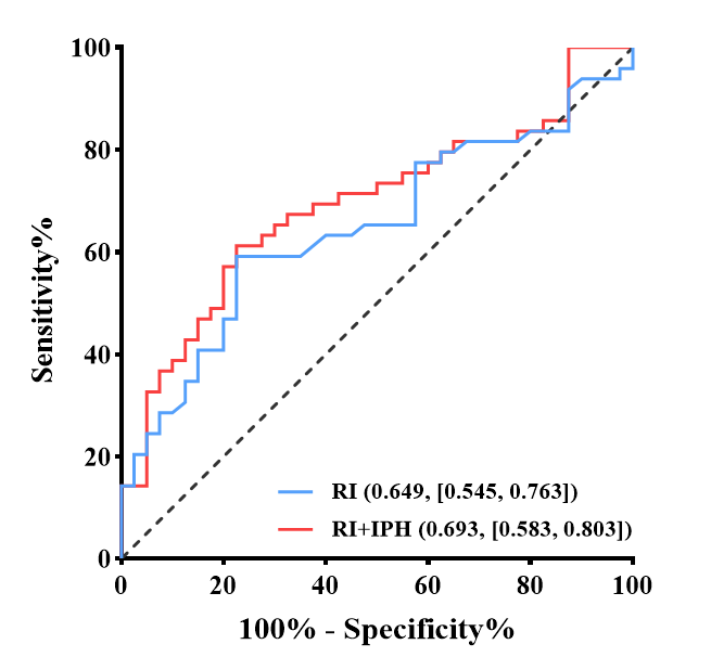
Figure 2: ROC curves of single RI and combined
parameter in distinguishing between patients with high and low ischemic stroke
recurrence risk.
DOI: https://doi.org/10.58530/2023/1511