1483
Multiparametric MRI–based Radiomics Analysis of Endometrial Carcinoma for Preoperative Risk stratification
Bai min1
1Radiology, Liaocheng People's Hospital, Liao'cheng, China
1Radiology, Liaocheng People's Hospital, Liao'cheng, China
Synopsis
Keywords: Uterus, Radiomics
Preoperative risk stratification of endometrial cancer (EC) is one of the important prognostic factors and is crucial for clinical treatment planning. We aimed to evaluate the predictive value of radiomics features based on multiparameter magnetic resonance imaging (MP-MRI) for preoperative risk stratification in patients with EC. The clinical and radiomic combined predictive models has a better performance than the model based on clinical characteristics. In the training set and the test set, the IDI and he reclassification improvement index of combined predictive models is higher than that of clinical model.Introduction
Preoperative risk stratification of endometrial cancer (EC) is one of the important prognostic factors and is crucial for clinical treatment planning. We aimed to evaluate the predictive value of radiomics features based on multiparameter magnetic resonance imaging (MP-MRI) for preoperative risk stratification in patients with EC.Abstract
A total of 104 patients with histopathologically proven EC with complete immunohistochemical information (including ER, PR, P53, and Ki-67). and preoperative pelvic MRI were retrospectively. Magnetic resonance imaging was performed using 3.0T system (Discovery MR750, GE Healthcare) scanners. The imaging protocols included transversal T1WI, T2WI with FS , diffusion-weighted imaging (DWI) and contrast enhanced T1WI. ITK-SNAP (www.itk-snap.org) software was used to perform three-dimensional tumor segmentation. The regions of interest were manually drawn along the tumor boundary on each slice. After tumor segmentation, all radiomics features were extracted using the pyradiomics plugin in python 3.7. Using the glmnet package in R language, a binary least absolute shrinkage and selection operator logistic regression analysis was used to screen the factors significantly related to risk stratification, and it was used as the radiomics label. Patients were divided into high or lowrisk groups based on the median risk score. We constructed two sets of different prediction models, one is a clinical prediction model based on clinical information-Model A, and the other is a combined model-Model B based on clinical risk factors and radiomics signatures. And provide nomogram as a quantification tool for visualization. This paper will evaluate the predictive performance of the predictive model from three aspects: Discrimination, Calibration and Clinical validity.All statistical analyses were performed using R 3.6.0 (http://www.R-project.org, 2019). All statistical results were two-tailed, and P values less than 0.05 indicated a statistically significant difference. A total of 358 features were extracted. After, 12 features were remained and selected for high-risk EC (formed as radiomics signature).We constructed two different clinical prediction models in the training set. The first one was model A based on clinical risk factorsKi67,ER,PR,MSI and myometrial invasion) .The second is model B, and the predictive factors include clinical risk factors and radiomic grade. Similarly, Model C and Model D are constructed on the validation set. And on the training set, the nomogram of the prediction model is constructed according to Model B.The AUC values of the training set and test set are both statistically significant, indicating that the prediction effect of model B is better. In the training set, the IDI was 0.1239, P<0.05; in the test set, the IDI was 0.1707, P<0.05. NRI value in the training set is 1.237, the 95% confidence interval is (0.84, 1.63), P<0.05; the NRI value in the test set is 1.39, and the 95% confidence interval is (0.90, 1.88). In evaluation of the calibration degree of the model of the training set and the test set, P value are greater than 0.05, indicating that the calibration is good. Decision curve analysis can estimate the clinical value of a clinical predictive model by quantifying the net benefit of this event by a threshold probability.Discussion
In this research, we constructed two sets of different prediction models based on MR images for preoperative risk stratifications in EC patients, one was a clinical prediction model based on clinical risk factors, and the other was a clinical and radiomic combined predictive models. It was found that clinical and radiomic combined predictive models for risk classification could discriminate between low risk and high risk EC groups. Reclassification improvement index and IDI showed that the clinical benefits of combined model were improved comparing with the clinical model alone, indicating that it could be an effective tool for clinical decision making in predicting preoperative risk stratification of EC.CDC analysis showed that the radiologists could have the higher net benefit with the aid of the radiomics model combined with clinical risk factors.Conclusion
Our findings indicated that radiomics models combined with clinical risk factors in predicting preoperative risk stratification of EC could be an additional noninvasive method with a good capability.Summary of main findings
Our findings indicated that radiomics models combined with clinical risk factors in predicting preoperative risk stratification of EC could be an additional noninvasive method with a good capability. The prediction effect of model B is better.Acknowledgements
No acknowledgement found.References
1.Colombo N, Creutzberg C, Amant F, et al. ESMO-ESGO-ESTRO Consensus Conference on Endometrial Cancer: Diagnosis, Treatment and Follow-up. Int J Gynecol Cancer. 2016; 26: 2-302.Chen J, Gu H, Fan W, et al. MRI-Based Radiomic Model for Preoperative Risk stratification in Stage I Endometrial Cancer. J Cancer. 2021;12(3):726-734.3. Sahin H, Sarioglu FC, Bagci M, et al. Preoperative Magnetic Resonance Volumetry in Predicting Myometrial Invasion, Lymphovascular Space Invasion, and Tumor Grade: Is It Valuable in International Federation of Gynecology and Obstetrics Stage I Endometrial Cancer? International journal of gynecological cancer: official journal of the International Gynecological Cancer Society. 2018; 28: 666-74.4.Xu X, Li H, Wang S, et al. Multiplanar MRI-Based Predictive Model for Preoperative Assessment of Lymph Node Metastasis in Endometrial Cancer. Front Oncol. 2019; 9: 1007Figures
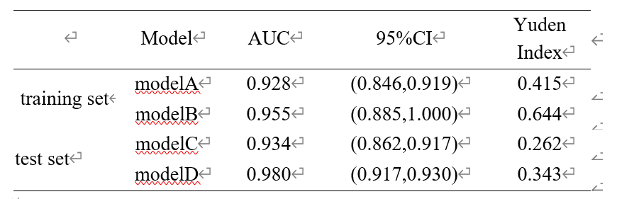
Comparison of different prediction models on training and test sets
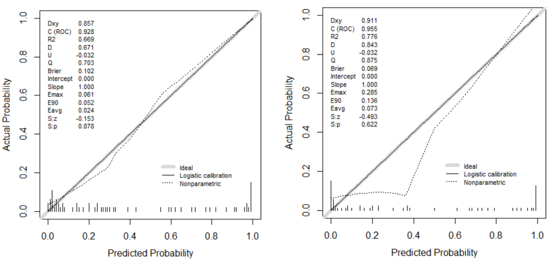
Fig1.Calibration curves of different prediction models in the training set

Fig2.Calibration curves for different prediction models in the test set
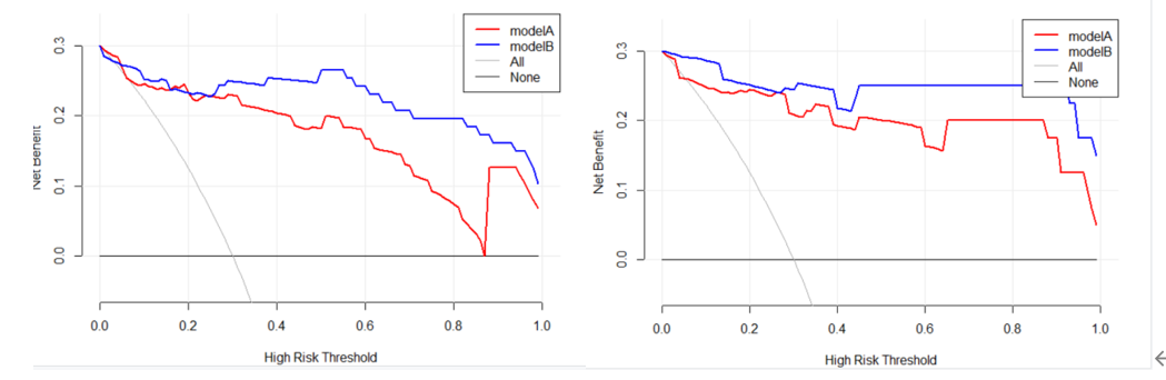
DCA decision curves for training and test sets. The left figure represents the DCA decision curve for the training set, and the right figure represents the DCA decision curve for the test set.
DOI: https://doi.org/10.58530/2023/1483