1458
CSF pseudo-diffusion spatial statistics for whole-brain voxel-wise analysis of CSF flow1Mallinckrodt Institute of Radiology, Washington University in St. Louis, St. Louis, MO, United States, 2Faculty of Health Science, McMaster University, Hamilton, ON, Canada, 3Institute for Informatics, Washington University in St. Louis, St. Louis, MO, United States
Synopsis
Keywords: Neurofluids, Diffusion/other diffusion imaging techniques, cerebrospinal fluid
Alterations in CSF flow patterns have been implicated in various brain disorders. At low b-values, diffusion-weighted imaging is sensitive to pseudorandom CSF flow. Here, we present CSF pseudo-diffusion spatial statistics (CΨSS), a whole-brain multi-subject voxel-wise analysis framework that exploits low-b-value diffusion-weighted imaging to study regional patterns of CSF flow. Using this technique, we show how brain atrophy and ventricular volumes affect regional CSF pseudorandom flow. In conclusion, CΨSS is a simple and effective approach for characterizing determinants of regional CSF pseudorandom flow.INTRODUCTION:
Cerebrospinal fluid (CSF) plays a crucial role in brain homeostasis and its clearance system. Alterations in CSF flow patterns have been implicated in various brain disorders1. Physiological modeling studies suggest that brain atrophy and ventricular configuration can affect CSF flow dynamics by changing intracranial anatomical constraints and fluid compliance. However, the inter-relation between intracranial anatomy and regional patterns of CSF flow remains elusive. While phase-contrast imaging and several other methods have been used to examine CSF flow, these imaging approaches are often limited to bulk CSF flow in specific brain regions and do not enable whole-brain characterization of complex pseudorandom movement of CSF. Low-b-value diffusion-weighted (DW) imaging has been recently proposed as an alternative quantitative approach for assessing CSF movement patterns2,3. Here, we present CSF pseudo-diffusion spatial statistics (CΨSS) that allows multi-subject whole-brain voxel-wise analysis of CSF movement patterns. In this study, we use CΨSS to determine the effects of brain atrophy and ventricular volumes on CSF pseudorandom flow.METHODS:
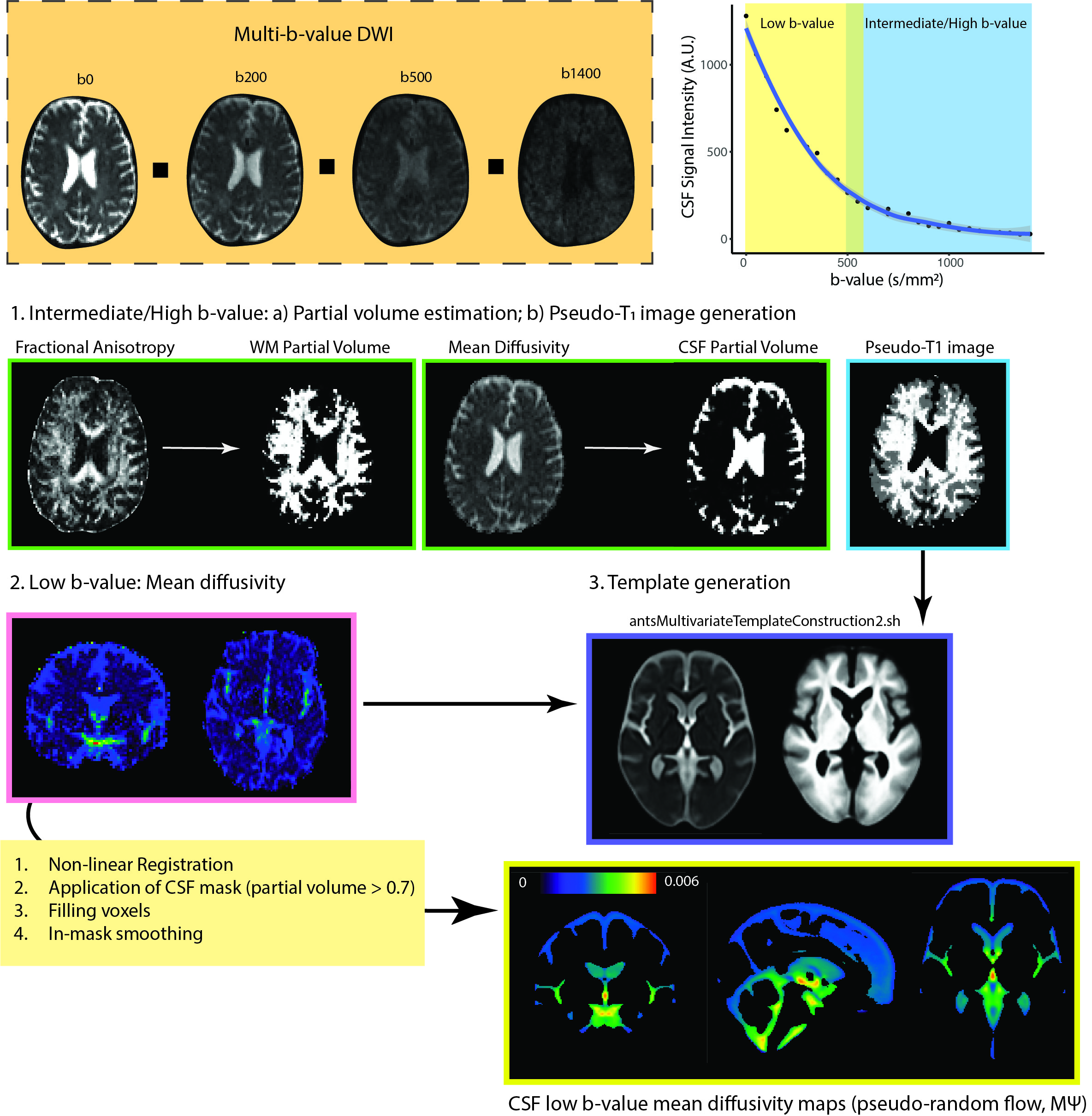
MRI acquisition: MRI data were collected as a part of the OASIS-3 study (n=197, 109 female, average age: 72 years [range: 46-92 years]; http://www.oasis-brains.org/)4. Whole brain echoplanar DW images were acquired on a 3T scanner (BioGraph mMR, Siemens, Erlangen, Germany). DW images were acquired with a multi-b-value protocol using the following imaging parameters: TE, 86 ms; TR, 10,300 ms; resolution, 2 × 2 × 2 mm3; field-of-view, 224 × 224 mm2; slice number, 80. Each diffusion gradient had a unique b-value (uniformly distributed between 0 and 1400 s/mm2; 26 volumes).CSF pseudo-diffusion spatial statistics: CSF pseudo-diffusion spatial statistics: DW images were corrected for Gibbs ring artifact and eddy current distortions. The diffusion tensor was fitted to the low-b-value DW images to generate low-b-value mean diffusivity (b: 0-550 s/mm2; 11 volumes) as a measure of pseudorandom flow magnitude (MΨ, mean pseudo-diffusion). The diffusion tensor model was also fitted into the intermediate/high b-value images were selected (b0; b: 500 - 1400 s/mm2). The resulting fractional anisotropy and mean diffusivity images were fed into a 2-tissue segmentation algorithm (Atropos, part of ANTs; http://stnava.github.io/ANTs/) to generate white matter and CSF partial volume maps5. Similar to gray matter-based spatial statistics6, partial volume maps were used to generate pseudo-T1-weighted images. MΨ and pseudo-T1 images were used to create study-specific templates using the antsMultivariateTemplateConstruction2.sh workflow (Figure 1). MΨ and CSF partial volume maps were then registered to the template space. The final mask was generated by keeping voxels with a CSF fraction > 0.7 in more than 65% of the subjects. Voxels with CSF fraction < 0.7 were then filled with the average of the surrounding satisfactory voxels. Voxel-wise permutation analyses were performed to investigate the effects of demographics, brain parenchymal fraction, and ventricular volumes on CSF low-b-value mean diffusivity.
RESULTS:
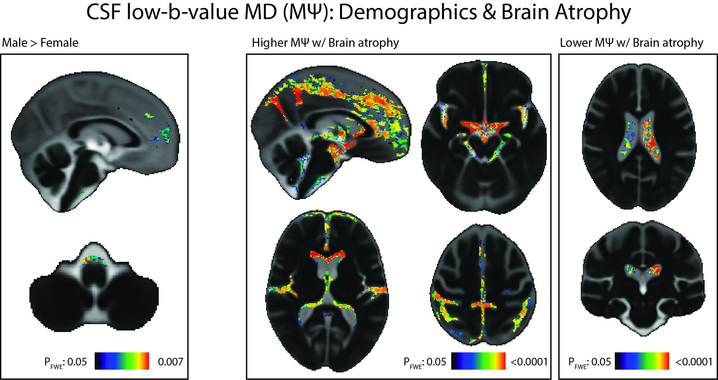
Demographics: Compared with females, males demonstrated higher CSF pseudorandom flow (MΨ) in the premedullary cistern (PFWE = 0.007) and frontal interhemispheric subarachnoid space (PFWE = 0.017; Figure 2). Accounting for brain atrophy, no age-related effect was observed on CSF pseudorandom flow.Brain atrophy: Brain atrophy (i.e., smaller brain parenchymal fraction) was associated with lower pseudorandom flow within the body of the lateral ventricles (PFWE <0.0001). In contrast, brain atrophy was associated with higher pseudorandom flow along the walls of the frontal horns of the lateral, third and fourth ventricles, Sylvian fissures and subarachnoid spaces along various sulci, cerebellopontine angles, and suprasellar, perimesencephalic, and prepontine cisterns (PFWE<0.0001; Figure 2).
Ventricular volumes: Larger fourth ventricle volume was associated with higher flow within the fourth ventricle (PFWE < 0.001), foramina of Luschka and Magendie (PFWE = 0.002), and the frontal horns of the lateral ventricles (PFWE = 0.004) and decreased retro-cerebellar CSF flow (PFWE = 0.02). Larger third ventricle volume was associated with higher pseudorandom flow within the foramina of Monro and adjacent to the massa intermedia in the third ventricle (PFWE < 0.001) and decreased flow in the dorsal extra-axial subarachnoid space (PFWE < 0.001, Figure 3). Larger lateral ventricle volume was associated with higher pseudorandom flow along the walls of the frontal horns of the lateral ventricles (PFWE = 0.001) and decreased pseudorandom flow in the body of the lateral ventricles (PFWE = 0.005) and dorsal extra-axial CSF (PFWE < 0.001; Figure 3).
DISCUSSION:
Our results indicate that brain atrophy preferentially facilitates flow along the basilar and peripheral subarachnoid spaces. Association of transventricular flow and flow across the foramina of Luschka and Magendie with larger fourth ventricle volume suggests that a more accommodating fourth ventricle directs the flow of the CSF into the ventricular system rather than the retro-cerebellar space. The paradoxical effects of larger lateral ventricle volumes on intraventricular flow (frontal horns vs. body) suggests that CSF flow within the lateral ventricles is compartmentalized and may be governed by distinct physiological mechanisms. Taken together, CΨSS is a simple and effective approach for characterizing determinants of CSF pseudorandom flow.Acknowledgements
Computations were performed using the facilities of the Washington University Center for High-Performance Computing, which was partially
funded by NIH grants 1S10RR022984-01A1 and 1S10OD018091-01.
References
1. Rasmussen, M. K., Mestre, H. & Nedergaard, M. Fluid transport in the brain. Physiol Rev 102, 1025–1151 (2022).
2. Bito, Y., Harada, K., Ochi, H. & Kudo, K. Low b-value diffusion tensor imaging for measuring pseudorandom flow of cerebrospinal fluid. Magn Reson Med 86, 1369–1382 (2021).
3. Harrison, I. F. et al. Non-invasive imaging of CSF-mediated brain clearance pathways via assessment of perivascular fluid movement with diffusion tensor MRI. Elife 7, (2018).
4. LaMontagne, P. J. et al. OASIS-3: Longitudinal Neuroimaging, Clinical, and Cognitive Dataset for Normal Aging and Alzheimer Disease. medRxiv 2019.12.13.19014902 (2019)
5. Avants, B. B., Tustison, N. J., Wu, J., Cook, P. A. & Gee, J. C. An open source multivariate framework for N-tissue segmentation with evaluation on public data. Neuroinformatics 9, 381–400 (2011).
6. Nazeri, A. et al. Functional Consequences of Neurite Orientation Dispersion and Density in Humans across the Adult Lifespan. Journal of Neuroscience 35, 1753–1762 (2015).
Figures