1455
Higher blood flow velocity pulsatility relates to increased interstitial fluid diffusivity - a potential proxy of high perivascular fluid flow1Department of Radiology & Nuclear Medicine, Maastricht University Medical Center, Maastricht, Netherlands, 2School for Mental Health and Neuroscience, Maastricht University, Maastricht, Netherlands, 3Department of Psychiatry & Neuropsychology, Maastricht University, Maastricht, Netherlands, 4Cardiovascular Research Institute Maastricht, Maastricht University, Maastricht, Netherlands, 5Department of Electrical Engineering, Eindhoven University of Technology, Eindhoven, Netherlands
Synopsis
Keywords: Neurofluids, Aging, Diffusion, pulsatility, waste clearance
Impaired cerebral waste clearance occurs in healthy ageing and various neurodegenerative diseases and is theorized to be due to compromised arterial pulsatility. Profiting from high-resolution 7 Tesla MRI, the current study investigated the association between pulsatility characteristics of a small artery in the basal ganglia (BG) with interstitial fluid (ISF) characteristics of the BG - as derived with intravoxel incoherent motion. This study found that an increased small vessel velocity pulsatility was related to higher ISF-diffusivity in the BG of an elderly sample. This increased ISF-diffusivity might represent increased perivascular fluid diffusivity, influencing the waste fluid transport out of these spaces.Introduction
Impaired cerebral clearance function could be due to changes in interstitial fluid (ISF) movement through the perivascular spaces (PVS), which is thought to dependent on arterial pulsatility1,2. Pulsatility characteristics are altered in (healthy) aging3, and in neurodegenerative diseases, such as Alzheimer’s disease4,5, where the clearance of toxic proteins (e.g., Amyloid-Beta) is also diminished6-10.7T MRI allows for high spatial resolution measurements of the blood flow velocity waveform in the small lenticulostriate arteries (LSAs)3,11, which are in close contact with the parenchyma in the basal ganglia (BG). The severity of vessel stiffening can be studied by measuring the blood flow velocity pulsatility of the LSAs3,12,13.
Moreover, MRI enables to noninvasively quantify ISF-characteristics using intravoxel incoherent motion imaging (IVIM)14,15. Spectral analysis of IVIM images has been used to identify an intermediate diffusion component between the two traditional components (i.e., microvascular and parenchymal), which is argued to both represent ISF-volume (fint) and ISF-diffusivity (Dint) in the parenchyma and within PVS14,15. Previous work has found higher fint and Dint values inside the PVS as compared to surrounding tissue, signaling increased ISF-volume and diffusivity16.
This study aims to gain more knowledge on how variations in LSA pulsatility relate to fint and Dint within the BG, a region with multiple PVS17. We foresee to find that stiffer arteries, reflected by higher velocity pulsatility, are associated with impairment of the surrounding tissue, represented by alterations in ISF-volume and/or ISF-diffusivity.
Methods
Subjects: Twenty-nine cognitively healthy elderly subjects were included in this study (Tab.1).MRI acquisition: All subjects underwent 7T MRI (Siemens Healthineers, Erlangen, Germany) using a 32-element channel phased-array head coil (Tab.2). Diffusion MR images (with a prototype IVIM sequence) and anatomical T1-weighted and T2-weighted images were acquired. A Time-Of-Flight angiogram was used to plan the 2D phase-contrast sequence, from which the velocity measurements of the largest LSA were retrieved.
Image analysis:
Anatomical images: Freesurfer (v5.1.0) was used to automatically segment anatomical T1-weighted images, with manual inspection. The BG masks were coregistered to native IVIM space (FLIRT,FSL v6.0.1)18.
PC-MRI: The largest LSA was processed to assess the blood flow velocity measures. After correction for background noise and aliasing, the vessel was segmented from the magnitude images. The pulsatility index (PI) of the LSA was calculated with $$$PI_{LSA}=\frac{v_{max} - v_{min}}{v_{mean}}$$$, where v represents velocity19.
IVIM: Diffusion trace images were calculated, and corrected for geometric susceptibility induced distortions (topup)20, head displacements and eddy currents (ExploreDTI v4.8.4)21.
IVIM data was analyzed in a voxel-wise manner with spectral analysis using non-negative least squares (NNLS)14,15, utilizing 80 logarithmically spaced basis functions (.1*10-3 to 200*10-3mm2/s) and adding a regularisation smoothing constraint (allowed misfit: 2-2.5%).
Dint was identified as 1.5*10-3 mm2/s<Diffusivity<4.0*10-3 mm2/s, and the contribution of the intermediate component to the total signal was determined by quantifying fint, while correcting for T1- and T2-relaxation effects14. The median Dint and fint values extracted from the BG ipsilateral to the postprocessed LSA.
Statistics: Spearman’s rho correlations were computed between fint, Dint and PILSA (IBM SPSS statistics v25). Partial Spearman correlations were conducted to investigate the influence of age, and subsequently of sex.
Results
Table 1 summarizes the sample characteristics and descriptive statistics of fint, Dint and PILSA. A significant positive association was found between PILSA and Dint (Rs=.399, p=.032)(Tab.3), where a higher PILSA relates to a higher Dint (Fig.1). This relation remained significant after age correction (Rs=.390, p=.040) and additional adjustment for sex (Rs=.413, p=.032).Discussion and conclusion
The current study found that a higher LSA velocity pulsatility is related to a higher ISF-diffusivity in the nearby tissue of the BG, in an elderly population.A higher LSA velocity pulsatility may reflect reduced damping of the upstream arterial vessel wall, caused by increased arterial stiffness. When perforating arteries momentarily dilate, the PVS become narrower, and the perivascular fluid is propelled forward, as the fluid is incompressible. When the vessel wall becomes stiffer, the short-term vessel dilation and narrowing of the PVS is reduced, resulting in on average more space (and time) for water molecules inside PVS to diffuse, resulting in increased ISF-diffusivity.
Furthermore, structural vessel wall alterations associated with increased stiffness may hinder the exchange of ISF from within PVS to ISF in the parenchyma and could propel to reduced clearance of waste products through fluid transport. However, more detailed research is needed to fully explain the interaction between pulsatility of perforating arteries and the condition of the surrounding tissue.
Although pulsatility characteristics are altered in healthy aging3, we found that the relation between pulsatility and ISF-diffusivity is independent of age. This urges for an independent, pathological relationship between altered vessel stiffness/compliance and ISF-diffusivity.
In healthy elderly, we did not find a relationship between fint and LSA pulsatiliy. Potentially, in later pathological stages of vessel wall alterations, perivascular fluid would be pushed downstream with larger pressure, where it might further damage the interface between the vessel wall and surrounding parenchyma and lead to PVS enlargement, resulting in increased ISF-volume (fint) and further impacting waste clearance function.
This study identified that reduced small vessel compliance is related to higher ISF-diffusivity, indicating altered ISF-movement within the PVS. Thereby, this study forwarded the IVIM-derived ISF-diffusivity as a potential non-invasive proxy of alterations in PVS fluid movement.
Acknowledgements
This research was supported by Alzheimer Nederland (research grant WE.03-2018-02). We would like to thank Siemens Healthineers (especially Thorsten Feiweier and Erik van den Bergh) for providing us with the prototype IVIM sequence.References
1. Iliff JJ, Wang M, Zeppenfeld DM, et al. Cerebral arterial pulsation drives paravascular CSF–interstitial fluid exchange in the murine brain. Journal of Neuroscience. 2013;33(46):18190-18199.
2. Jessen NA, Munk ASF, Lundgaard I, Nedergaard M. The glymphatic system: a beginner’s guide. Neurochemical research. 2015;40(12):2583-2599.
3. Schnerr RS, Jansen JF, Uludag K, et al. Pulsatility of Lenticulostriate Arteries Assessed by 7 Tesla Flow MRI—Measurement, Reproducibility, and Applicability to Aging Effect. Frontiers in physiology. 2017;8:961.
4. Jellinger KA. The pathology of ischemic-vascular dementia: an update. Journal of the neurological sciences. 2002;203:153-157.
5. Rivera-Rivera LA, Schubert T, Turski P, et al. Changes in intracranial venous blood flow and pulsatility in Alzheimer’s disease: A 4D flow MRI study. Journal of Cerebral Blood Flow & Metabolism. 2017;37(6):2149-2158.
6. Tarasoff-Conway JM. Clearance systems in the brainimplications for Alzheimer disease. Nature Reviews Neurology. 2015;11(8):457.
7. Rasmussen MK, Mestre H, Nedergaard M. The glymphatic pathway in neurological disorders. The Lancet Neurology. 2018;17(11):1016-1024.
8. Bakker EN, Bacskai BJ, Arbel-Ornath M, et al. Lymphatic clearance of the brain: perivascular, paravascular and significance for neurodegenerative diseases. Cellular and molecular neurobiology. 2016;36(2):181-194.
9. Reeves BC, Karimy JK, Kundishora AJ, et al. Glymphatic system impairment in Alzheimer’s disease and idiopathic normal pressure hydrocephalus. Trends in molecular medicine. 2020;26(3):285-295.
10. Joseph CR. Novel MRI Techniques Identifying Vascular Leak and Paravascular Flow Reduction in Early Alzheimer Disease. Biomedicines. 2020;8(7):228.
11. Kang CK, Park CA, Lee DS, et al. Velocity measurement of microvessels using phase‐contrast magnetic resonance angiography at 7 tesla MRI. Magnetic resonance in medicine. 2016;75(4):1640-1646.
12. Arts T, Onkenhout LP, Amier RP, et al. Non-Invasive Assessment of Damping of Blood Flow Velocity Pulsatility in Cerebral Arteries With MRI. Journal of Magnetic Resonance Imaging. 2022;55(6):1785-1794.
13. Geurts LJ, Zwanenburg JJM, Klijn CJM, Luijten PR, Biessels GJ. Higher Pulsatility in Cerebral Perforating Arteries in Patients With Small Vessel Disease Related Stroke, a 7T MRI Study. Stroke. 2019;50(1):62-68.
14. Wong S, Backes W, Drenthen G, et al. Spectral Diffusion Analysis of Intravoxel Incoherent Motion MRI in Cerebral Small Vessel Disease. Journal of Magnetic Resonance Imaging. 2019.
15. van der Thiel MM, Freeze WM, Verheggen IC, et al. Associations of increased interstitial fluid with vascular and neurodegenerative abnormalities in a memory clinic sample. Neurobiology of Aging. 2021;106:257-267.
16. van der Thiel MM, Roos NA, Drenthen GS, et al. On the origin of a potential clearance marker: The contribution of enlarged perivascular fluid diffusion to a 7T IVIM interstitial fluid proxy. Paper presented at: Proc 31st Annual Meeting ISMRM 2022; London.
17. Wardlaw JM, Benveniste H, Nedergaard M, et al. Perivascular spaces in the brain: anatomy, physiology and pathology. Nature Reviews Neurology. 2020:1-17.
18. Jenkinson M, Bannister P, Brady M, Smith S. Improved optimization for the robust and accurate linear registration and motion correction of brain images. Neuroimage. 2002;17(2):825-841.
19. Gosling R, King D. The role of measurement in peripheral vascular surgery: arterial assessment by Doppler-shift ultrasound. In: SAGE Publications; 1974.
20. Andersson JL, Skare S, Ashburner J. How to correct susceptibility distortions in spin-echo echo-planar images: application to diffusion tensor imaging. Neuroimage. 2003;20(2):870-888.
21. Leemans A, Jeurissen B, Sijbers J, Jones D. ExploreDTI: a graphical toolbox for processing, analyzing, and visualizing diffusion MR data. Paper presented at: Proc Intl Soc Mag Reson Med2009.
Figures
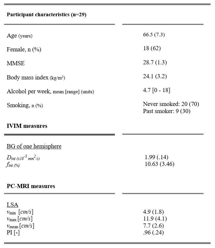
Table 1. This table displays the sample characteristics and descriptive statistics of the IVIM and the PC-MRI measures. Mean (standard deviation) are reported unless stated otherwise.
Abbreviations: IVIM = Intravoxel incoherent motion, Dint = Interstitial fluid diffusion, fint = Interstitial fluid fraction, MMSE = Mini-Mental State Examination, PC-MRI = Phase contrast MRI, LSA = Lenticulostriate artery, v = Velocity, PI = Pulsatility index.
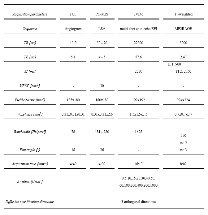
Table 2. This table summarizes the acquisition parameters of the sequences used within the study.
Abbreviations: LSA = Lenticulostriate artery, TOF = time-of-flight, PC-MRI = Phase contrast MRI, IVIM = intravoxel incoherent motion, TR = repetition time, TE = echo time, VENC = velocity encoding.
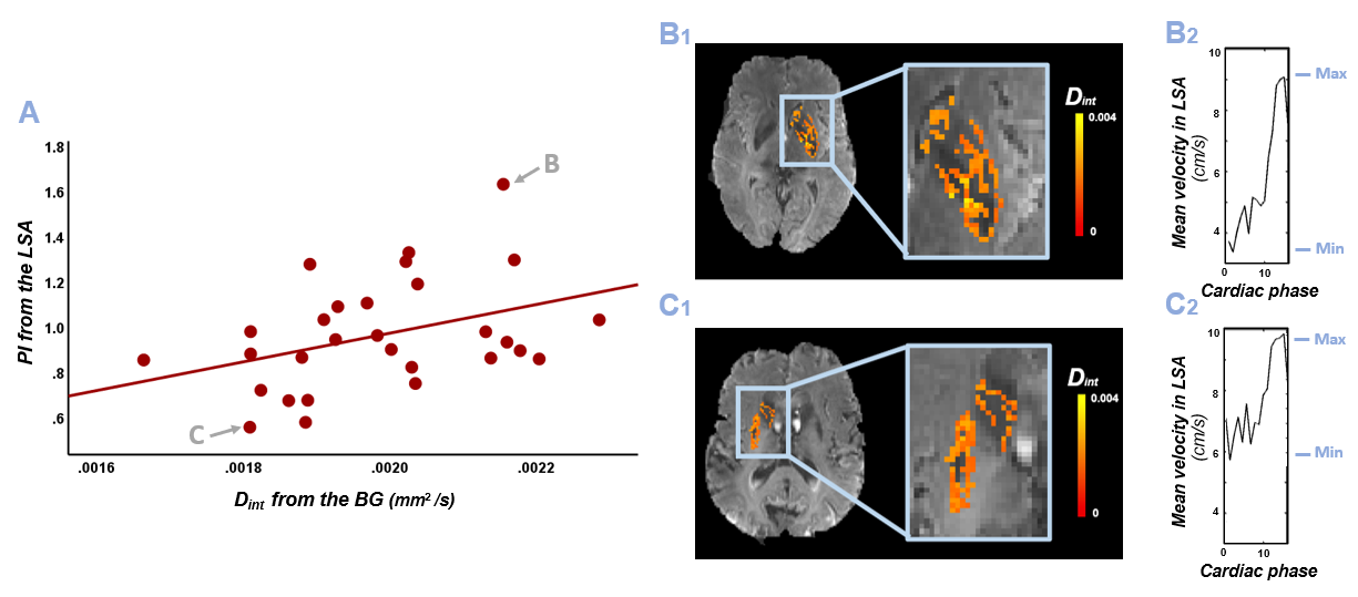
Arrows point to the subjects used to present exemplary Dint maps and LSA velocity profiles over the cardiac phase, i.e., from a subject (69y, female) with a high Dint (B1) and PI (B2) and from a subject (77y, male) with a low Dint (C1) and PI (C2). The minimum and maximum velocity are indicated in blue (B2-C2).
Abbreviations: Dint = Interstitial fluid diffusion, LSA = Lenticulostriate artery, PI = Pulsatility index, BG = Basal ganglia.
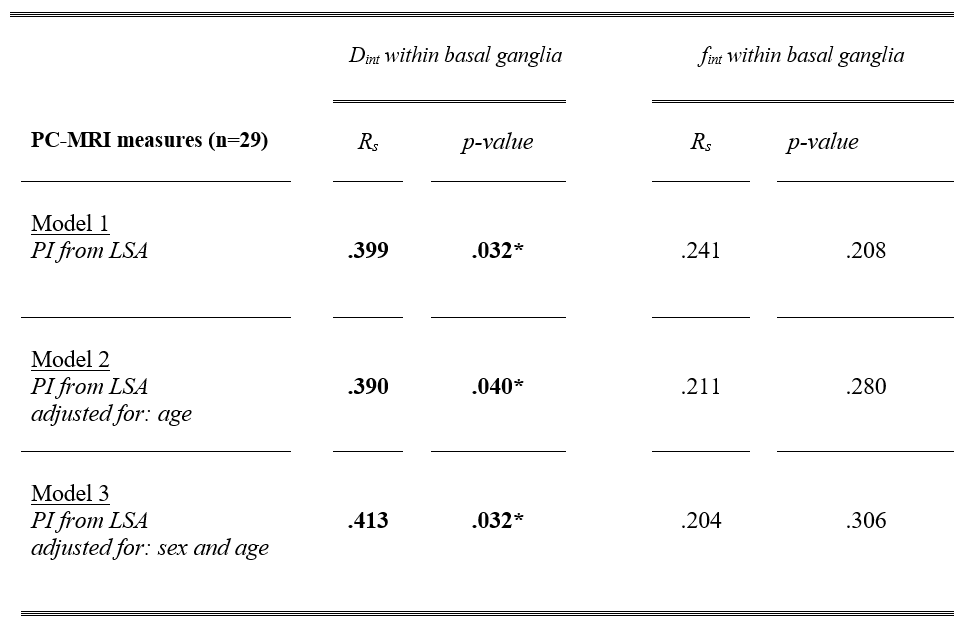
Table 3. Spearman’s rho correlations of the PC-MRI derived PI from the largest LSA and the IVIM values (Dint and fint) from the ipsilateral basal ganglia (Model 1). Partial Spearman’s rho correlations additionally adjusting for age (Model 2) and additionally for sex (Model 3). * p < 0.05.
Abbreviations: IVIM = Intravoxel incoherent motion, Dint = Interstitial fluid diffusion, fint = Interstitial fluid fraction, PC-MRI = Phase contrast MRI, LSA = Lenticulostriate artery, PI = Pulsatility index, Rs = Spearman’s rho correlation.