1449
Intravoxel Incoherent Motion Diffusion-weighted MR Imaging and IDEAL-IQ for Assessment of Treatment Response in Multiple Myeloma
Yanhua Tang1, Li Wang2, and Yichen Ma3
1Radiology, Beijing Chao-Yang Hospital, Capital Medical Universit, Beijing, China, 2Beijing Chao-Yang Hospital, Capital Medical Universit, Beijing, China, 3Beijing Chao-Yang Hospital, Capital Medical University, Beijing, China
1Radiology, Beijing Chao-Yang Hospital, Capital Medical Universit, Beijing, China, 2Beijing Chao-Yang Hospital, Capital Medical Universit, Beijing, China, 3Beijing Chao-Yang Hospital, Capital Medical University, Beijing, China
Synopsis
Keywords: Bone, Diffusion/other diffusion imaging techniques, intravoxel incoherent motion, diffusion-weighted imaging, iterative decomposition of water and fat with echo asymmetry and least-squares estimation quantitation sequence (IDEAL-IQ), multiple myeloma, treatment response
This study explored the feasibility of IVIM DWI and IDEAL-IQ parameters in evaluating the treatment response of patients with multiple myeloma (MM) who received systemic treatment within 6 months. The results showed that within 6 months after treatment, the changes of D and f values of IVIM DWI in deep-responders before and after treatment were significantly different from those in non-deep responders.This indicates that the changes of D and f values of IVIM DWI may be helpful to evaluate the short-term (within 6 months) therapeutic response of myeloma, while the evaluation of IDEAL-IQ on short-term therapeutic response needs further study.Introduction
Multiple myeloma is a malignant hematoma characterized by abnormal proliferation of plasma cells, mainly involving bone marrow(1). According to the recent International Myeloma Working Group standards, MRI can be used to detect the focal lesions in patients with myeloma(2, 3). However, bone marrow magnetic resonance imaging evaluation provides not only morphological information, but also additional functional information about bone marrow such as water diffusivity and micro-capillary perfusion through the application of diffusion-weighted imaging sequence, so as to improve the overall performance of MRI(4). Recent years, the perfusion parameters of intravoxel incoherent motion (IVIM) diffusion-weighted imaging (DWI) have been widely reported as promising biomarkers for tumor angiogenesis assessment(5). However, these parameters are not frequently used in the application of the musculoskeletal diseases(6). It has been reported that IVIM DWI is helpful to differentiate benign and malignant vertebral tumors(7), and is positively correlated with the changes of bone marrow maximum enhancement after treatment in MM patients(8). Therefore, IVIM DWI may provide useful information for the qualitative and follow-up of MM lesions. The iterative decomposition of water and fat with echo asymmetry and least-squares estimation quantitation sequence (IDEAL-IQ) recently was reported as one of the most convenient and accurate techniques to quantify bone marrow fat (BMF)(9). The infiltration of myeloma could change the proportion of fat content in bone marrow.(10) Accordingly, the purpose of our study was to determine the feasibility of IVIM and IDEAL-IQ parameters for assessment of treatment response in MM patients with systemic therapy in six months.Method
Seventy-two stage-IIImyeloma patients were enrolled in this prospective study between January 2018 and May 2022 with local research ethics committee approval, and requirement for written informed consent was waived. All patients underwent spine IVIM DWI sequence with 9 b values(0, 25, 50, 75, 100, 200, 400, 600, 800 and 1000 sec/mm2)before and after systemic therapy. Finally, 38 patients received the examinations in six months were enrolled in the study. The response was defined according to the most recent International Myeloma Working Group criteria for MM, and the patients were divided in to deep (complete response or very good partial response) and non-deep responders (partial response, minimal response, stable disease, or progressive disease).The variations in apparent diffusion coefficient (ADC) values, the IVIM parameters (perfusion fraction [f], molecular diffusion coefficient [D], and perfusion-related D [D*]) values and the IDEAL-IQ parameters(R*, fat fraction [FF]) were calculated and compared between deep responders and non-deep responders before and after treatment with Kruskal-Wallis univariate ANOVA analysis. Bland-Altman tests were used to compare the reproducibility of IVIM parameters measurements.Result
Among 38 cases, 14 patients were classified as deep-responders and 24 patients as non-deep responders. The D and f value changes between pre- and post-treatment in the deep responders were significantly different from those in the non-deep responders (P= 0.035, and P= 0.019, respectively,<0.05). Receiver operating characteristic analysis of change in D and f in the two groups indicated that 0.316 ×10⁻³mm2/s increase in D, and 9.475% increase in f correctly identified good response to treatment with 53.3% sensitivity and 78.3% specificity(area under the curve, AUC, 0.704) of D, and 73.3% sensitivity and 73.9% specificity (AUC, 0.728) of f, respectively. There was no significant change in ADC , D*, R*, and FF value in the two groups before and after treatment (P= 0.988, P= 0.709,P= 0.378 and P= 0.317, respectively, >0.05) in six months.Discussion
We found the D changes could help assess the treatment response in MM. The D value increase 0.2425×10⁻³mm2/s values with 53.3% sensitivity and 78.3% specificity (AUC, 0.773) could identify good response to treatment. These could be explained by early necrosis and edema which made reduced cellularity and decrease of angiogenesis of the bone marrow following treatment in good response MM patients(11). The progression of multiple myeloma is accompanied by the increase of the number of bone marrow plasma cells and micro-vessel density(12), which leads to the free water diffusion restriction. It’s reported that a moderate correlation of D value with the marrow cellularity and plasma cell percentage(10).We found the f values in the deep responders significantly increased after treatment, and 9.475% increase could identify good response to treatment with 73.3% sensitivity and 73.9% specificity (AUC, 0.728) in f. Chen Y(7)found that the f value of malignant tumor in bone was lower than that of acute benign compression fractures and tuberculous spondylitis. This may support our result of f value in good responders before treatment was lower than that after treatment.Cui Ren et al.(13)found FF value of healthy and active group in patients with ankylosing spondylitis was not significantly different. Also it’s reported (11)that 20 weeks after treatment, the fat component increased in good response MM patients. This may support our result that the FF value change before and after the therapy in 6 months also was not significantly different in the deep and non-deep responder groups.Conclusion
The D and the f value changes in IVIM DWI may help for the assessment of treatment response in myeloma in six months.Acknowledgements
References
1. Mehta A. Multiple myeloma. Hematology. 2015;20(1):58-9.2. Hillengass J, Usmani S, Rajkumar SV, et al. International myeloma working group consensus recommendations on imaging in monoclonal plasma cell disorders. The Lancet Oncology. 2019;20(6):e302-e12.3. Cowan AJ, Green DJ, Kwok M, et al. Diagnosis and Management of Multiple Myeloma: A Review. JAMA. 2022;327(5):464-77.4. Dutoit JC, Verstraete KL. MRI in multiple myeloma: a pictorial review of diagnostic and post-treatment findings. Insights into imaging. 2016;7(4):553-69.5. Wu H, Liang Y, Jiang X, et al. Meta-analysis of intravoxel incoherent motion magnetic resonance imaging in differentiating focal lesions of the liver. Medicine. 2018;97(34):e12071.6. Koutoulidis V, Papanikolaou N, Moulopoulos LA. Functional and molecular MRI of the bone marrow in multiple myeloma. The British journal of radiology. 2018;91(1088):20170389.7. Chen Y, Yu Q, La Tegola L, et al. Intravoxel incoherent motion MR imaging for differentiating malignant lesions in spine: A pilot study. European journal of radiology. 2019;120:108672.8. Bourillon C, Rahmouni A, Lin C, et al. Intravoxel Incoherent Motion Diffusion-weighted Imaging of Multiple Myeloma Lesions: Correlation with Whole-Body Dynamic Contrast Agent-enhanced MR Imaging. Radiology. 2015;277(3):773-83.9. Ji Y, Hong W, Liu M, Liang Y, Deng Y, Ma L. Intervertebral disc degeneration associated with vertebral marrow fat, assessed using quantitative magnetic resonance imaging. Skeletal Radiol. 2020;49(11):1753-63.10. Jo A, Jung JY, Lee SY, et al. Prognosis Prediction in Initially Diagnosed Multiple Myeloma Patients Using Intravoxel Incoherent Motion-Diffusion Weighted Imaging and Multiecho Dixon Imaging. Journal of magnetic resonance imaging : JMRI. 2020.11. Messiou C, Giles S, Collins DJ, et al. Assessing response of myeloma bone disease with diffusion-weighted MRI. The British journal of radiology. 2012;85(1020):e1198-203.12. Hillengass J, Bauerle T, Bartl R, et al. Diffusion-weighted imaging for non-invasive and quantitative monitoring of bone marrow infiltration in patients with monoclonal plasma cell disease: a comparative study with histology. British journal of haematology. 2011;153(6):721-8.13. Ren C, Zhu Q, Yuan H. Mono-exponential and bi-exponential model-based diffusion-weighted MR imaging and IDEAL-IQ sequence for quantitative evaluation of sacroiliitis in patients with ankylosing spondylitis. Clin Rheumatol. 2018;37(11):3069-76.Figures
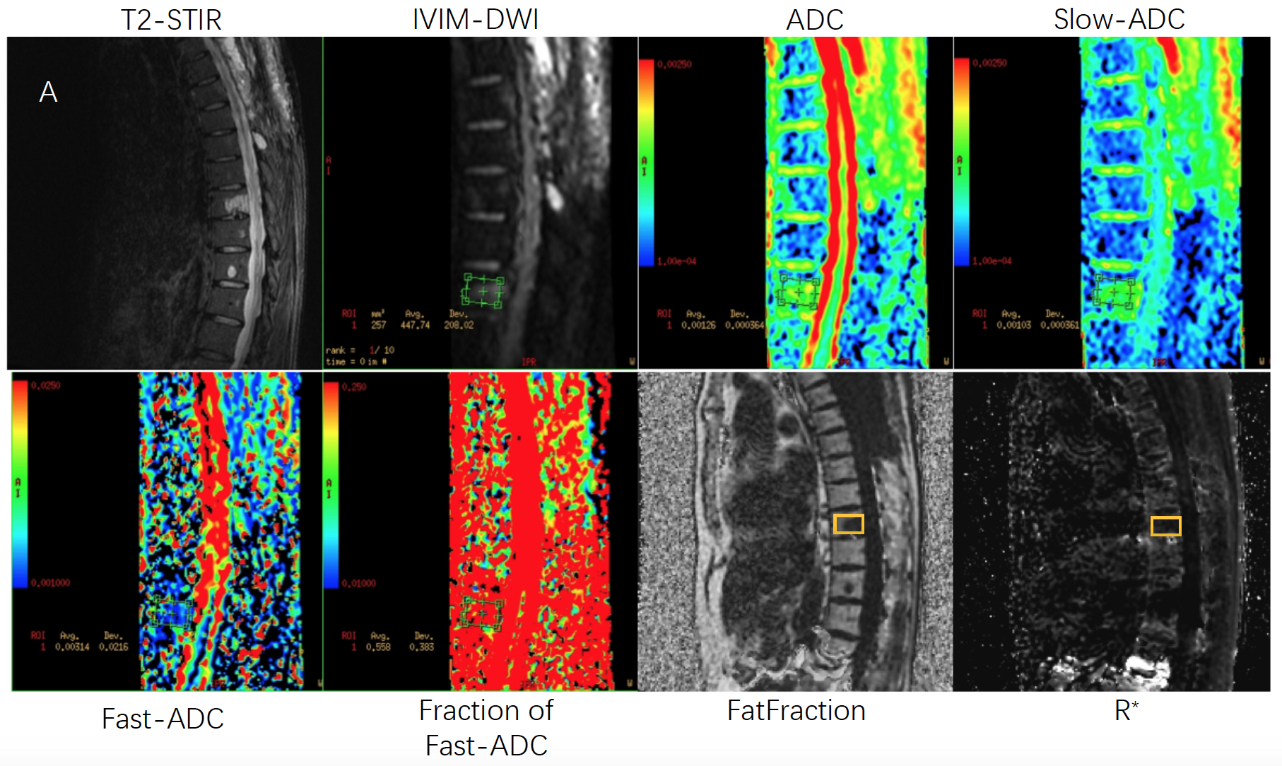
Figure A: A 42-year-old man with a Durie-Salmon Stage III MM before treatment. The thoracic vertebral focal lesion is seen at IVIM MR DWI and IDEAL-IQ. The ROI is put on the maximum lesion of the vertebrae, and the maximum area of the largest lesion is drawn and recorded.
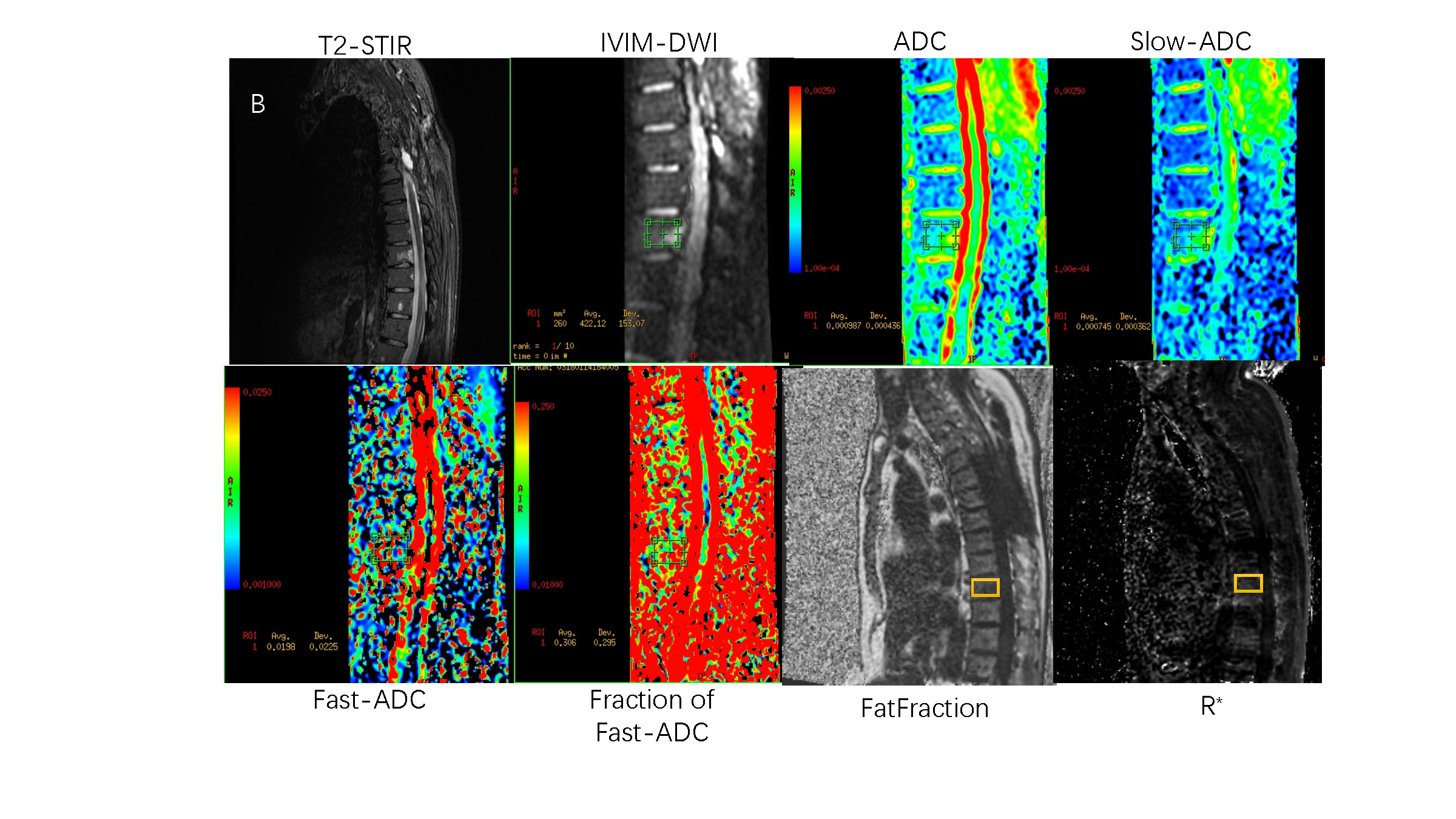
Figure B: The same patient with IVIM MR DWI and IDEAL-IQ examination 2.5 months after treatment. The ROI is put on the maximum lesion of the vertebrae, and the maximum area of the largest lesion is drawn and recorded.
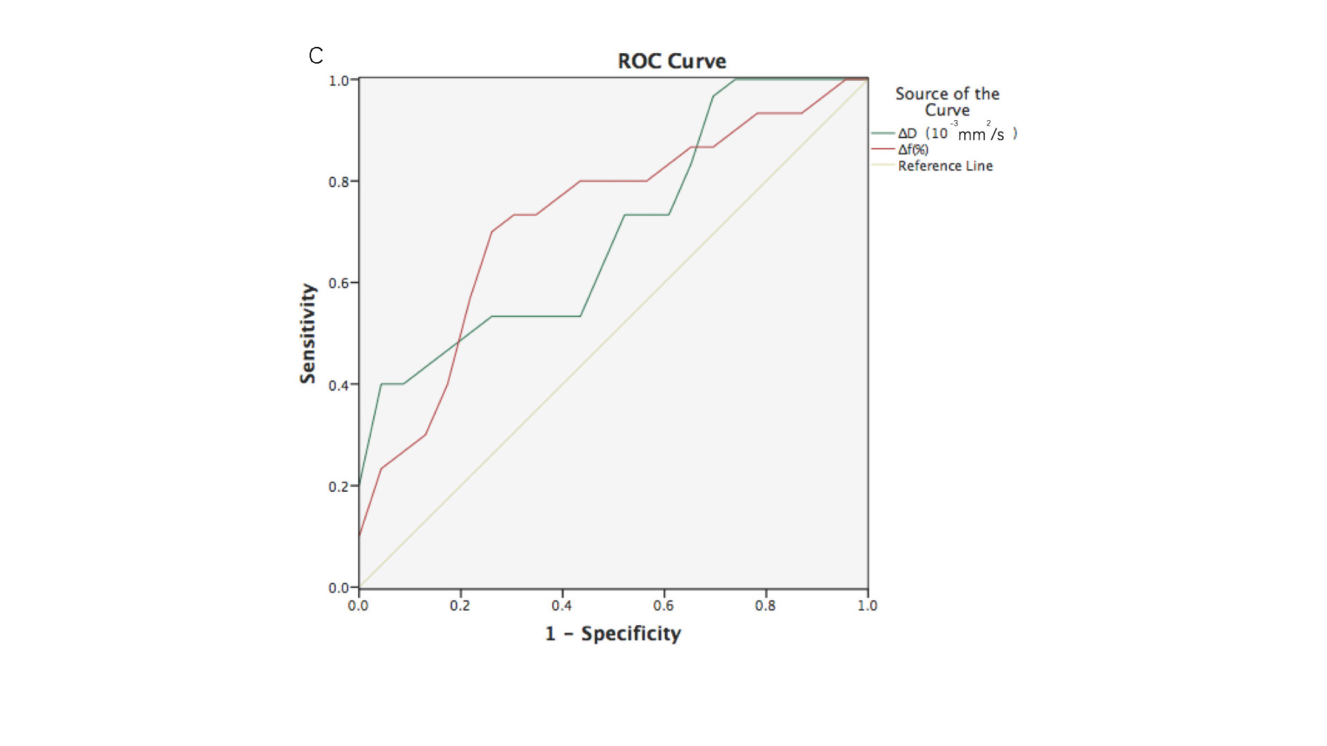
Figure C: Comparison of the receiver operating characteristic(ROC) curves of the ∆D(Dafter treatment-Dbefore treatment) (green line) and ∆f(fafter treatment-fbefore treatment) (red line) in assessment the good response to treatment.
DOI: https://doi.org/10.58530/2023/1449