1437
Volumetric Dynamic Imaging for Functional Kinematic Assessment of the Wrist1The Bernard and Irene Schwartz Center for Biomedical Imaging, Department of Radiology, New York University Grossman School of Medicine, New York, NY, United States, 2Department of Computer Science and Engineering, New York University Tandon School of Engineerin, Brooklyn, NY, United States
Synopsis
Keywords: Joints, MSK, Wrist
Dynamic MRI can be useful for evaluation of wrist instability. However, most available real-time MRI methods are either limited due to their 2D nature or provide only low temporal resolution and insufficient image quality. Here, we propose a novel approach for volumetric dynamic wrist examination by assembling 2D real-time data into 3D snapshots using MRI-visible markers. The method has been demonstrated for ulnar-radial deviation using a flexible wrist coil and 3D-printed support platform for guiding motion. Future work will use a high-resolution static MRI as morphological prior to segment bones on the dynamic volumes and allow for quantitative kinematic assessment.Introduction
Static MRI examination of the wrist, as performed routinely in clinical practice, provides excellent spatial resolution and contrast for characterizing bone and soft tissue1. However, initial stages of wrist instability often manifest only during active motion and do not show visible abnormalities on routine static examinations. Therefore, dynamic examination is desirable for patients experiencing pain during movement of the wrist, due to snapping or sudden changes in the intercarpal alignment2,3. Real-time MRI techniques have been proposed for accomplishing this task2. However, the 2D nature of most real-time methods makes it difficult to capture out-of-plane translations or rotations of the carpal bones. Existing dynamic 3D MRI methods, on the other hand, do not reach the required temporal resolution and image quality to properly capture sudden and rapid motion abnormalities. Here, we describe a novel approach for dynamic volumetric wrist examination by assembling 2D real-time data into 3D snapshots. We used a custom-developed wrist platform, which ensures consistent repetitive movement and includes MRI-visible markers for automatic alignment of the 2D slices. The technique was demonstrated for continuous ulnar-radial deviation.Methods
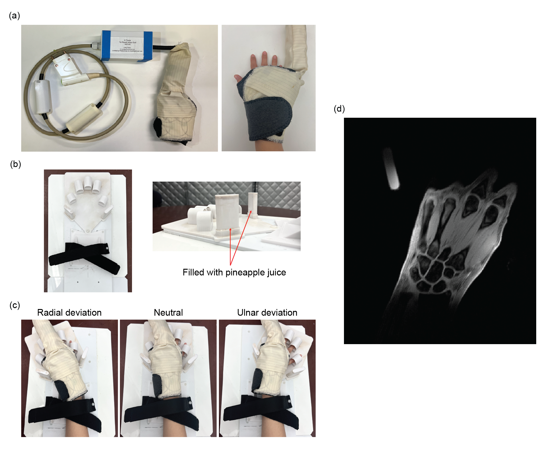
Coil and Platform DesignA custom-tailored wrist coil was created by repurposing a "blanket" coil that was built previously in our center4,5. The wrist coil was designed to wrap tightly around the wrist in the fashion of a medical support brace. The coil has eight high-impedance elements (ø=6cm) that are geometrically arranged in 2 rows of 4 elements. A support platform was designed with a 3D printer to guide the wrist movement and ensure consistency over multiple repetitions. The forearm is immobilized using Velcro straps and cushions, enforcing that only the wrist joint moves during maneuvers (Fig 1b/c). Two tubes were integrated into the moving part of the platform and filled with pineapple juice, whose bright MR signal (Fig 1d) was used to track the wrist position during motion.
Experiments
To demonstrate feasibility, we scanned healthy volunteers (after obtaining informed consent) on a clinical 3T scanner (MAGNETOM Prisma, Siemens Healthineers). Volunteers were asked to perform continuous ulnar-radial deviation whenever gradient noise could be heard. Dynamic data were acquired with an RF-spoiled 2D FLASH sequence, which uses radial sampling with interleaved acquisition order over five successive images6. Each interleave included 13 equidistant angular projections covering 360°. The acquisition scheme was repeated 50 times for each slice to properly capture the continuous motion. Spectral fat suppression was performed prior to each repetition. 24 slices were sequentially acquired in coronal orientation. Relevant parameters included: FOV 220x220mm2, resolution 1x1mm2, slice thickness 2mm, FA 4°, TR/TE 3.69/2.21ms, BW 500Hz/px, total duration 5:35min.
Image Reconstruction
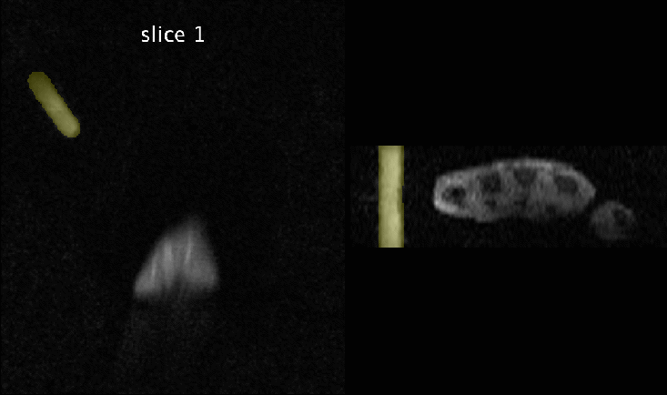
Dynamic images were reconstructed slice-by-slice using the GRASP algorithm7, which applies a total-variation constraint along the time dimension. 13 projections were combined into each image frame, resulting in a temporal resolution of 48ms/frame. To fuse the dynamic 2D images into a dynamic volume, the position marker was segmented in each image using a U-Net8. The Jaccard similarity index was calculated for segmentation masks of each frame from the different slices. Frames with the highest similarity score were assigned to the same wrist position and stacked into a 3D volume.
Results
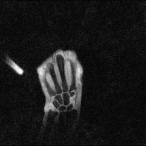
For reference, Fig 1d shows a static high-resolution scan acquired with a radial 3D GRE sequence using the 8-channel wrist coil, which demonstrates the high SNR provided by the coil.Fig 2 shows a real-time movie of the continuous ulnar-radial deviation maneuver in one slice, depicting motion of the carpal bones with high accuracy. The position marker is visible in each frame and allows identifying the angular position of the wrist.
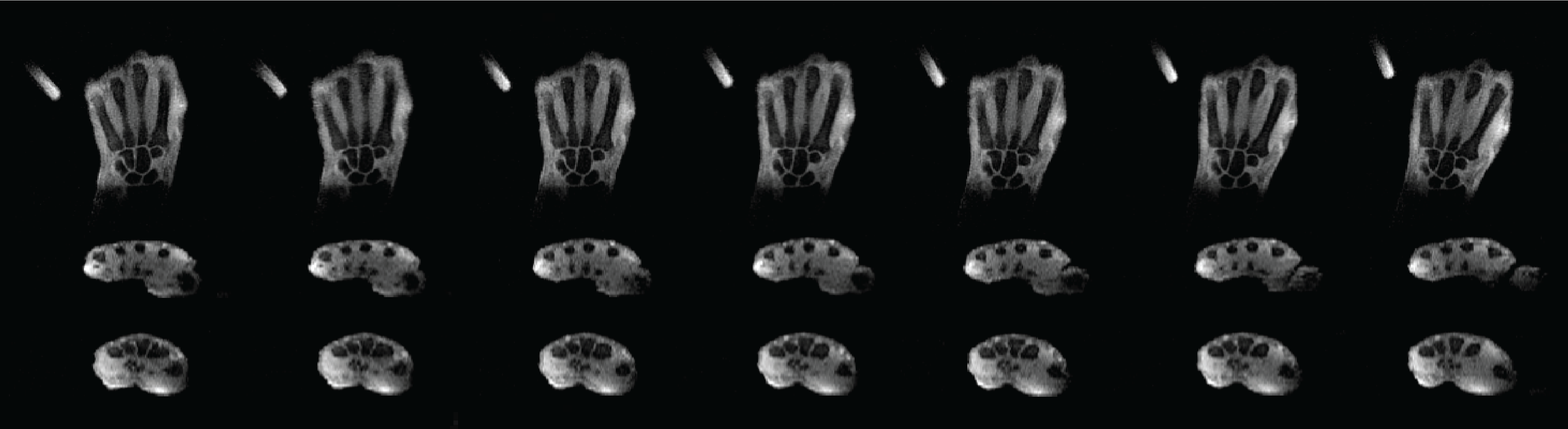
The automatic U-Net segmentation and resulting slice alignment is shown in Fig 3 for one angular position. After the alignment, the position marker (yellow color) remains in a consistent position for all slices. Fig 4 shows the fused dynamic volume for different wrist positions. Carpal bones and metacarpal bones are well-aligned, as seen especially in the two axial reformats, which demonstrates the efficiency of the proposed marker-based alignment procedure.
Discussion
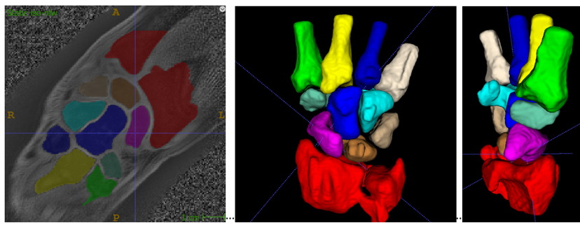
Here, we described a new idea to obtain dynamic 3D wrist images using a 2D real-time sequence and position markers to align images from different slices. The approach uses a 3D-printed platform that can be positioned freely in the scanner to maximize patient comfort and does not require additional optical or electronical sensors. Future work will focus on extracting the trajectories of carpal bones during continuous movement. Given the high contrast obtained with fat suppression and the low anisotropicity compared to previous approaches, individual carpal bones can be segmented in each dynamic volume using tools such as itkSNAP9. To improve the accuracy of the kinematic assessment, we plan to utilize a static high-resolution scan (similar to Fig 1d) for generating a 3D model of the wrist bones (Fig 5), which can then be used as strong morphological prior for determining the shape and location of individual bones in the dynamic volumes10. To this end, a next step will be to find a poly-rigid transformation model for mapping the reference carpal bone shapes onto the dynamic volumes to obtain dynamic series of binary segmentations for each carpal bone, which represent the kinematic motion to be analyzed.Acknowledgements
This work was supported in part by NIH R21 AR080325 and performed under the rubric of the Center for Advanced Imaging Innovation and Research (CAI2R, www.cai2r.net), an NIBIB National Center for Biomedical Imaging and Bioengineering (NIH P41 EB017183).References
1. Vassa R, Garg A, Omar IM. Magnetic resonance imaging of the wrist and hand. Polish Journal of Radiology. 2020 Aug 26;85(1):461-88.
2. Frahm J, Voit D, Uecker M. Real-time magnetic resonance imaging: radial gradient-echo sequences with nonlinear inverse reconstruction. Investigative Radiology. 2019 Dec 1;54(12):757-66.
3. Shaw CB, Foster BH, Borgese M, Boutin RD, Bateni C, Boonsri P, Bayne CO, Szabo RM, Nayak KS, Chaudhari AJ. Real-time three-dimensional MRI for the assessment of dynamic carpal instability. PloS one. 2019 Sep 19;14(9):e0222704.
4. Zhang B, Cloos MA, Yang J, Nguyen TD, Brown R. Ultra-flexible 3T HIC Receive Array for Carotid Imaging. In2019 International Conference on Electromagnetics in Advanced Applications (ICEAA) 2019 Sep 9 (pp. 0459-0464). IEEE.
5. Ngyuen T, Zhang B, Wen Y, Brown R. A lightweight and ultra-flexible "blanket" coil design for carotid artery wall imaging. Proceedings of the International Society for Magnetic Resonance in Medicine Annual Meeting and Exhibition, 2019, Montreal, Canada. p. 2079.
6. Zhang S, Block KT, Frahm J. Magnetic resonance imaging in real time: advances using radial FLASH. J Magn Reson Imaging. 2010 Jan;31(1):101-9.
7. Feng L, Grimm R, Block KT, Chandarana H, Kim S, Xu J, Axel L, Sodickson DK, Otazo R. Golden‐angle radial sparse parallel MRI: combination of compressed sensing, parallel imaging, and golden‐angle radial sampling for fast and flexible dynamic volumetric MRI. Magnetic resonance in medicine. 2014 Sep;72(3):707-17.
8. Ronneberger O, Fischer P, Brox T. U-net: Convolutional networks for biomedical image segmentation. In International Conference on Medical image computing and computer-assisted intervention 2015 Oct 5 (pp. 234-241). Springer, Cham.
9. Yushkevich PA, Piven J, Hazlett HC, et al. User-guided 3D active contour segmentation of anatomical structures: Significantly improved efficiency and reliability. NeuroImage 2006;31:1116–1128.
10. Abbas B, Fishbaugh J, Petchprapa C, Lattanzi R, Gerig G. Analysis of the kinematic motion of the wrist from 4D magnetic resonance imaging. In: undefined. Vol. 10949. Proc.SPIE; 2019. doi: 10.1117/12.2513131.
Figures