1418
Radial Stack-of-Stars Abdominal MRI at 7 Tesla1Department of Medical Imaging, Radboud University Medical Center, Nijmegen, Netherlands, 2Department of Radiology, NYU Langone Health, New York, NY, United States
Synopsis
Keywords: High-Field MRI, Body, Radial MRI
Abdominal MRI at 7T is sensitive to transmit field inhomogeneities and motion-induced artifacts. Transmit inhomogeneities have previously been addressed using time-interleaved acquistion of modes (TIAMO), providing uniform flip angles across a large field-of-view in the body. Meanwhile, the radial stack-of-stars sequence has been shown to be well-suited for motion-corrected MRI. In this work, TIAMO and motion-corrected radial MRI were combined to create abdominal images of three volunteers at high spatial resolutions. Results showed homogeneous transmit fields in all volunteers with excellent image quality at very high resolution. However, low-resolution scans suffered from artifacts due to gradient non-linearities and residual motion.
Introduction
It has been shown that radial k-space sampling can greatly reduce the sensitivity of abdominal MRI to ghosting artifacts.1 Using radial stack-of-stars 3D gradient-echo acquisition, diagnostic MR images can be obtained during free breathing, which is of high value for pediatric patients or subjects who experience difficulties holding breath. However, free-breathing scans still suffer from motion blur. To reduce such blurring and increase apparent image resolution, various motion-correction techniques have been proposed.2The effective image resolution of radial stack-of-stars MRI depends on the efficacy of the motion-correction employed and on the intrinsic image SNR. Since SNR scales with B0, image resolution can be increased by acquiring MR data at high magnetic field strengths.3 However, ultra-high field (UHF) MRI introduces several technical challenges, including constraints in the (local) amount of deposited radiofrequency (RF) power, a large water-lipid frequency difference, and inhomogeneity of the RF transmit field.
With parallel-transmit (pTx) capabilities, the RF field can be homogenized within a relatively small area through static RF shimming with a multi-transmit coil. For a larger field-of-view, this process can be performed pragmatically using a method referred to as “time-interleaved acquisition of modes” (TIAMO).4 TIAMO exploits two complementary RF shim settings of a pTx coil array, which are played out in an interleaved fashion to greatly improve the overall homogeneity of the transmit field. Meanwhile, the large water-lipid shift at 7T can cause blurring in images acquired with radial sampling. This fat blur can be circumvented using water excitation, lipid suppression, or Dixon-wise data processing of multiple echoes.
In this work, we aimed to acquire abdominal images with (very) high resolution at 7T during free breathing. To this end, we developed a radial stack-of-stars sequence combined with TIAMO RF shimming. Moreover, water excitation and retrospective motion correction were included in the acquisition and data processing to prevent motion and fat blur.
Methods
TIAMO was integrated into a pTx-capable radial stack-of-stars multi 3D gradient-echo (mGRE) sequence.5 Prior to each scan, a calibration scan (FLASH) was played out and two complementary phase settings for the 8 transmit coil elements were calculated using an offline Matlab routine.6 Exploiting the large chemical shift difference between water and lipids, we used an asymmetric low-bandwidth ($$$\approx$$$950 Hz) slab-selective pulse for water excitation (Figure 1). This pulse provided water excitation by shifting the resonance frequencies of lipid signal outside of the imaged volume, while requiring very little peak RF power. To prevent any remaining lipid signal from folding into the FOV, data were acquired in coronal orientation. An FID-navigator readout of 32 samples at every TR was integrated into the sequence for respiratory motion detection via principal component analysis. The k-space data were binned on a single-spoke basis in accordance with the respiratory signal.7 40% of the data nearest to the exhalation phase were selected for reconstruction, while the rest of the data were discarded.Data sets were acquired of three healthy volunteers (ages 31, 36, and 48; 1 female), after obtaining informed consent, on a 7T whole body system (MAGNETOM Terra, Siemens Healthineers, Erlangen, Germany) with an 8-channel Tx/Rx body-array coil (Tesla Dynamic Coils, Zaltbommel, Netherlands). Parameters of the radial stack-of-stars sequence varied between volunteers (Table 1) and images were reconstructed using the inverse non-uniform fast Fourier transform (iNUFFT). Since the two TIAMO modes have complementary transmit coil profiles, data from both modes were merged into 16 “virtual” coil elements, as if all data were obtained simultaneously.4 The 16 complex-valued coil images were combined by calculating the root sum-of-squares.
Results
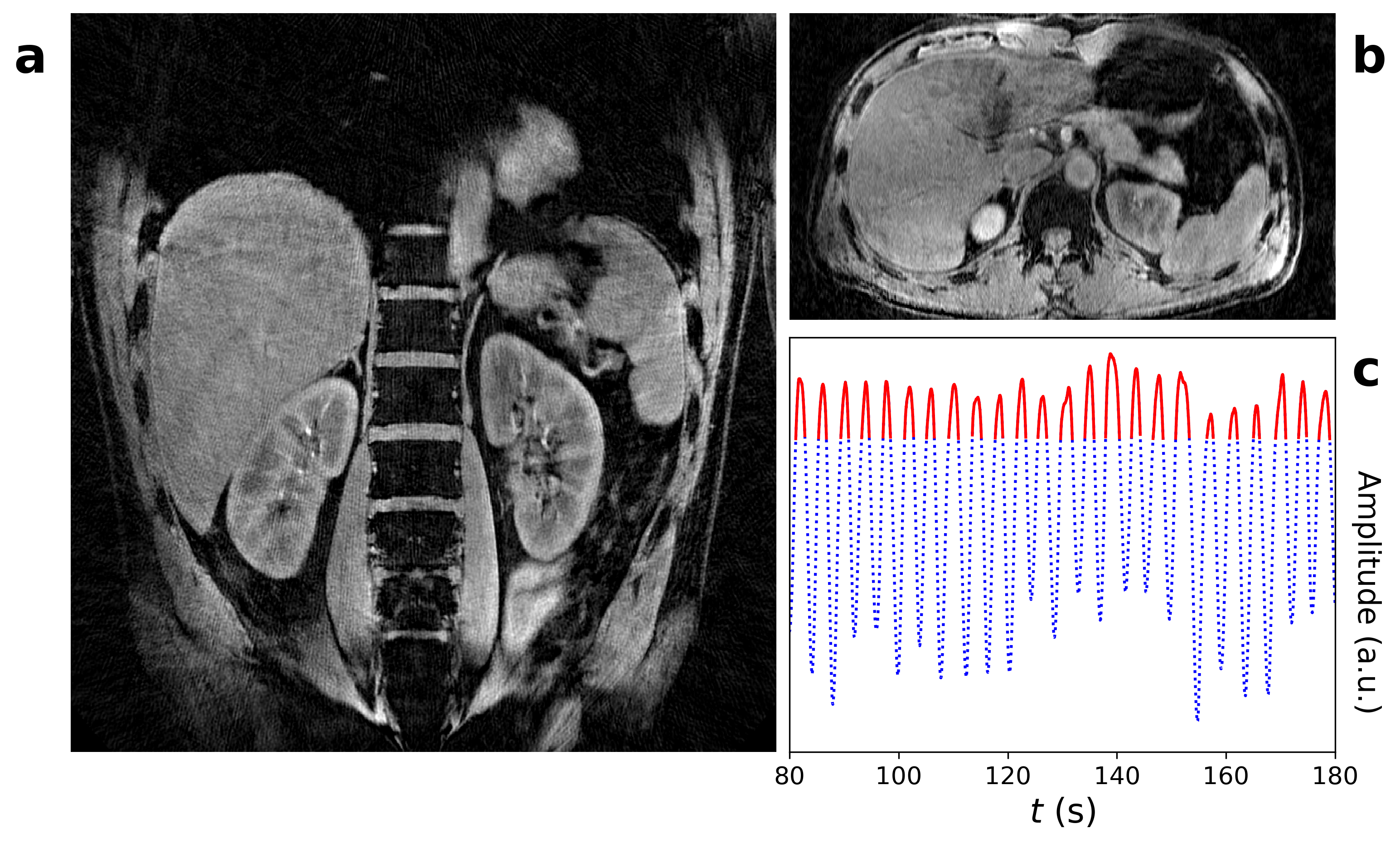
Reconstructed images of volunteer 1 contained a high SNR and a sharp depiction of the spine, kidneys, and adrenal glands (Figure 2). However, significant deformations were visible at the superior/inferior edges due to non-linearities of the imaging gradients, as well as streaks at the liver dome due to residual motion. Images of volunteer 2 showed a clear change in T2*-contrast across echoes, but contained severe streaking artifacts due to hyperintense signal regions at the borders of the FOV (Figure 3). Images of volunteer 3 showed motion-corrected radial stack-of-stars at very high resolution (0.8x0.8x1.0 mm3) with high SNR. A sharply delineated liver edge indicated successful motion-correction with minor residual motion blur. In all three volunteers, the RF transmit field appeared homogeneous, indicating successful RF shimming with TIAMO.Discussion
Preliminary results from the modified radial stack-of-stars sequence showed that abdominal MR images can be obtained at 7T with high resolutions which exceed capabilities of current clinical systems. In three presented cases, a homogeneous apparent RF transmit field was generated using the TIAMO technique. However, the studied protocol was sensitive to residual motion and gradient non-linearities at the borders of the FOV. Future work will be focused on (1) reducing these artifacts caused by gradient non-linearities, while also (2) shortening the RF pulse duration, and (3) improving motion-correction methods to increase scan efficiency and reduce residual motion artifacts.Conclusion
We showed preliminary results of radial stack-of-stars MR imaging at 7T with homogeneous transmission across the full FOV. Motion-corrected ultra-high field radial stack-of-stars MRI may enable abdominal imaging at unprecedented spatial resolutions.Acknowledgements
The body array coil used in this work was constructed with support from the European Fund for Regional Development EFRO OP-2014-2023-Oost [PROJ-01009].References
- Block KT, Chandarana H, Milla S, et al. Towards Routine Clinical Use of Radial Stack-of-Stars 3D Gradient-Echo Sequences for Reducing Motion Sensitivity. J Korean Soc Magn Res Med. 2014;18(2):87-106.
- Feng L. Golden-Angle Radial MRI: Basics, Advances, and Applications. J Magn Reson Imaging. 2022;56(1):45-62.
- Vaughan JT, Snyder CJ, DelaBarre LJ, et al. Whole-Body Imaging at 7T: Preliminary Results. Magn Reson Med. 2009;61:244-248.
- Orzada S, Maderwald S, Poser BA, et al. RF Excitation Using Time Interleaved Acquisition of Modes (TIAMO) to Address B1 Inhomogeneity in High-Field MRI.
- Schulz J, Kraff O, Quick H, et al. A Software-based TIAMO approach to enable high resolution large FOV body imaging at 7T ultra-high field. Proceedings of the 31st Annual Meeting of ISMRM, #2090, 2022.
- Brunheim S, Gratz M, Johst S, et al. Fast and Accurate Multi-Channel B$$$_1^+$$$ Mapping Based on the TIAMO Technique for 7T UHF Body MRI. Magn Reson Med. 2018;79(5):2652-2664.
- Maatman IT, Ypma S, Kachelrieß M, et al. Eliminating limits of spatiotemporal resolution in radial stack-of-stars imaging using FID navigators and single-readout binning. Proceedings of the 31st Annual Meeting of ISMRM, #2347, 2022.
Figures