1413
Substantially Improved Receive Sensitivity of Human Whole-Brain MRI at 7T using a High-Permittivity Material (HPM) Slurry-Filled Helmet1Center of Magnetic Resonance Research (CMRR), Department of Radiology, University of Minnesota, Minneapolis, MN, United States, 2Department of Biomedical Engineering, University of Minnesota, Minneapolis, MN, United States, 3Department of Engineering Science and Mechanics, Pennsylvania State University, University Park, PA, United States, 4Center for NMR Research, Department of Neurosurgery and Radiology, College of Medicine, Pennsylvania State University, Hershey, PA, United States
Synopsis
Keywords: High-Field MRI, New Devices, High Permittivity Material (HPM), High Dielectric Constant (HDC) Material
Novel methods such as high-permittivity materials (HPM) and metasurfaces have improved RF coil transmission efficiency and receive field (B1-) sensitivity for MRI applications at ultrahigh field. One of the recent studies, which applied ceramic HPM helmet with the permittivity of 100, showed significant improvement in signal-to-noise ratio (SNR). Motivated from the previous studies with various forms of HPM at 3T and 7T, this study introduces an easy and accessible method with an HPM slurry helmet to largely improve imaging quality of human brain MRI at 7T, which could improve B1- field by 57% and SNR by 47%.
Introduction
Human brain MRI scans at ultrahigh fields (UHF) with high spatiotemporal resolution have been beneficial for scientists to study the functionality, connectivity, and metabolism of the human brain. In addition, applications of various novel techniques such as high-permittivity materials (HPM) and metasurfaces have shown passive B1 shimming effects and improvements in transmission efficiency and receive sensitivity, especially for UHF MRI applications1- 5. The recent human brain studies at 7T showed average sensitivity improvement by 21% with a low-loss ceramic HPM helmet and relatively low permittivity (εr≈100)1. Since it is challenging to fabricate low-loss ceramic HPM helmets with a curved shape conforming to the human head, the accessibility to low-loss HPM applications for whole-brain MRI is limited. This study investigates a novel approach using an HPM slurry (a mixture of BaTiO3 and deionized water) filled into a desired 3D-printed helmet-shaped shell, which can largely improve the quality of human whole-brain MRI at 7T.Methods
BaTiO3 powder was mixed with deionized water to prepare the HPM slurry (εr≈200). Two sets of 2-piece helmet-shaped shells were 3D-printed. One set of helmet shells was filled with the HPM slurry, while the other was kept empty as the control condition. The 16-channel 1H transmitters with 32-channel receivers were assembled with a 2-piece helmet-shaped shell and tuned/matched for conditions with the HPM slurry (HPM) and without (control) (Figure 1). T1-weighted images and spin density images were collected, and the noise level was measured to calculate signal-to-noise ratios (SNR). Actual flip-angle images (AFI) and spin-echo echo-planar images (SE-EPI) with various RF pulse voltages were collected to calculate transmission (B1+) and reception (B1-) fields under loaded/unloaded conditions with the HPM slurry. B1+ was calculated with the method by Van de Moortele et al.6,7, and B1- was estimated by voxel-wise sinusoidal fitting from SE-EPI with various pulse voltages. Informed consent was gathered from the subjects to conduct human brain scans with both the HPM slurry and control helmets at 7T.Results
Anatomical images in Figure 2C-D show background noise level was 6.8% higher in the HPM condition than in the control, in contrast, the signal intensity in the T1-weighted and spin-density anatomic MRI was significantly increased using the HPM helmet (Figure 2A-B). The representative B1+ maps in sagittal orientation show similar field distribution and strength in Figure 3. Figure 4 shows the B1- maps and representative 1D B1- profiles to compare the differences between the HPM and control conditions. Figure 5 shows the B1- ratio maps and profiles between the conditions with HPM and control across the human head. Average B1- ratios under the HPM and the control conditions were calculated from the central region of interest (ROI), peripheral ROI, and ROI of each B1- image slice, and they are summarized in Figure 5C. The average B1- the ratio of the HPM over the control conditions in whole-brain was 1.57, which means about 57% improvement in B1- (i.e. receive sensitivity). The average B1- ratio from central ROI and peripheral ROI show 51% and 57% B1- improvement in the HPM condition. The average SNR improvement with the HPM was 47% from all slices after considering the small (6.8%) increase in the noise level.Discussion
The noise level with the HPM slurry helmet was 6.8% higher than the control. This suggests HPM helmet could contribute as an additional noise source resulting from the deionized water mixed with BaTiO3 powder having a high dielectric loss. Even with similar transmission efficiency and homogeneity, the HPM slurry helmet shows significant B1- and SNR improvements. A recent study reported 21% sensitivity improvement using a low-loss ceramic helmet with εr≈1001; the simulation result suggests that a ceramic HPM helmet with optimal permittivity of 200 would result in higher SNR improvement in peripheral than the central regions1, which was confirmed by the experimental measurements of the present study. We also observed more B1- improvements in regions near the curved surface in the superior part of the helmet shells. The geometric difference between the HPM slurry-filled shell used in this study and the previous studies could introduce the regional difference in B1- ratios. Additionally, B1+ maps also showed a small improvement in similar brain areas (marked with red circles in Figure 3), and this could result in the difference in the B1- ratio in central and peripheral regions.Conclusion
In conclusion, we successfully fabricated and tested the 3D-printed helmet filled with an HPM slurry, which demonstrated large receive sensitivity and SNR improvements across the whole human brain. We observed a 47% SNR gain which exceeds the 21% gain using the low-loss ceramic HPM helmet with relatively low permittivity as reported in the liturature1, and the SNR gain could reach double in some regions of the brain (Figure 5). The HPM slurry-filled helmet with a relatively high permittivity provides a robust and low-cost RF engineering solution for improving human brain MRI at 7T. The RF coil performance could be further improved by using a low-loss ceramic helmet with similar geometry and permittivity as applied in this study.Acknowledgements
This work was supported in part by NIH grants of U01 EB026978 and P41 EB027061.References
1. Lakshmanan, K., Carluccio, G., Walczyk, J., Brown, R., Rupprecht, S., Yang, Q. X., Lanagan, M. T., & Collins, C. M. (2021). Improved whole-brain SNR with an integrated high-permittivity material in a head array at 7T. Magn Reson Med, 86(2), 1167-1174.
2. Luo, W., Lanagan, M. T., Sica, C. T., Ryu, Y., Oh, S., Ketterman, M., Yang, Q. X., & Collins, C. M. (2013). Permittivity and performance of dielectric pads with sintered ceramic beads in MRI: early experiments and simulations at 3 T. Magn Reson Med, 70(1), 269-275.
3. Schmidt, R., Slobozhanyuk, A., Belov, P., & Webb, A. (2017). Flexible and compact hybrid metasurfaces for enhanced ultra high field in vivo magnetic resonance imaging. Sci Rep, 7(1), 1678.
4. Sica, C. T., Rupprecht, S., Hou, R. J., Lanagan, M. T., Gandji, N. P., Lanagan, M. T., & Yang, Q. X. (2020). Toward whole-cortex enhancement with an ultrahigh dielectric constant helmet at 3T. Magn Reson Med, 83(3), 1123-1134.
5.Yang, Q. X., Wang, J., Wang, J., Collins, C. M., Wang, C., & Smith, M. B. (2011). Reducing SAR and enhancing cerebral signal-to-noise ratio with high permittivity padding at 3 T. Magn Reson Med, 65(2), 358-362.
6. Van de Moortele P., Ugurbil K. Very Fast Multi Channel B1 Calibration at High Field in the Small Flip Angle Regime. Proc Intl Soc Mag Reson Med 2009; 17: 367
7. Van de Moortele PF, Akgun C, Adriany G, Moeller S, Ritter J, Collins CM, Smith MB, Vaughan JT, Ugurbil K. B1 destructive interferences and spatial phase patterns at 7T with a head transceiver array coil. Magn Reson Med 2005; 54 (6):1503-18.
Figures
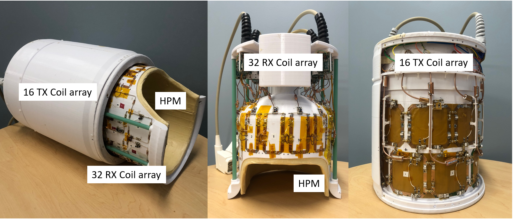
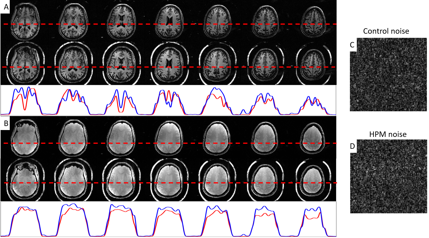
Figure 2 (A) T1-weighted images and (B) spin density images in axial orientation collected with empty helmet shells (control, top) and HPM slurry helmet shells (bottom) in the same scale and representative 1D profiles along the red dotted lines. (C, D) Representative background noise maps from the control and the HPM slurry condition, which had overall average noise levels from the control and the HPM slurry condition of 0.589 and 0.629, respectively, thus, the noise ratio of HPM/control was 1.068.
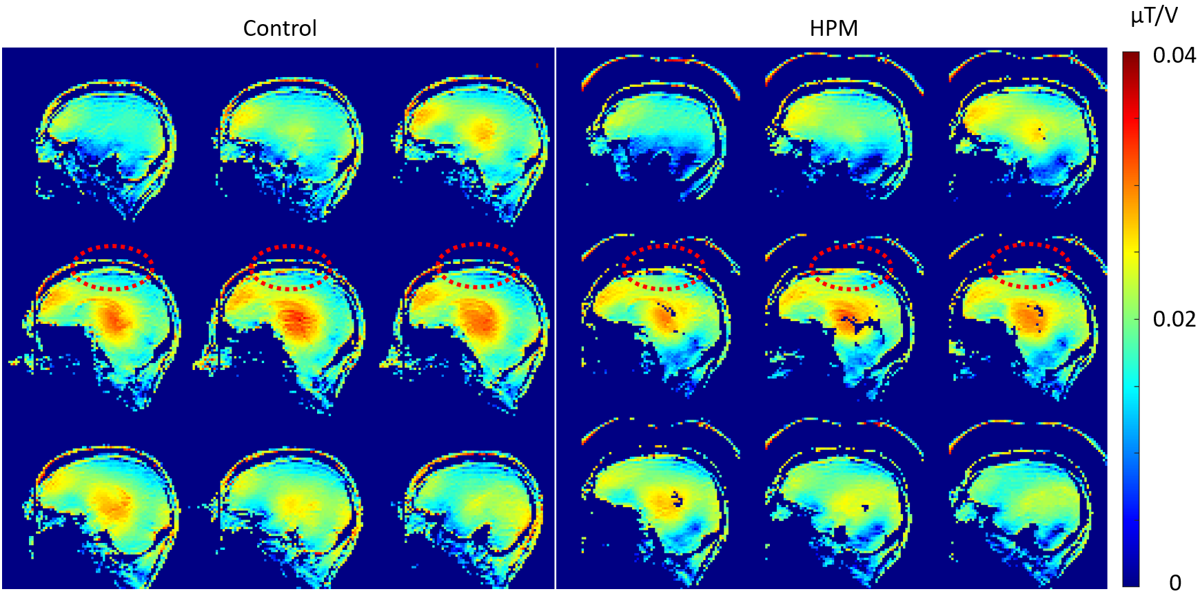
Figure 3 B1+ maps for the control condition (left) and the HPM slurry helmet condition (right) with the same scale. Both B1+ maps showed similar transmission field strength and distribution. Red dotted circles show a slight difference in the central region of the brain, which possibly introduced a regional difference in B1- ratios for superior imaging slices.
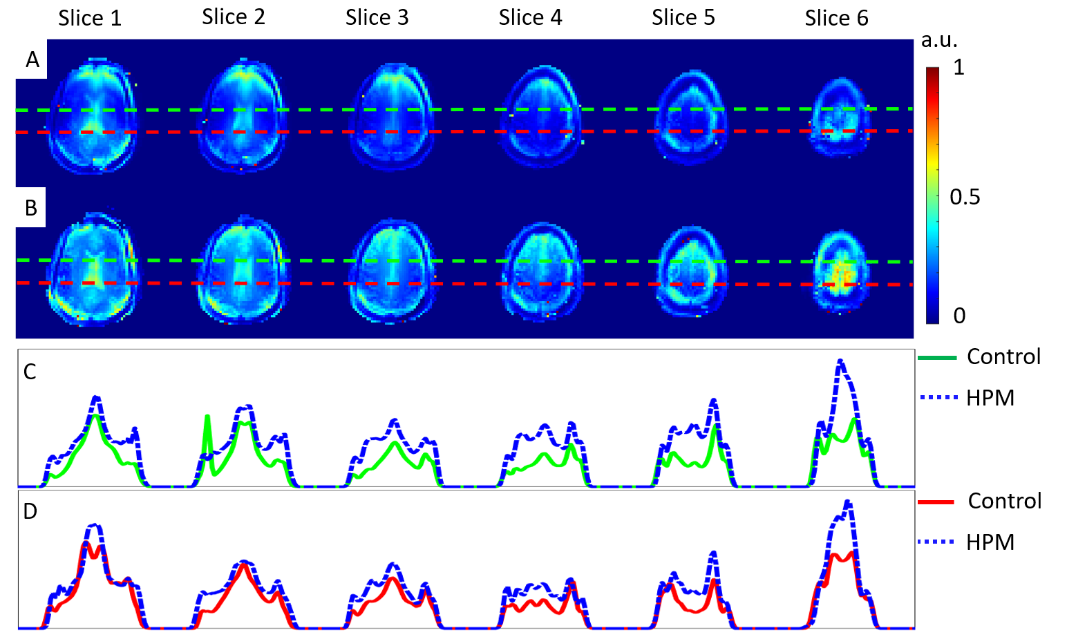
Figure 4 (A, B) Representative B1- maps (axial orientation) estimated by the sinusoidal curve fitting method with multiple SE-EPIs acquired at various RF pulse voltages under the control condition (A) and the HPM slurry helmet condition (B). (C, D) 1D B1- profiles along the green and red dotted line shown in (A) and (B). The B1- profiles along the green dotted lines are shown in (C) and the B1- profiles along the red dotted lines are shown in (D).
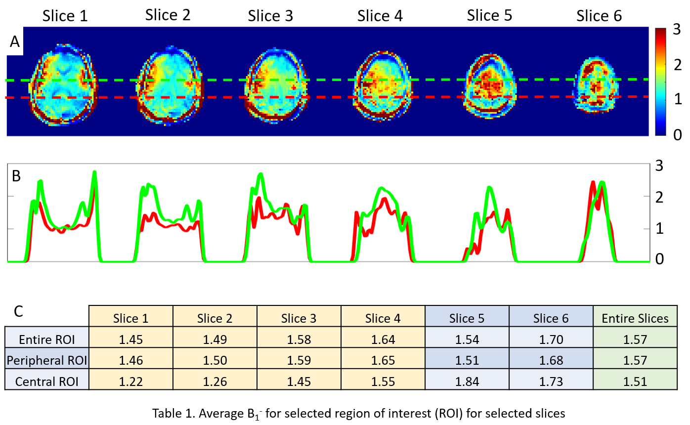
Figure 5 (A) The B1- ratio maps of the HPM B1- maps over the control B1- maps shown in Figure 4. (B) 1D B1- ratio profile drawn along the green and red dotted lines shown in (A). The color of the profile follows the color of the dotted lines in (A). (C) Average B1- ratio calculated from different regions of interest (ROI) in the human brain. The overall B1- improvement using the HPM slurry helmet can reach 57%. Blue boxes show the average B1- ratio from two superior imaging slices.