1410
Direct abdominal vein thrombus imaging (DATI): a contrast free black-blood MR technique for the diagnosis of abdominal vein thrombosis1The First People’s Hospital of Qinzhou, Qinzhou, China, 2School of Biomedical Engineering, Guangzhou Medical University, Guangzhou, China, 3Siemens Healthineers, Shanghai, China
Synopsis
Keywords: Vessels, Thrombo-Embolic
Abdominal vein thrombosis (AVT) is a significant cause of morbidity. Accurate diagnosis of AVT is relevant for treatment proper decision-making. CTV or MRV requires the use of contrast medium, which may lead to patient's renal failure or allergic reaction. To address this issue, we sought to develop a direct AVT imaging (DATI) technique which is free of contrast medium. The technique is based on a respiratory navigating SPACE sequence with DANTE black-blood preparation and evaluated preliminarily on 19 AVT patients at 3.0T. Experiment results show that DATI can provide definitive thrombus detection for the diagnosis of AVT.PURPOSE
AVT is a common disease, of which portal vein thrombosis (PVT) is the most common in clinic. Although PVT can be asymptomatic in some patients with cirrhosis, it can manifest with life-threatening complications such gastrointestinal bleeding, development or abrupt worsening of ascites or hepatic encephalopathy [1]. In addition, the presence of malignant thrombus is an absolute contraindication to liver transplant, due to the low survival and high post-transplant recurrence [2]. Therefore, accurate evaluation of AVT is of great clinical importance for appropriate treatment selection [3]. At present, diagnostic imaging techniques, such as CTV and MRV, together with clinical and laboratory findings, are often relied upon for thrombus discrimination, but they almost need to use contrast medium. Recently, an MR black-blood thrombus imaging (BTI) technique has been developed and successfully used for the diagnosis of deep vein thrombosis and cerebral venous thrombosis [4-6]. Inspired by this, we sought to develop a direct AVT imaging (DATI) technique which is a black-blood and free of contrast medium for the diagnosis of AVT.METHODS
Experiment: The DATI technique is based on a respiratory navigating 3D variable-flip-angle turbo-spin-echo sequence with a black-blood preparation, i.e delay alternating with nutation for tailored excitation (DANTE). The technique was first optimized on 5 healthy subjects (age 24.6 ± 5.9 years) to reduce the respiratory motion artefacts and enhance the abdominal venous blood flow signal suppression a 3T system (Skyra, Siemens, Germany) with a standard 18-channel coils positioned anteriorly and an integrated spine coil located posteriorly. After the optimization completed, the technique with optimized imaging parameters was then applied on 19 suspected AVT patients (51.7±9.8 years, 17male, 2 female) who were completed CTV within 24h. DATI parameters included: FOV=236mm×294mm, matrix=204×256, slice thickness=2mm, bandwidth=977Hz/pixel, TR=1100 ms, TE=20ms, turbo factor = 80. Image analysis: All images were loaded to a workstation (Leonardo, Siemens Healthineers) for analysis. Two radiologists independently evaluated randomized images and gave image quality and diagnostic confidence scores (1-poor, 4-excellent) for DATI and CTV, without knowledge of each subject’s information. The accuracy (ACC), sensitivity (SE), specificity (SP), positive predictive value (PPV), negative predictive value (NPV) of CTV were calculated using CTV as reference. The diagnostic agreement between DATI and CTV as well as the interobserver agreement were conducted using Cohen κ test.Results
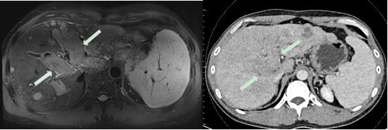
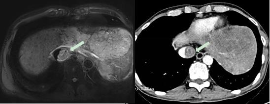
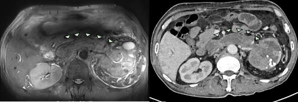
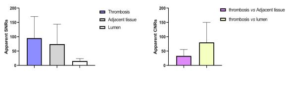
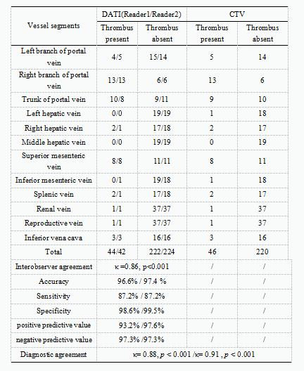
Representative images obtained by DATI and CTV from three AVT patients were shown in Figure 1&3. The thrombi can be visualized on DATI images directly, which is consistent with those detected by CTV in terms of the thrombus size and position. Image quantitative analysis results demonstrated that DATI can provide adequate thrombus signal intensity and the contrast between the thrombus to black-blood venous lumen for the diagnosis of AVT (Figure 4),and offers good-to-excellent image quality (reader1/reader2: 3.26 ±0.73/3.21 ±0.63, κ =0.74) and excellent diagnostic confidence (reader1/reader2: 3.73 ±0.45/3.63 ±0.60, κ = 0.75) for the diagnosis of AVT. The interobserver agreements of DATI were showed in all the patients regarding the presence or absence of thrombus (κ =0.84, p<0.001). There is a good consistency between the two readers for Diagnosis of DATI and CTV (κ =0.88,κ =0.91 p<0.001). According to the CTV consensus reading, a total of 266 venous segments were observed in 19 patients and 46 (17.2%) of them were found the thrombus (Table 1). Taking CTV as reference, DATI has high accuracy (97.0%), SE (87.2%), SP (99.1%), PPV (95.4%), NPV (97.3%).Conclusion
DATI can provide excellent venous blood signal suppression and definitive thrombus detection for the diagnosis of AVT without the use of a contrast agent.Acknowledgements
No acknowledgement found.References
[1] PRIMIGNANI M. Portal vein thrombosis, revisited [J]. 1878-3562 (Electronic)):
[2] EASL Clinical Practice Guidelines: Management of hepatocellular carcinoma [J]. 1600-0641 (Electronic)):
[3] LAU W Y, WANG K, ZHANG X P, et al. A new staging system for hepatocellular carcinoma associated with portal vein tumor thrombus [J]. 2304-3881 (Print)):
[4] XIE G, CHEN H, HE X, et al. Black-blood thrombus imaging (BTI): a contrast-free cardiovascular magnetic resonance approach for the diagnosis of non-acute deep vein thrombosis [J]. Journal of cardiovascular magnetic resonance : official journal of the Society for Cardiovascular Magnetic Resonance, 2017, 19(1): 4.
[5] CHEN H, HE X, XIE G, et al. Cardiovascular magnetic resonance black-blood thrombus imaging for the diagnosis of acute deep vein thrombosis at 1.5 Tesla [J]. Journal of cardiovascular magnetic resonance : official journal of the Society for Cardiovascular Magnetic Resonance, 2018, 20(1): 42.
[6] YANG Q, DUAN J, FAN Z, et al. Early Detection and Quantification of Cerebral Venous Thrombosis by Magnetic Resonance Black-Blood Thrombus Imaging [J]. Stroke, 2016, 47(2): 404-409.
Figures