1376
A dual-stage partially interpretable neural network for joint suppression of bSSFP banding and flow artifacts in non-phase-cycled cine imaging1The Institute of Medical Imaging Technology, School of Biomedical Engineering, Shanghai Jiao Tong University (SJTU), Shanghai, China
Synopsis
Keywords: Machine Learning/Artificial Intelligence, Artifacts, Banding artifacts, Flow artifacts
bSSFP cine imaging suffers from banding and flow artifacts in the region of off-resonance. Suppressing one kind of artifacts may evoke the other kind. For example, phase cycling suppresses banding artifacts, yet its acquisition at multiple frequency offsets often evokes flow artifacts. Here, we develop a partially interpretable neural network for jointly suppressing banding and flow artifacts without phase cycling. Based on a single cine image, the method generates an artifact-corrected image and a voxel-identity map, which guides the artifact suppression and improves its interpretability. Preliminary investigation shows that the method reduces banding and flow artifacts without introducing new artifacts.
Introduction
bSSFP cine imaging suffers from banding and flow artifacts in regions whose frequency offset equals an odd multiple of 1/TR1,2. The former causes signal hypoenhancement and the latter causes signal hyperenhancement3,4. Previous methods mainly focus on suppressing one of these two artifacts5–7. However, suppressing one type of artifacts may evoke the other type of artifacts8. For example, phase cycling is commonly employed to mitigate banding artifacts9,10. However, since phase cycling acquires data at multiple frequency offsets, the chance of incurring flow artifacts is considerably increased rather than reduced11,12. Joint suppression of banding and flow artifacts has been rarely reported13.In this work, we propose a novel approach for jointly suppressing bSSFP cine banding and flow artifacts based on deep learning, which has shown promise for numerous artifact reduction tasks14–16. The proposed method uses only a single cine image as the input and generates two outputs, including an artifact-corrected image and a voxel-identity map, which shows the artifact identification at each voxel to provide a partial interpretation of the correction.
Methods
Model architectureThe overall network consists of two sub-networks (Figure 1): a Voxel Identification (VI) sub-network to recognize the artifact identity at each voxel and an Artifact Suppression (AS) sub-network to suppress the artifacts. Both sub-networks use a U-net architecture as the backbone. The VI sub-network takes a single cine image as the input, and outputs a number at each voxel, which is valued near 1 if the voxel is in a dark band, near 0 if in a flow artifact, and near 0.5 if artifact-free. The AS sub-network takes two images as the input. The first image is a partially corrected image generated by multiplying the raw image with its voxel-identity map, which hyperenhances the banding voxels and hypoenhances the flow voxels. The second image is an “artifact mask” generated by thresholding of the voxel-identity map. The AS sub-network composites a U-net with a Feature Pyramid Module (FPM), which was previously introduced to integrate multi-scale features from a U-net17,18.
Training and testing
Twenty healthy subjects (8 male, age 24±2) were imaged in a 3T scanner (uMR790, United Imaging Healthcare) after providing written informed consent. Cine imaging was performed with short- and long-axis views and 12 frequency offsets uniformly sampling between -1/(2*TR) and 1/(2*TR). The sequence parameters were bandwidth/flip angle/acquisition matrix/slice thickness/TR/=1000Hz/60°/224×199/8mm/2.98ms. All images were interpolated, cropped, and normalized to have a resolution of 1.5mm and an image size of 192×192.
Data from 10 subjects were used for training, including 360 short-axis, 24 two-chamber, and 24 four-chamber cine movies. The VI sub-network was trained before the AS sub-network. The label for training the VI sub-network is an image averaged from the central 5 frequency offsets divided by each phase-cycled image, followed by a sigmoid. The label for the AS sub-network is the averaged image. The loss for the VI and AS sub-network is a linear combination of the reconstruction loss, measured by MSE, and perceptual loss, whose definition was previously introduced19,20.
Three networks were trained, including the proposed dual-stage network, a FPM U-net, and the same cascaded network as the proposed method but with an end-to-end training. Testing was performed in the remaining 10 subjects. Correction accuracy was evaluated by NRMSE and SSIM via paired t-test.
Results
Figure 2 shows artifact identification results of the VI sub-network. The first image has a frequency offset of 0Hz, resulting in nearly no artifacts in the heart region. The second image has a frequency offset of 1/(2TR), resulting in strong banding and flow artifacts. The output of the VI sub-network well agreed with the label image.Figure 3 shows the artifact suppression results of the three networks in two subjects. In the first subject, the dual-stage network outperformed the other two networks with more accurate recovery of the septal wall. In the second subject, all 3 networks yielded good reconstructions, yet the phase-cycled combination image showed motion blurring due to slice mis-registration between different breath-holds, entailed by the phase-cycled acquisition.
Figure 4 shows the quantitative comparison of the three networks over the subjects. The dual-stage network had a slightly lower NRMSE (0.2755±0.0573) than the FPM U-net (0.2797±0.0594, p=0.002), and slightly higher SSIM (0.8126±0.0758) than both the cascaded U-net (0.8097±0.0733, p=0.0005) and FPM U-net (0.8107±0.0751, p=0.0007).
Figure 5 shows generalizability of the dual-stage network in a) long-axis movies which have only a limited amount of training data, and b) short-axis images with parameter values not used by the training data. Notice the excellent suppression of banding artifacts in the abdomen and subcutaneous fat, and of flow artifacts in the aorta. The voxel-identity map showed location of various artifacts, rendering the correction results interpretable.
Conclusions
The preliminary results suggest that the network is able to jointly reduce banding and flow artifacts in bSSFP cine imaging. Importantly, the method does not increase scan time or introduce other caveats, such as motion and flow artifacts. Furthermore, the artifact identity map provides an explanation for the correction performed by the AS sub-network, adding confidence to the result analysis.Acknowledgements
No acknowledgement found.References
1. Scheffler K, Lehnhardt S. Principles and applications of balanced SSFP techniques. Eur Radiol 2003; 13:2409–2418.
2. Schär M, Kozerke S, Fischer SE, Boesiger P. Cardiac SSFP imaging at 3 Tesla. Magn Reson Med 2004; 51:799–806.
3. Storey P, Li W, Chen Q, Edelman RR. Flow artifacts in steady-state free precession cine imaging. Magn Reson Med 2004; 51:115–122.
4. Bieri O, Scheffler K. Fundamentals of balanced steady state free precession MRI. J Magn Reson Imaging 2013; 38:2–11.
5. Zur Y, Wood ML, Neuringer LJ. Motion-insensitive, steady-state free precession imaging. Magn Reson Med 1990; 16:444–459.
6. Vasanawala SS, Pauly JM, Nishimura DG. Linear combination steady-state free precession MRI. Magn Reson Med 2000; 43:82–90.
7. Bieri O, Scheffler K. Flow compensation in balanced SSFP sequences. Magn Reson Med 2005; 54:901–907.
8. Markl M, Pelc NJ. On flow effects in balanced steady-state free precession imaging: Pictorial description, parameter dependence, and clinical implications. J Magn Reson Imaging 2004; 20:697–705.
9. Bangerter NK, Hargreaves BA, Vasanawala SS, Pauly JM, Gold GE, Nishimura DG. Analysis of multiple-acquisition SSFP. Magn Reson Med 2004; 51:1038–1047.
10. Elliott AM, Bernstein MA, Ward HA, Lane J, Witte RJ. Nonlinear averaging reconstruction method for phase-cycle SSFP. Magn Reson Imaging 2007; 25:359–364.
11. Datta A, Nishimura DG, Baron CA. Banding-free balanced SSFP cardiac cine using frequency modulation and phase cycle redundancy. Magn Reson Med 2019; 82:1604–1616.
12. Robb JS, Hu C, Peters DC. Interleaved, undersampled radial multiple-acquisition steady-state free precession for improved left atrial cine imaging. Magn Reson Med 2020; 83:1721–1729.
13. Datta A, Nishimura DG, Baron CA. BMART-Enabled Field-Map Combination of Projection-Reconstruction Phase-Cycled SSFP Cardiac Cine for Banding and Flow-Artifact Reduction. ArXiv210211428 Phys 2021
14. Kim KH, Park S-H. Artificial neural network for suppression of banding artifacts in balanced steady-state free precession MRI. Magn Reson Imaging 2017; 37:139–146.
15. Defazio A, Murrell T, Recht M. MRI Banding Removal via Adversarial Training. Advances in Neural Information Processing Systems, 2020; 7660–7670
16. Xie K, Gao L, Lu Z, et al. Inpainting the metal artifact region in MRI images by using generative adversarial networks with gated convolution. Med Phys n/a
17. Lin T-Y, Dollár P, Girshick R, He K, Hariharan B, Belongie S. Feature Pyramid Networks for Object Detection. 2017 IEEE Conference on Computer Vision and Pattern Recognition (CVPR), 2017; 936–944
18. Xiao T, Liu Y, Zhou B, Jiang Y, Sun J. Unified Perceptual Parsing for Scene Understanding. Computer Vision – ECCV 2018, Cham, 2018; 432–448
19. Simonyan K, Zisserman A. Very Deep Convolutional Networks for Large-Scale Image Recognition. 2015
20. Dosovitskiy A, Brox T. Generating Images with Perceptual Similarity Metrics based on Deep Networks. Advances in Neural Information Processing Systems, 2016;
Figures
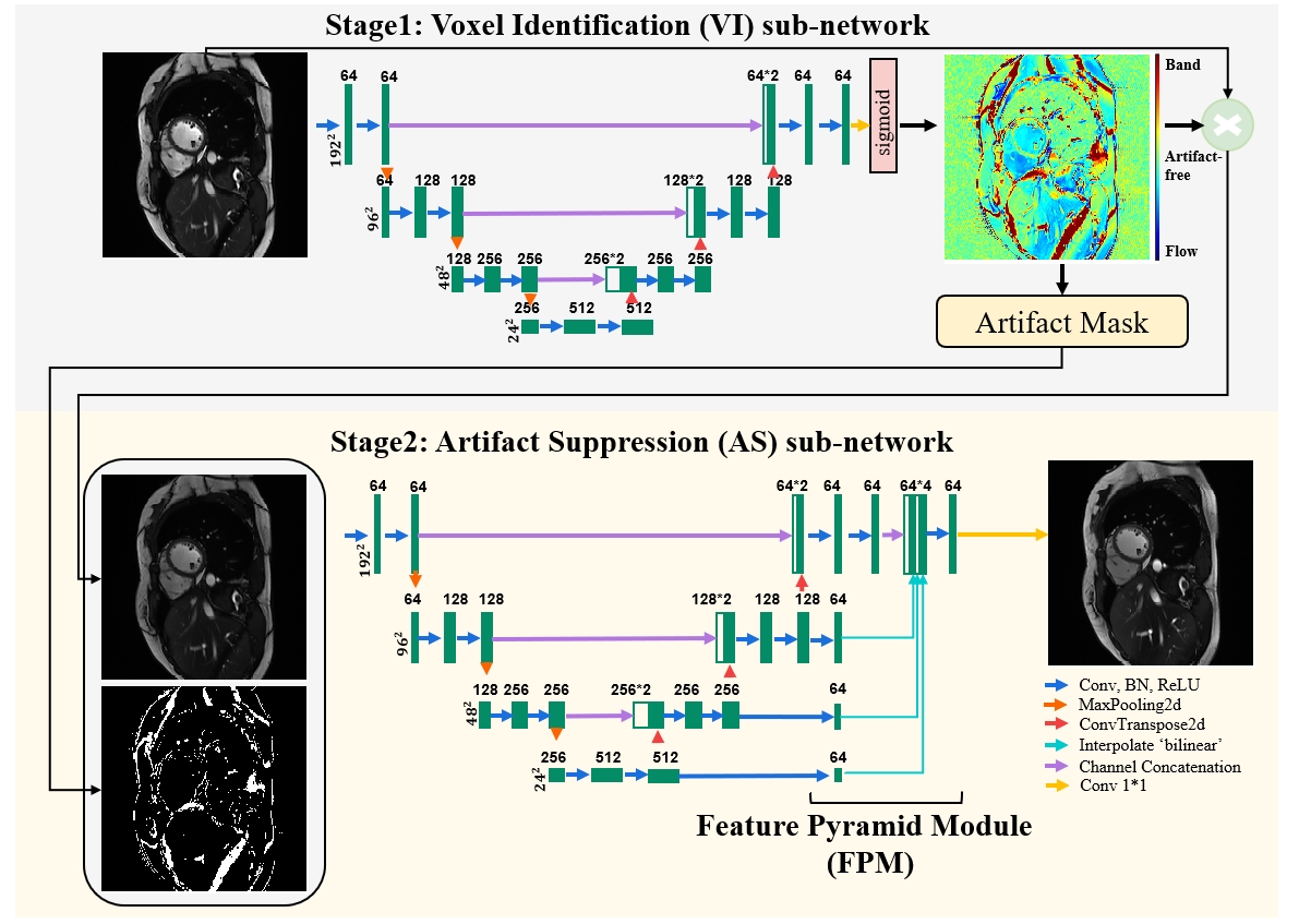
Figure1. The scheme of the proposed method to suppress banding and flow artifacts. The VI sub-network takes a single cine image as the input, and outputs a number at each voxel, which is valued near 1 if the voxel is in a dark band, near 0 if in a flow artifact, and near 0.5 if artifact-free. The AS sub-network takes a partially corrected image and an “artifact mask” as the input, which provide artifact contextual and identity information, respectively. A feature pyramid module is used to integrate the multi-scale features from the U-net to generate the final artifact-corrected image.
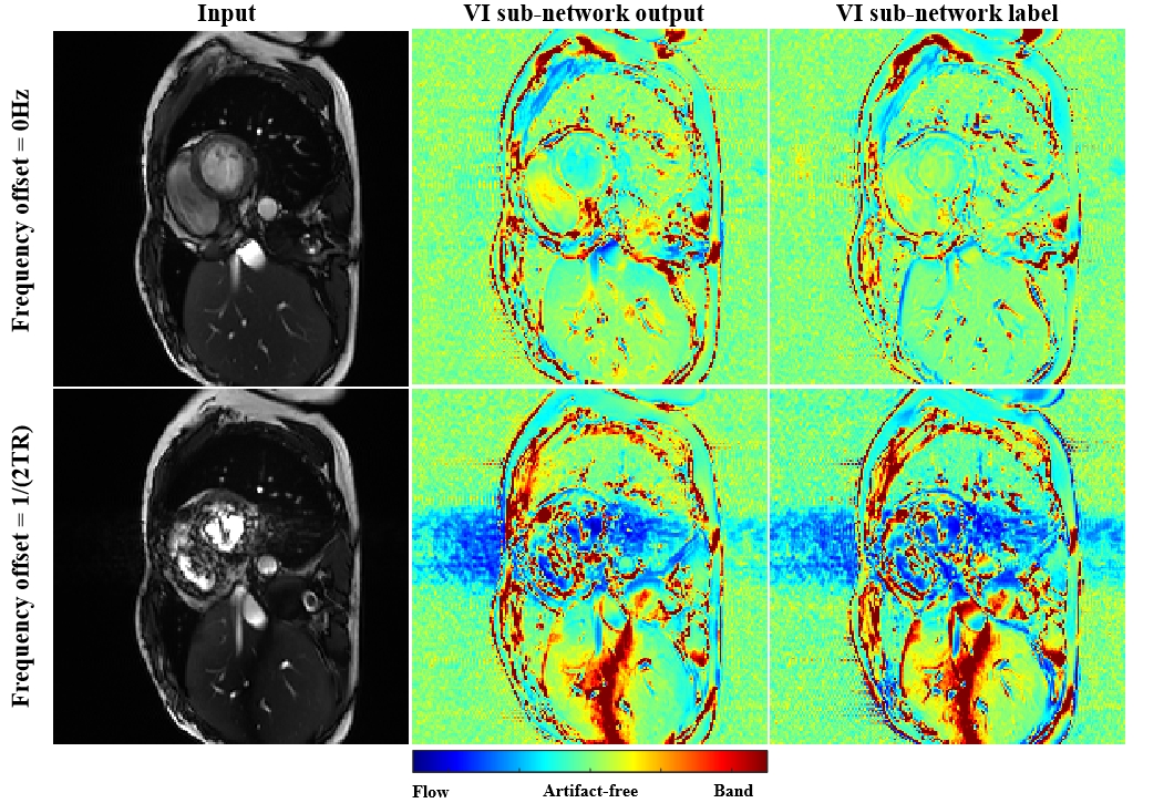
Figure 2. The performance of the voxel identification sub-network. Column 1 shows the end-systolic frame of the input cine movies, acquired at a frequency offset of 0Hz and 1/(2TR), respectively. Column 2 shows the predicted voxel-identity map, which well agreed with the training label shown in Column 3. The label is generated by dividing the phase-cycled combination image by the incident cine image. Thus, higher values indicate banding and lower values indicate flow artifacts. The identity map guides the following artifact suppression and improves its interpretability.
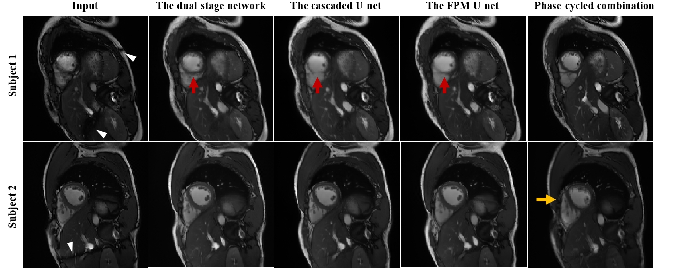
Figure 3. Comparison of the dual-stage method, the cascaded U-net, the FPM U-net, and phase-cycled combination. In subject 1, relatively strong banding and flow artifacts appeared in the heart. The dual-stage method achieved an improved recovery of the septum (red arrows) compared with the other two methods. In subject 2, while all methods achieved similar qualities, the phase-cycled combination had motion blurring (yellow arrows) due to acquisition of the images in multiple breath-holds. The white arrow heads point to the banding artifacts in the abdomen and subcutaneous fat.
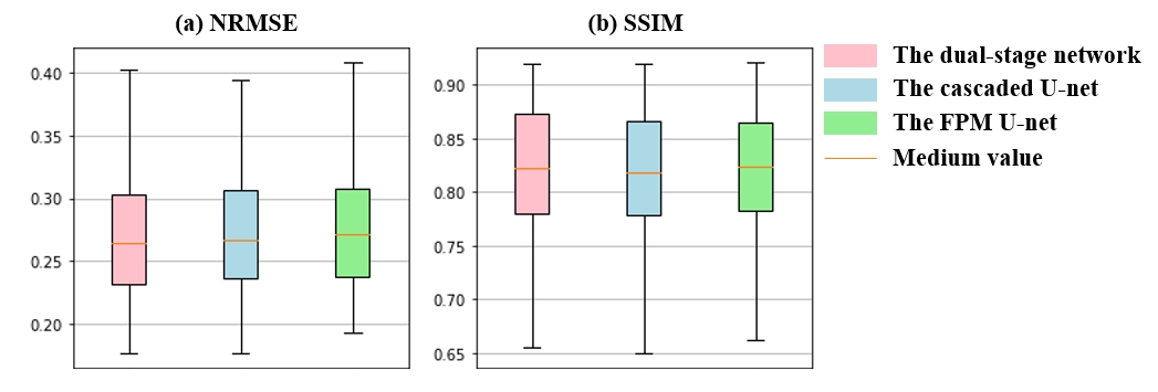
Figure 4. Quantitative comparison of the three deep learning methods. The dual-stage method resulted in slightly lower NRMSE (0.2755±0.0573) than the FPM U-net (0.2797±0.0594,p=0.0020), and slightly higher SSIM (0.8126±0.0758) than the cascaded U-net (0.8097±0.0733, p=0.0005) and FPM U-net (0.8107±0.0751, p=0.0007).
Figure 5. The generalizability of the dual-stage network in a) long-axis movies which have only a limited amount of training data, and b) short-axis images with sequence parameters not used by the training data. Notice that the 2-chamber and 4-chamber images were trained with less than 5 percent of overall data. The banding artifacts in the abdomen and subcutaneous fat, and the flow artifacts in the aorta were well suppressed by the dual-stage method. The voxel-identity map provides an explanation for the correction performed by the AS sub-network.