1350
Rapid PD, T2, and T2* Mapping with 2in1-RARE-EPI and Model-Based Reconstruction in Multiple Sclerosis1Berlin Ultrahigh Field Facility (B.U.F.F), Max Delbrück Center for Molecular Medicine in the Helmholtz Association, Berlin, Germany, 2Department of Physics, Humboldt University of Berlin, Berlin, Germany, 3Digital Health - Machine Learning Research Group, Digital Health Center, Hasso Plattner Institute, University of Potsdam, Potsdam, Germany, 4Experimental and Clinical Research Center (ECRC), a joint cooperation between the Charité Medical Faculty and the Max Delbrück Center for Molecular Medicine in the Helmholtz Association, Berlin, Germany
Synopsis
Keywords: Relaxometry, Multiple Sclerosis
One obstacle of established quantitative resonance imaging methods is the excessive scan time. This study examines the use of radially-sampled 2in1-RARE-EPI in conjunction with model-based reconstruction methods for accelerated and simultaneous PD, T2, and $$$T_{2}^{*}$$$ mapping. We demonstrate that this approach facilitates a substantial decrease in acquisition time (01:40 vs. 07:12 min) of 2in1-RARE-EPI without impairing the quality of the parametric maps. Our patient data demonstrate the applicability of model-based reconstructed 2in1-RARE-EPI for T2 and $$$T_{2}^{*}$$$ mapping of multiple sclerosis lesions.
INTRODUCTION
Quantitative magnetic resonance imaging (qMRI) adds diagnostic value to the assessment of brain pathologies such as multiple sclerosis (MS)1. Recent studies showed that T2 and $$$T_{2}^{*}$$$ mapping are viable imaging markers to monitor disease progression at an early stage of MS2-3. A major obstacle of clinical qMRI is the long acquisition time of currently established methods that increases with the number of MR parameters to be acquired.To overcome this problem, our group recently developed a hybrid radially-sampled 2in1-RARE-EPI technique tailored for simultaneous T2 and $$$T_{2}^{*}$$$ mapping and demonstrated its applicability in MS patients4. Model-based reconstruction (MBreco) is conceptually appealing for the estimation of MR parameters from undersampled k-space data5-8. Recognizing this opportunity this work combines 2in1-RARE-EPI with MBreco methods to accelerate the estimation of PD, T2, and $$$T_{2}^{*}$$$ maps. Our results demonstrate a synergistic effect, resulting in substantially reduced acquisition times.
METHODS
2in1-RARE-EPI comprises a RARE module followed by an EPI module and acquires complementary spokes per echo train, facilitating retrospective undersampling and reconstruction using MBreco. Measurements involving healthy subjects and MS patients were performed on a SkyraFit 3T system (Siemens, Erlangen, Germany) using a 32-channel head coil (FOV 232x232 mm2, matrix size 256x256, excitations=200, TR=2000 ms, bandwidth 610=Hz/pixel, ETLRARE/ETLEPI=14/18, ESPRARE/ESPEPI=6.5/2.3 ms). For validation, standard multi-echo spin-echo (MSE) and multi gradient-echo (MGRE) techniques were used with geometries and timings identical to the 2in1-RARE-EPI acquisitions, but with ETLMSE/ETLMGRE=15/12 and number of excitations=256.For the 2in1-RARE-EPI data, the TE images were reconstructed using regridding with linear density compensation and a magnitude sum-of-squares channel combination using BART9. Conventional pixelwise fitting of the magnitudes of the TE images (2in1-RARE-EPI, MSE, MGRE) was performed using linear least-squares approach to obtain T2 and $$$T_{2}^{*}$$$ maps.
The 2in1-RARE-EPI data was retrospectively undersampled from 200 to 34 spokes per echo. Then, PD and T2 maps were calculated from the RARE module, while $$$T_{2}^{*}$$$ and B0 maps were calculated from the EPI module, using the BART implementations of the MBreco strategies7-8. For both MBreco methods, 10 Newton iterations were used. Since MBreco is an iterative non-convex problem, providing reasonable initial guesses of the parameters helps the MBreco to converge to a suitable solution5,7-8. Hence, we performed a compressed sensing reconstruction with wavelet regularization10 to the last RARE echo using BART, and used the resulting image as an initial guess for the initial magnetization in the $$$T_{2}^{*}$$$ calculation. This is a well-motivated guess since the signal only starts experiencing $$$T_{2}^{*}$$$ decay after the last RARE echo. As an initial guess for $$$R_{2}^{*}=1/T_{2}^{*}$$$, the $$$R_{2}=1/T_{2}$$$ map obtained from the RARE module was utilized. Finally, the B0 map was initialized from the first three EPI echoes using a three-point model-based water/fat separation7-8,11.
RESULTS
Figure 1 shows T2 and $$$T_{2}^{*}$$$ maps of a healthy volunteer using the three acquisitions and the two mapping techniques. The adoption of the MBreco methods for 2in1-RARE-EPI provides comparable results to the pixelwise fitting approach while dramatically decreasing acquisition time by a factor of 4.Figure 2 shows the absolute error of the 2in1-RARE-EPI $$$T_{2}/T_{2}^{*}$$$ maps with/without MBreco relative to the conventional MSE/MGRE reference maps, and the 2in1-RARE with MBreco relative to the conventional mapping. For T2, a mean absolute percentage error (MAPE) of ~10% was found. For $$$T_{2}^{*}$$$, MAPE increases to ~20%. It stands to reason that this error was mainly caused by poor B0 shimming which manifests itself as an artifact covering the frontal horn of the lateral ventricles in the $$$T_{2}^{*}$$$ maps (figure1, second row) derived from the MGRE reference, 2in1-RARE-EPI and MBreco 2in1-RARE-EPI.
Beyond the T2 and $$$T_{2}^{*}$$$, PD and B0 maps obtained for a healthy subject, synthetic T2- and $$$T_{2}^{*}$$$-weighted images were derived from the quantitative maps (figure 3).
Figure 4 highlights T2 and $$$T_{2}^{*}$$$ maps of the brain of an MS patient. The mean and standard deviations obtained for an ROI (7x7 pixels) covering the MS lesion reveal that 2in1-RARE-EPI with MBreco allows for correct quantification.
Figure 5 shows the impact of different initialization strategies in the convergence of the iterative $$$T_{2}^{*}$$$ estimation using MBreco. The RARE module provides reasonable initial maps for the initial magnetization (as the last TE image) and $$$T_{2}^{*}$$$ (as the estimated T2), which facilitates a rapid convergence to a reasonable solution after only four iterations of MBreco.
DISCUSSION AND CONCLUSIONS
This work demonstrates that the acquisition time of 2in1-RARE-EPI, can be drastically reduced (01:40 min vs 07:12 min) using MBreco without affecting the quality of the parametric maps. With this benefit, our work provides a technical foundation to support the implementation of quantitative mapping in routine practice, and for conducting broader clinical studies on the potential use of T2 and $$$T_{2}^{*}$$$ as imaging biomarkers of neuroinflammatory and neurodegenerative diseases. The potential range of clinical applications for MBreco 2in1‑RARE‑EPI for T2 and $$$T_{2}^{*}$$$ mapping extends well beyond MS to several other pathologies in the brain and other target organs. Recognizing the spin‑physics of 2in1‑RARE‑EPI, our approach can also be adapted to support simultaneous PD, T2, $$$T_{2}^{*}$$$ and temperature mapping.Acknowledgements
This study has received funding in part (T.N., T.W.E., J.M.M.) from the European Research Council (ERC) under the European Union's Horizon 2020 research and innovation program under grant agreement No 743077 (ThermalMR).
References
1. C. Granziera, et.al., Quantitative magnetic resonance imaging towards clinical application in multiple sclerosis. Brain, vol. 144, p. 1296–1311, 2021.
2. G.Bonnier, et. al., Advanced MRI unravels the nature of tissue alterations in early multiple sclerosis. Annals of clinical and translational neurology, vol. 1, p. 423–432, 2014.
3. A. I. Blazejewska, et. al., Increase in the iron content of the substantia nigra and red nucleus in multiple sclerosis and clinically isolated syndrome: a 7 Tesla MRI study. Journal of Magnetic Resonance Imaging, vol. 41, p. 1065–1070, 2015.
4. C. J. J. Herrmann, et. al., Simultaneous T2 and mapping of multiple sclerosis lesions with radial RARE-EPI. Magnetic resonance in medicine, vol. 86, p. 1383–1402, 2021.
5. K. T. Block, et. al., Model-based iterative reconstruction for radial fast spin-echo MRI. IEEE transactions on medical imaging, vol. 28, p. 1759–1769, 2009.
6. T. Hilbert, et. al., Accelerated T2 mapping combining parallel MRI and model-based reconstruction: GRAPPATINI. Journal of Magnetic Resonance Imaging, vol. 48, p. 359–368, 2018.
7. X. Wang, et. al., Physics-based reconstruction methods for magnetic resonance imaging. Philosophical Transactions of the Royal Society A, vol. 379, p. 20200196, 2021.
8. Z. Tan, et. al., Free-Breathing Liver Fat and R2* Mapping: Multi-Echo Radial FLASH and Model-based Reconstruction (MERLOT)".
9. M. Uecker, et. al., Berkeley advanced reconstruction toolbox. in Proc. Intl. Soc. Mag. Reson. Med, 2015.
10. M. Lustig, et. al., Sparse MRI: The application of compressed sensing for rapid MR imaging. Magnetic Resonance in Medicine, vol. 58, p. 1182–1195, 2007.
11. Z. Tan, et. al., Dynamic water/fat separation and inhomogeneity mapping—joint estimation using undersampled triple-echo multi-spoke radial FLASH. Magnetic Resonance in Medicine, vol. 82, p. 1000–1011, 2019.
Figures
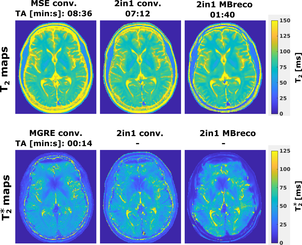
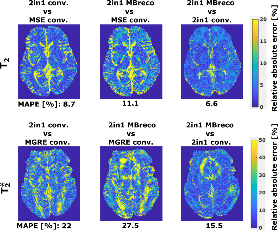
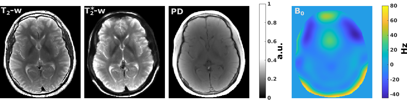
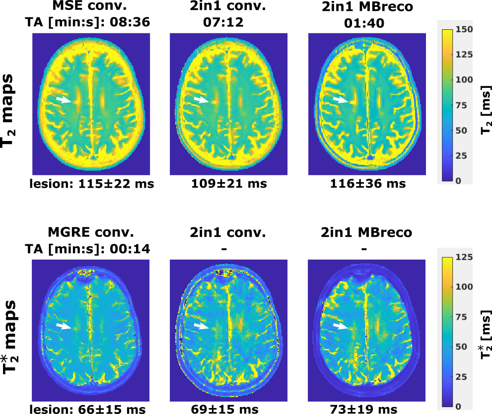
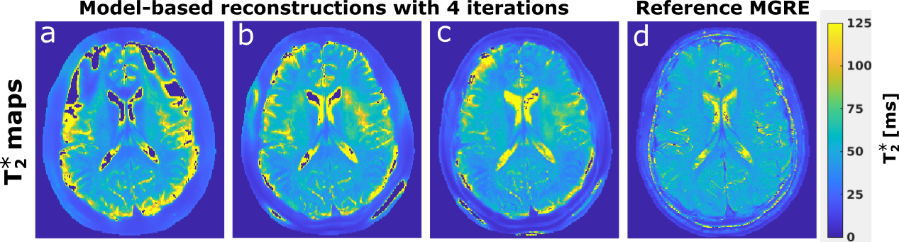
Figure 5. Impact of using initial maps on the convergence of the iterative MBreco for $$$T_{2}^{*}$$$ mapping. In (a), all maps were initialized as zero. In (b), M0 and B0 were initialized using a three-point model-based water/fat separation8, and $$$R_{2}^{*}$$$ as zero. In (c), the RARE module provided reasonable initial guesses for M0 (as the last TE image) and $$$T_{2}^{*}$$$ (as the estimated T2), and B0 was initialized as in (b). With the latter approach, a reasonable solution is obtained after only four iterations.