1348
Spiral-based multi-contrast imaging protocol for a full brain MR exam in 2 minutes1Center for Biomedical Imaging Research, Department of Biomedical Engineering, School of Medicine, Tsinghua University, Beijing, China, 2MR Clinical Science, Philips Health Technology (China), Beijing, China
Synopsis
Keywords: Multi-Contrast, Brain
Fast multi-contrast brain exams are highly desirable in clinical practice. Spiral sampling has high efficiency in terms of spatial encoding. Thus it holds great potential for many MRI applications. In this work, we optimized a fast multi-contrast brain imaging protocol based on multi-shot spiral acquisitions. Six contrasts (T1W-FLAIR, T2W, PDW, T2*W, T2W-FLAIR, DWI) and apparent diffusion coefficient (ADC) maps with an in-plane resolution of 1.0 mm2 for a full brain MR exam can be obtained in about 2 minutes.Introduction
Fast multi-contrast brain exams are highly desirable in clinical practice 1-9. Recently, EPI-based protocols for multi-contrast brain imaging were proposed in several previous studies 4,6-9. Compared to EPI, spiral imaging yields an approximately isotropic point spread function and has higher efficiency in terms of spatial encoding 10-12. Thus, it holds great potential for many MRI applications. In this work, we optimized a 2D spiral-based multi-contrast brain imaging protocol for T1-weighted FLAIR (T1W-FLAIR), T2-weighted (T2W), proton-density-weighted (PDW), T2*-weighted (T2*W), T2-weighted FLAIR (T2W-FLAIR), diffusion-weighted imaging (DWI) and apparent diffusion coefficient (ADC) mapping with an in-plane resolution of 1.0 mm2 in about 2 minutes. The acquisition and reconstruction framework presented here offer a new choice for fast brain examinations.Methods
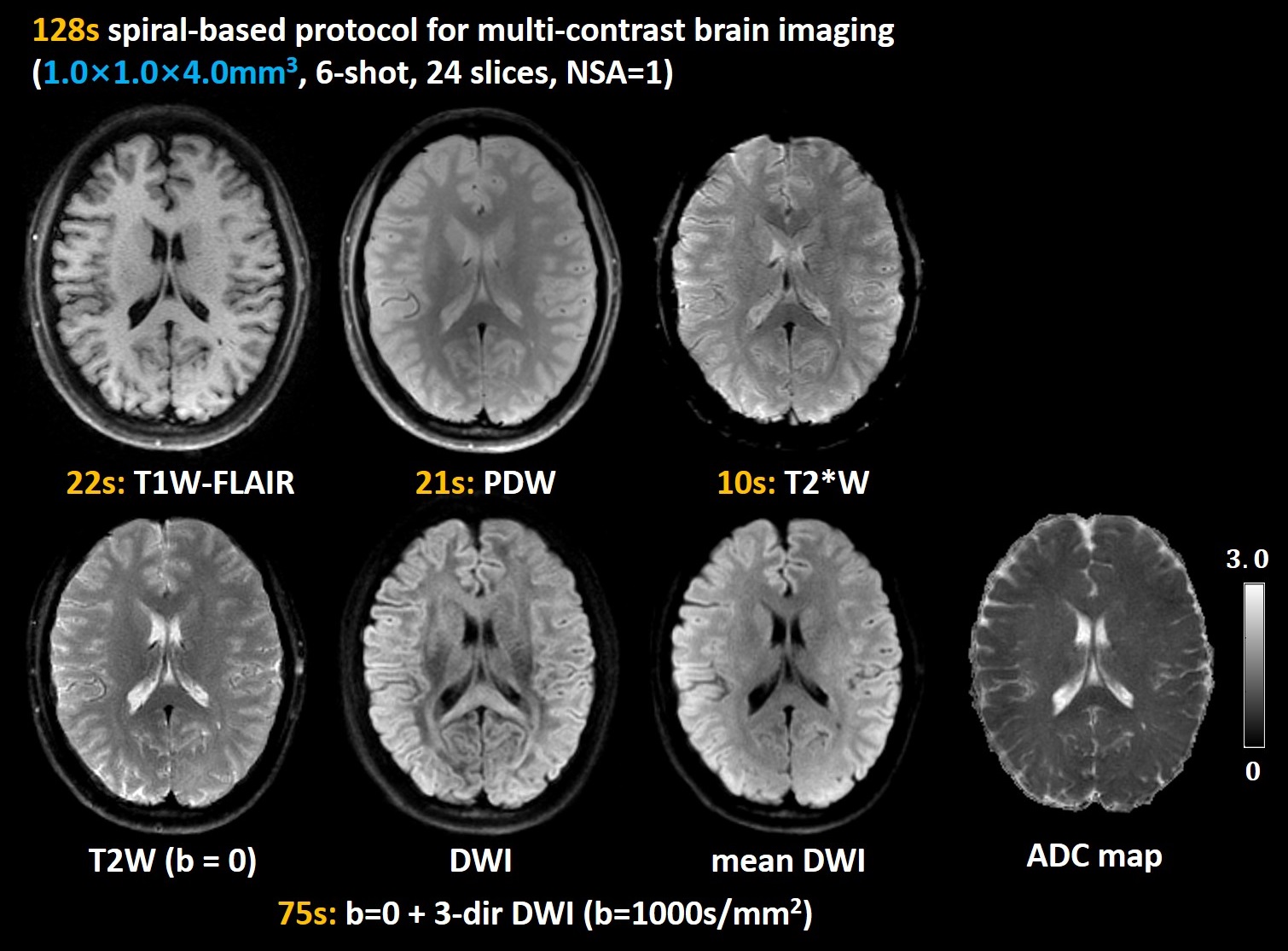
This study was approved by the local Institutional Review Board and written informed consent was obtained from all participants. All MRI data were acquired on an Ingenia CX 3.0T scanner (Philips Healthcare, Best, The Netherlands) with a 32-channel head coil. Two healthy volunteers were recruited to participate in the following experiments, respectively. The detailed 2D acquisition parameters were listed in the Table 1.Experiment 1, 6-shot protocol: FOV=220×220mm2, resolution=1.0×1.0×4.0mm3, acquisition matrix=220×220, spiral readout duration=27.0ms, 24 axial slices with a gap of 1mm cover the whole brain. The protocol includes T1W-FLAIR, T2W, PDW, T2*W, 3-direction DWI with b value=1000 s/mm2. The total scan time is 128s.
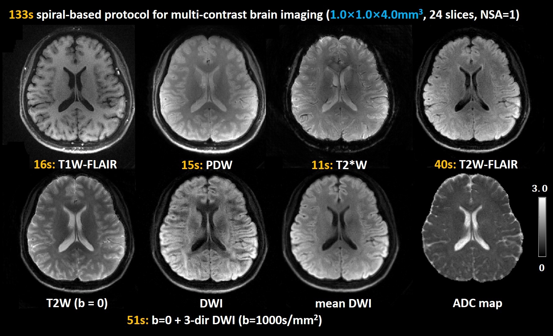
Experiment 2, 4-shot protocol: FOV=210×210mm2, resolution=1.0×1.0×4.0mm3, acquisition matrix=212×212, spiral readout duration=26.0ms, 24 axial slices with a gap of 1mm cover the whole brain. The protocol includes T1W-FLAIR, T2W, T2W-FLAIR, PDW, T2*W, 3-direction DWI with b value=1000 s/mm2. The total scan time is 133s. To mitigate off-resonance artifacts and maintain comparable SNR, under-sampling along the radial direction 13 is adopted to increase the radial spacing of the spiral trajectories so that the 4-shot spiral readouts can be reduced. The under-sampling factor is 1.25. Note that the T2*W images are acquired using a 6-shot full-sampling acquisition, from which the sensitivity maps can be calculated to reconstruct other images acquired by the 4-shot under-sampled acquisitions.
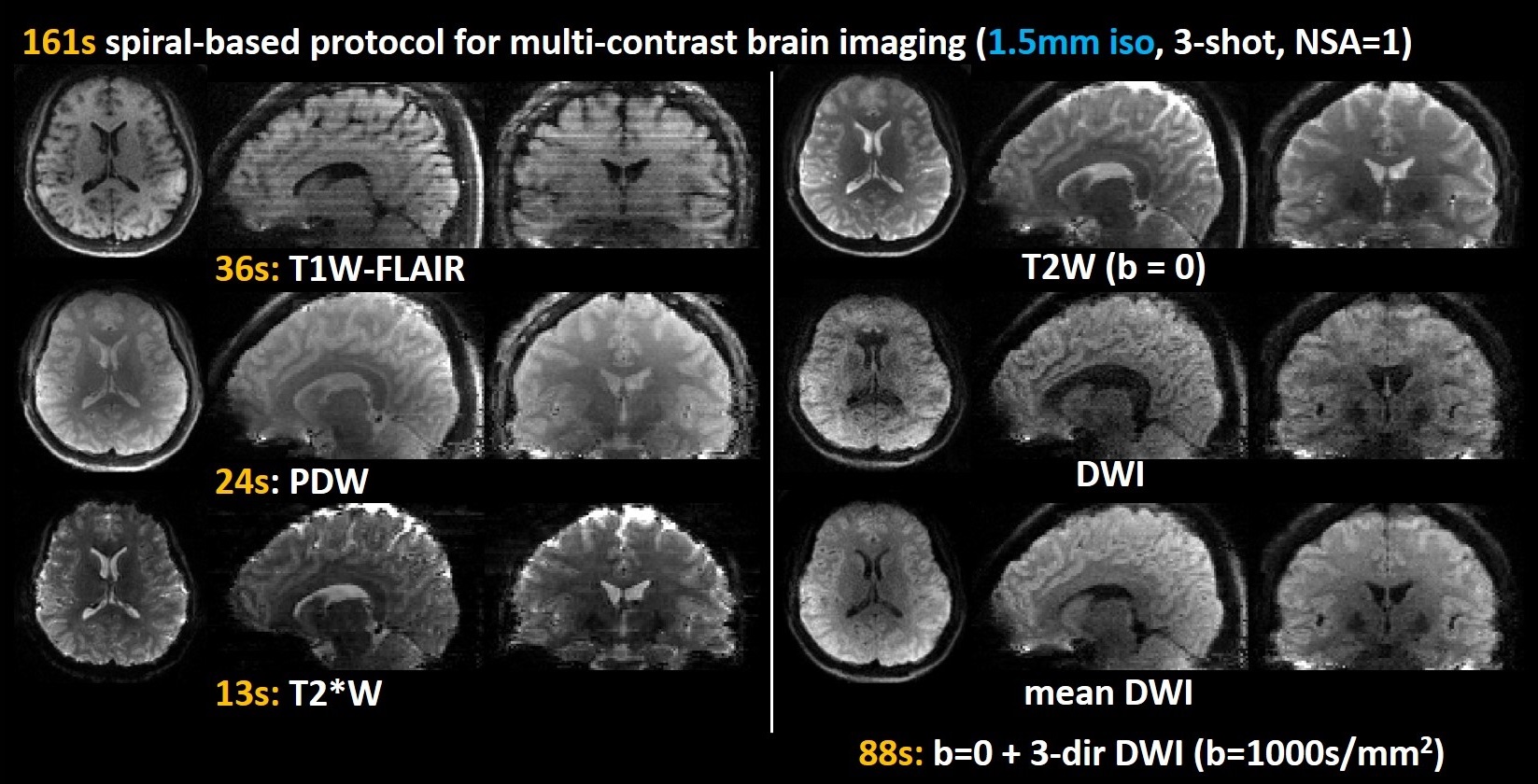
Experiment 3, 3-shot protocol: FOV=210×210×104mm3, 1.5mm isotropic resolution, matrix=140×140, spiral readout duration=27.0ms, 69 axial slices. The protocol includes T1W-FLAIR, T2W, PDW, T2*W, 3-direction DWI with b value=1000 s/mm2. The total scan time is 161s.
In all experiments, Spectral Presaturation with Inversion Recovery (SPIR) technique was used to suppress fat signals. T1W-FLAIR and T2W-FLAIR images are acquired using an inversion-recovery (IR) -prepared spin-echo spiral acquisitions (Figure 1). In this study, the imaging parameters were optimized to achieve similar spatial resolution and image contrast compared to the clinical standard. The image reconstruction was implemented off-line. Additionally, low-resolution field maps acquired using a multi-echo GRE sequence were used for deblurring.
Results and Discussion
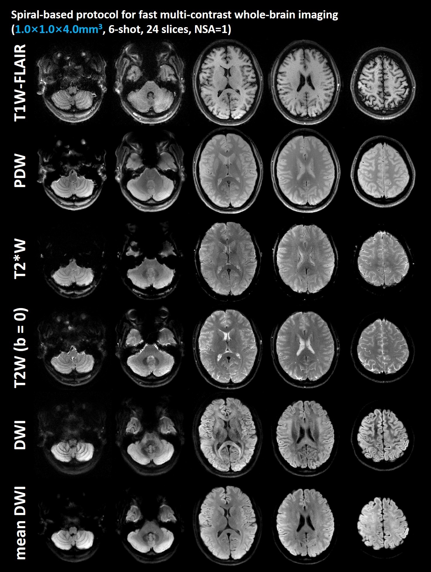
Figure 2 shows the T1W-FLAIR, T2W, PDW, T2*W, DW images with the same resolution acquired by the 6-shot spiral acquisitions from Experiment 1. Five slices are shown here. The in vivo results of the proposed protocol demonstrate satisfactory image quality, proper tissue contrast, and high spatial resolution.For Experiment 1, T1W-FLAIR, T2W, PDW, T2*W, DWI, mean DWI and ADC map from a slice of the healthy subject S1 are shown in the Figure 3. The total scan time of this 6-shot protocol is 128s.
Figure 4 shows the multi-contrast images including T1W-FLAIR, T2W, PDW, T2*W, T2W-FLAIR, DWI, mean DWI and ADC map from a slice of the healthy subject S2 in Experiment 2. With under-sampling factor of 1.25, no visual SNR degradation is observed. The total scan time of this 4-shot protocol is 133s. The acquisition time of T2W-FLAIR is up to 40s due to its long TR. Mean DWI provides similar tissue contrast to T2W-FLAIR, and it has a higher SNR. In addition, magnetization transfer contrast (MTC) preparation module can be used to improve T2W-FLAIR’s image contrast 7. In addition, multi-contrast images and 6-direction DW images with higher resolution can be acquired within 3 minutes.
The reconstructed multi-contrast images with 1.5-mm isotropic resolution are shown in Figure 5. Axial, coronal and sagittal planes of T1W-FLAIR, T2W, PDW, T2*W, DW images and mean DWI are shown here. The total scan time of this 3-shot protocol is 161s. For this set of the spiral images, automatic deblurring algorithm was used for off-resonance correction 14. Simultaneous multi-slice (SMS) techniques can be used to further improve the scan efficiency.
Conclusion
In this work, we optimized a fast multi-contrast brain imaging protocol based on multi-shot spiral sampling. Six contrasts (T1W-FLAIR, T2W, PDW, T2W-FLAIR, T2*W, DWI and mean DWI) and ADC maps with an in-plane resolution of 1.0×1.0 mm2 can be obtained in about 2 minutes. Further systematic clinical studies are warranted to clarify its diagnostic ability.Acknowledgements
No acknowledgement found.References
1. Polak D, Cauley S, Huang SY, et al. Highly-accelerated volumetric brain examination using optimized wave-CAIPI encoding. J Magn Reson Imaging. 2019;50(3):961-974.
2. Supada P, Thomas W, Susie H, et al. Ultrafast Brain MRI: Clinical Deployment and Comparison to Conventional Brain MRI at 3T. Journal of Neuroimaging 2016;26(5):503-510
3. Ryu KH, Choi DS, Baek HJ, et al. Clinical feasibility of 1-min ultrafast brain MRI compared with routine brain MRI using synthetic MRI: a single center pilot study. J Neurol. 2019 Feb;266(2):431-439.
4 Skare S, Sprenger T, Norbeck O, et al. A 1-minute full brain MR exam using a multicontrast EPI sequence. Magn Reson Med. 2018;79(6):3045-3054.
5. Nael K, Khan R, Choudhary G, et al. Six-minute magnetic resonance imaging protocol for evaluation of acute ischemic stroke: pushing the boundaries. Stroke 2014;45(7):1985-1991.
6. Lo W, Setsompop K, Liao C, et al. A comprehensive distortion-free 2-minute brain MR examination using BUDA and Wave-CAIPI. In Proceedings of the 28th Annual Meeting of ISMRM. 2020; 0294.
7. Conklin J, Clifford B, Bollmann S, et al. A comprehensive multi-shot EPI protocol for high-quality clinical brain imaging in 3 minutes. In Proceedings of the 28th Annual Meeting of ISMRM. 2020; 0300.
8. Clifford B, Conklin J, Huang S, et al. Clinical evaluation of an AI-accelerated two-minute multi-shot EPI protocol for comprehensive high-quality brain imaging. In Proceedings of the 29th Annual Meeting of ISMRM. 2021; 0661.
9. Wang Y, Dong Z, Hu Z, et al. Multicontrast Distortion-free MRI Using PSF-EPI. In Proceedings of the 27th Annual Meeting of ISMRM. 2019; 1251.
10. Wilm BJ, Barmet C, Gross S, et al. Single-shot spiral imaging enabled by an expanded encoding model: Demonstration in diffusion MRI. Magn Reson Med. Jan 2017;77(1):83-91.
11. Kasper L, Engel M, Barmet C, et al. Rapid anatomical brain imaging using spiral acquisition and an expanded signal model. Neuroimage. Mar 2018;168:88-100.
12. Kasper L, Engel M, Heinzle J, et al. Advances in spiral fMRI: A high-resolution study with single-shot acquisition. Neuroimage. Feb 1 2022;246:118738.
13. Li G, Ye X, Xin Shao, et al. Four-shot Navigator-free Spiral Acquisition Strategy for High-resolution Diffusion Imaging. In: Proceedings of the 29th Annual Meeting of ISMRM. 2021. p1328.
14. Chen W, Meyer CH. Fast automatic linear off-resonance correction method for spiral imaging. Magn Reson Med. 2006;56(2):457-462.
Figures
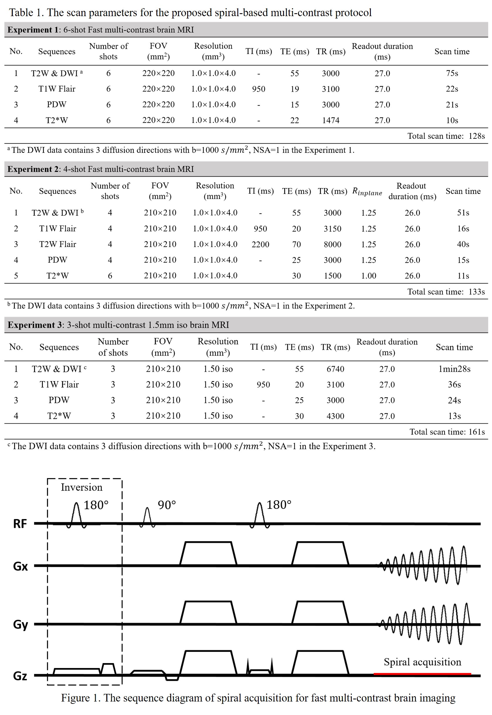
Table 1: The detailed acquisition parameters.
(I) Protocol 1: 6-shot acquisition. T1W-FLAIR (22s), PDW (21s), T2*W (10s), T2W+DWI (75s). The total scan time is 128s.
(II) Protocol 2: 4-shot acquisition. T1W-FLAIR (16s), T2W-FLAIR (40s), PDW (15s), T2*W (11s), T2W+DWI (51s). The total scan time is 133s.
(III) Protocol 3: 3-shot multi-contrast acquisition with 1.5mm isotropic resolution. T1W-FLAIR (36s), PDW (24s), T2*W (13s), T2W+DWI (88s). The total scan time is 161s.
Figure 1: The sequence diagram of spiral-based fast multicontrast brain imaging.