1334
A Pilot Evaluation of TSE MVXD based IVIM in Characteristics and Diagnosis of Nasopharyngeal Carcinoma1Department of Radiology, Guangdong Provincial People’s Hospital, Guangdong Academy of Medical Sciences, Guangzhou, Guangdong Province, China, Guangzhou, China, 2Guangdong Provincial Key Laboratory of Artificial Intelligence in Medical Image Analysis and Application, Guangdong Provincial People's Hospital, Guangdong Academy of Medical Sciences, Guangzhou, China, Guangzhou, China, 3MSC Clinical & Technical Solutions, Philips Healthcare, China, Shenzhen, China
Synopsis
Keywords: Diffusion/other diffusion imaging techniques, Contrast Mechanisms, Turbo Spin-echo; Multivane-XD; Intravoxel Incoherent Motion; Contrast-enhanced imaging; Nasopharyngeal carcinoma
Diagnosis of Nasopharyngeal Carcinoma (NPC) remains challenge since the contrast-enhanced T1-weighted imaging (CE-T1WI) may lead to harmful impact by the accumulation of contrast agent in patients and its potential adverse reactions. TSE MVXD DWI based IVIM, a considerable alternative, was performed on 32 NPC patients with significant tumors to evaluate the relation with CE-T1WI. As the result showed, the TSE MVXD DWI based IVIM had significantly relation with CE T1WI. It is potentially a promising and valuable non-invasive method in the detection of NPC.Introduction
According to the current guidelines, MRI with contrast-enhanced T1-weighted imaging (CE-T1WI) sequence is recommended for the long-term follow-up in patients with NPC [1-3]. However, the CE-T1WI, a gadolinium-based method, may lead to harmful impact by the accumulation of contrast agent [4]. Besides, potential adverse reactions (e.g. nausea, headaches, and irritation) may occur in patients. Thus, an alternative for predicting enhancement in CE-T1WI without gadolinium is needed.Intravoxel incoherent motion (IVIM), which could reflect the regional blood flow and micro vessel density, could be a non-contrast-enhanced technique to predict enhancement in CE-T1WI [5,6]. It has been used in NPC patients in recent studies [7]. Hence, it may be one of the solutions. But due to the B0 inhomogeneity of nasopharyngeal region, it limits the application of IVIM, and during the long examination time, patients will more probably move, leading to motion artifacts. TSE MVXD DWI showed distortion free, fewer motion artifacts and higher image quality[8]. Meanwhile, it had provided better diagnosis for prostate cancer [9]. To address these problems, the combination of Multivane-XD (TSE MVXD) and TSE DWI for IVIM is considered in the present study.
Methods
Thirty-two patients (23 males, 9 females; mean ± standard deviation age, 48.7 ± 13.5 years; range, 29–79 years) with NPC who were found significant tumors were included in the analysis. The TSE MVXD DWI based IVIM images of them were used to evaluate the relationship with CE-T1WI.All imaging was performed on a 3.0 T Philips Ingenia CX scanner with a 16-channel head and neck combined coil.
The TSE MVXD DWI based IVIM protocol was as follows: voxel size, 1.81.84 mm3; intersection gap, 1mm; repetition time/echo time, 2400 ms/123 ms; field of view, 195195 mm2; TSE factor, 15; MultiVane factor, 170%; SENSE factor, 2.5; scan time, 13min50s.
The gadolinium (gadopentetate dimeglumine; Magnevist; Bayer HealthCare, Berlin, Germany) was intravenously injected at 0.2 ml/kg body weight and 1.0 ml/s. The CE-T1WI sequence was started approximately 3 to 5 min after the injection, while the IVIM protocols were finished before the contrast.
The IVIM parameters were calculated using segmented biexponential analysis. All IVIM analysis were calculated using in IMAge/enGINE MRI Diffusion Toolbox [10].
The enhancement ratios were calculated using the images of T1WI and CE-T1WI [5]. The CE-T1WI images were registered to T1WI images. The enhancement ratio was calculated as the Eq. (1).
Enhance ratio =((SICE-T1WI)-(SIT1WI))/(SIT1WI) (1)
where SICE-T1WI was defined as the mean signal intensity of the ROIs in CE-T1WI, and SIT1WI defined as the mean signal intensity of the ROIs in T1WI.
A ROI was manually placed on the tumor in each image to include as much of the tumor area as possible except large vessels and visually large necrotic areas [11]. CE-T1WI images were used as references to determine the tumor areas, and the ROIs were copied to the corresponding areas on the TSE MVXD DWI based IVIM and T1WI images, which was shown as Figure 1.
Normality of the distribution was tested using the Shapiro-Wilk test. Student’s t-test was used to compare means of the normally distributed variables, while the Wilcoxon-rank test was used to compare the median of the skewed variables. The correlation coefficients were described as Pearson r values. Multivariate linear regression analysis was performed for the determination of independent TSE MVXD DWI based IVIM predictors of CE-T1WI. All statistical tests were two-sided, and P values < 0.05 were considered statistically significant. All statistical analyses were performed with statistical software GraphPad Prism (version 6.0; GraphPad Software, San Diego, California, USA) and R version 3.4.1 (R Foundation, Vienna, Austria).
Results
The correlations between the enhancement ratio and IVIM parameters were assessed using Pearson’s r values, which was shown in Figure 2. The correlation was observed between f and enhancement ratio (r=0.539, P<0.001; Fig. 2C). However, statistically significant correlation was not observed between enhanced ratio and any other IVIM parameter (all P > 0.05). In Figure 3, the multivariate regression analysis revealed that f was the independent IVIM predictors of enhancement ratio in patients with NPC.Discussion
As the result showed, TSE MVXD DWI based IVIM had the potential to be an alternative approach to CE-T1WI in the detection of NPC. The main reasons were mentioned below. Firstly, the f derived from TSE MVXD DWI based IVIM was significantly related with enhancement ratios, and it was the significant IVIM predictor for the enhancement in tumors in patients with NPC. The higher f represents the higher regional blood flow and micro vessel density [6].What’s more, the TSE MVXD DWI based IVIM showed lower distortion, fewer motion artefacts and higher image quality, but the scan time largely increases and consequently causes motion artifacts. Fortunately, the use of the radial k-space sampling in MVXD could be used for the correction of phase, rotation, translation, and weighting to reduce spatial inconsistencies [15].Conclusion
The TSE MVXD DWI based IVIM had desired image quality, and the IVIM parameters were significantly related with the enhancement ratio of CE-T1WI. It showed the potential value to be an alternative to the contrast-enhancement imaging in patients with NPC.Acknowledgements
No acknowledgement found.References
1 Tang LL, Chen YP, Chen CB et al (2021) The Chinese Society of Clinical Oncology (CSCO) clinical guidelines for the diagnosis and treatment of nasopharyngeal carcinoma. Cancer Commun (Lond) 41:1195-1227. doi:10.1002/cac2.12218
2 Bossi P, Chan AT, Licitra L et al (2021) Nasopharyngeal carcinoma: ESMO-EURACAN Clinical Practice Guidelines for diagnosis, treatment and follow-up(dagger). Ann Oncol 32:452-465. doi:10.1016/j.annonc.2020.12.007
3 Simo R, Robinson M, Lei M, Sibtain A, Hickey S (2016) Nasopharyngeal carcinoma: United Kingdom National Multidisciplinary Guidelines. J Laryngol Otol 130:S97-S103. doi:10.1017/S0022215116000517 4 McDonald RJ, McDonald JS, Kallmes DF et al (2015) Intracranial gadolinium deposition after contrast-enhanced MR imaging. Radiology 275:772-782.
5 Rogers HJ, Verhagen MV, Shelmerdine SC, Clark CA, Hales PW (2019) An alternative approach to contrast-enhanced imaging: diffusion-weighted imaging and T1-weighted imaging identifies and quantifies necrosis in Wilms tumour. Eur Radiol 29:4141-4149. doi:10.1007/s00330-018-5907-z
6 Lemke A, Laun FB, Simon D, Stieltjes B, Schad LR (2010) An in vivo verification of the intravoxel incoherent motion effect in diffusion‐weighted imaging of the abdomen. Magn Reson Med 64:1580-1585. 7 Noij DP, Martens RM, Marcus JT et al (2017) Intravoxel incoherent motion magnetic resonance imaging in head and neck cancer: A systematic review of the diagnostic and prognostic value. Oral Oncol 68:81-91. doi:10.1016/j.oraloncology.2017.03.016
8 Meier-Schroers M, Marx C, Schmeel FC et al (2018) Revised PROPELLER for T2-weighted imaging of the prostate at 3 Tesla: impact on lesion detection and PI-RADS classification. Eur Radiol 28:24-30. doi:10.1007/s00330-017-4949-y
9 Akamine Y, Okuaki T, Goshima S et al (2018) Reduced distortion in prostate DWI by using split echo type TSE-DWI (SPLICE) with MultiVane acquisition. International Society of Magnetic Resonance and Medicine
10 Yang M, Yan Y, Wang H (2019) IMAge/enGINE: a freely available software for rapid computation of high-dimensional quantification. Quant Imaging Med Surg 9:210-218. doi:10.21037/qims.2018.12.03
11 Jia QJ, Zhang SX, Chen WB et al (2014) Initial experience of correlating parameters of intravoxel incoherent motion and dynamic contrast-enhanced magnetic resonance imaging at 3.0 T in nasopharyngeal carcinoma. Eur Radiol 24:3076-3087. doi:10.1007/s00330-014-3343-2
12 Mikayama R, Yabuuchi H, Sonoda S et al (2018) Comparison of intravoxel incoherent motion diffusion-weighted imaging between turbo spin-echo and echo-planar imaging of the head and neck. Eur Radiol 28:316-324. doi:10.1007/s00330-017-4990-x
13 Pieper S, Halle M, Kikinis R (2004) 3D Slicer2004 2nd IEEE International Symposium on Biomedical Imaging: Nano to Macro (IEEE Cat No 04EX821), pp 632-635 Vol. 631
14 Ferreira T, Rasband W (2012) ImageJ user guide. ImageJ/Fiji 1:155-161.
15 Pipe JG, Gibbs WN, Li Z, Karis JP, Schar M, Zwart NR (2014) Revised motion estimation algorithm for PROPELLER MRI. Magn Reson Med 72:430-437.
Figures
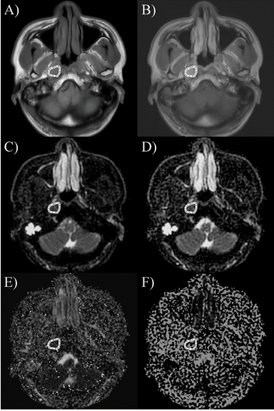
The ROIs of the T1WI, CE-T1WI and IVIM maps
A) T1WI image; B) CE-T1WI image; C) ADC map; D) D map; E) f map; F) D* map;
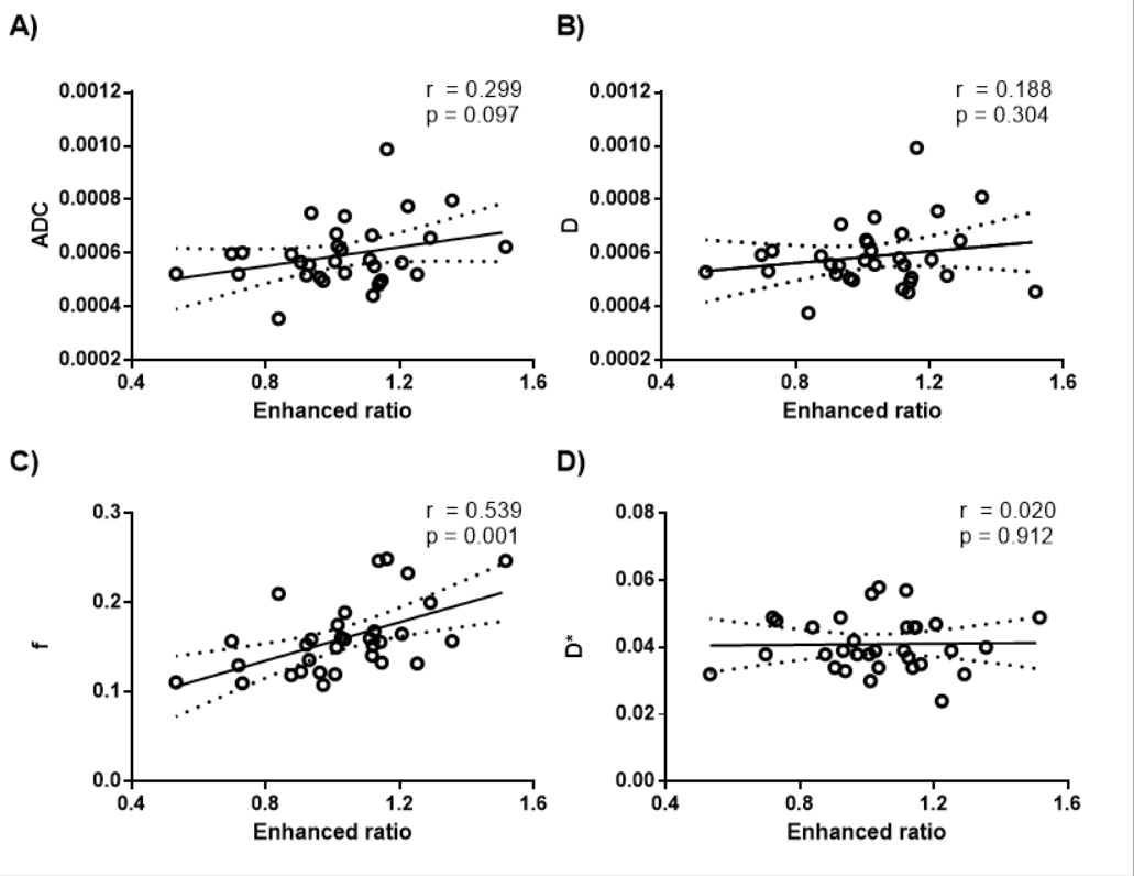
The Correlation between enhanced ratio and IVIM parameters
A) The correlation between enhanced ratio and ADC; B) The Correlation between enhanced ratio and D; C) the correlation between enhanced ratio and f; D) the correlation between enhanced ratio and D*;
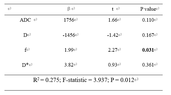
Multivariate linear regression of the TSE MVXD DWI based IVIM parameters with T1 enhanced ratio
Values in bold indicate significance. ADC, Apparent diffusion coefficient; D, Pure molecular diffusion; f, Perfusion fraction; D*, Perfusion related diffusion;