1329
Comparison of Image Quality and ADC Measurement between DWIs with and without Reverse Encoding Distortion Correction in Head and Neck Tumors
Hirotaka Ikeda1, Yoshiharu Ohno1,2, Kaori Yamamoto3, Maiko Shinohara3, Masato Ikedo3, Masao Yui3, Akiyoshi Iwase4, Minami Furuta1, Yuki Obama1, Hiroyuki Nagata2, Takahiro Ueda1, Yoshiyuki Ozawa1, and Hiroshi Toyama1
1Radiology, Fujita Health University School of Medicine, Toyoake, Japan, 2Joint Research Laboratory of Advanced Medical Imaging, Fujita Health University School of Medicine, Toyoake, Japan, 3Canon Medical Systems Corporation, Otawara, Japan, 4Fujita Health University Hospital, Toyoake, Japan
1Radiology, Fujita Health University School of Medicine, Toyoake, Japan, 2Joint Research Laboratory of Advanced Medical Imaging, Fujita Health University School of Medicine, Toyoake, Japan, 3Canon Medical Systems Corporation, Otawara, Japan, 4Fujita Health University Hospital, Toyoake, Japan
Synopsis
Keywords: Cancer, Head & Neck/ENT
We hypothesize that RDC is useful for image quality and diagnostic performance improvements on DWI with b value at 1500 s/mm2 in suspected prostatic cancer patients, although there was little influence of RDC on DWI at in vitro study. The purpose of this study was to determine the influence of RDC for ADC measurement at in vitro study and its’ utility for improving image quality and diagnostic performance of malignant from benign head and neck tumors on DWI as in vivo study.Introduction
Diffusion-weighted imaging (DWI) is one of the key sequences for management of head and neck tumors and has been generally obtained by means of single-shot echo-planar imaging (EPI) as the current standard sequence (1-3). However, a major disadvantage of single-shot EPI is that it is considerably prone to artifacts, particularly susceptibility artifacts at tissue interfaces and image blurring, which even tend to increase at higher field strengths. Therefore, several approaches for DWI such as parallel transmit EPI or readout-segmented multi-shot EPI, periodically rotated overlapping parallel lines with enhanced reconstruction (PROPELLER), multiplexed sensitivity encoding or reverse-polarity gradient, etc. have been tested for improving image quality and reducing artifacts due to various causes (4-7). In 2022, Canon Medical Systems Corporation introduces and clinically sets reverse encoding distortion correction (RDC) for body DWI with applying deep learning reconstruction (DLR). However, no major reports are not assessed the capability of RDC for improving image quality and influence for ADC measurement accuracy on prostatic DWI at in vitro or in vivo studies. We hypothesize that RDC is useful for image quality and diagnostic performance improvements on DWI with b value at 800 s/mm2 in suspected head and neck tumor patients, although there was little influence of RDC on DWI at in vitro study. The purpose of this study was to determine the influence of RDC for ADC measurement at in vitro study and its’ utility for improving image quality and diagnostic performance of malignant from benign head and neck tumors on DWI as in vivo study.Materials and Methods
For in vitro study, the quantitative diffusion phantom (High Precision Devices, Inc, Boulder, CO) developed by NIST/ QIBA consists of 13 vials filled with varying concentrations of polyvinylpyrrolidone in aqueous solution was scanned by DWIs with and without RDC (i.e. RDC DWI and DWI) to evaluate ADC measurement accuracy in this study. In addition, 48 suspected head and neck tumor patients underwent DWI at b value as 0 and 800 s/mm2 with and without RDC at a 3T MR system and pathological examinations as in vivo study. According to the pathological examination results, 21 malignant and 27 benign tumors were in this study. On in vitro study, ADCs at each phantom on DWI with and without RDC were determined and correlated with standard reference by Pearson’s correlation analysis. In addition, mean difference of ADC between each DWI and standard reference was compared each other by paired t-test. On in vivo study, deformation ratios (DRs) were determined as tumor area ratio between each DWI and T2-weighted image. To compare ADC measurement accuracy between RDC DWI and DWI, ADC values of tumor and spinal cord were determined by ROI measurements. To compare the capability for image quality and ADC measurement between RDC DWI and DWI, DR and ADC were compared between two methods by paired t-test. For determination of relationship between ADCs from RDC DWI and DWI, ADC values were correlated with Spearman's correlations. Then, ROC-based positive test was performed to determine each feasible threshold value for differentiating malignant from benign head and neck tumors. Finally, sensitivity, specificity and accuracy were compared each other by McNemar's test.Results
Representative cases are shown in Figures 1. On in vitro study, Pearson’s correlation of ADC between each DWI and standard reference was determined as significant and excellent (DWI: r=0.999, p<0.0001; RDC DWI: r=1.0, p<0.0001). There were no significant differences of ADC between RDC DWI and DWI at each phantom (p>0.05). On in vivo study, compared results of DRs and ADCs of tumor and spinal cord between RDC DWI and DWI are shown in Figure 2. DRs and ADCs of tumor and spinal cord were significant differences between both DWIs (p<0.05). On Spearman’s correlations of ADC in tumor and spinal cord between RDC DWI and DWI, there were significant and good correlations of tumor (ρ=0.97, p<0.0001) and spinal cord (ρ=0.92, p<0.0001) between DWI and RDC DWI. Diagnostic performance of each DWI with feasible threshold value is shown in Figure 3. There were no significant differences of sensitivity, specificity and accuracy between DWI and RDC DWI (p>0.05).Conclusion
Reverse encoding distortion correction (RDC) can improve the image quality of diffusion weighted images in patients with head and neck tumor, although there was no significant influence for ADC measurement at in vitro and in vivo studies.Acknowledgements
This study was technically and financially supported by Canon Medical Systems Corporation.References
- Thoeny HC, De Keyzer F, King AD. Diffusion-weighted MR imaging in the head and neck. Radiology. 2012;263(1):19-32.
- Widmann G, Henninger B, Kremser C, Jaschke W. MRI Sequences in Head & Neck Radiology - State of the Art. Rofo. 2017;189(5):413-422.
- Abdel Razek AAK, Elsebaie NA, Gamaleldin OA, AbdelKhalek A, Mukherji SK. Role of MR Imaging in Head and Neck Squamous Cell Carcinoma. Magn Reson Imaging Clin N Am. 2022;30(1):1-18.
- Chen X, Xian J, Wang X, et al. Role of periodically rotated overlapping parallel lines with enhanced reconstruction diffusion-weighted imaging in correcting distortion and evaluating head and neck masses using 3 T MRI. Clin Radiol. 2014; 69(4):403-409.
- Seeger A, Batra M, Süsskind D, Ernemann U, Hauser TK. Assessment of uveal melanomas using advanced diffusion-weighted imaging techniques: value of reduced field of view DWI ("zoomed DWI") and readout-segmented DWI (RESOLVE). Acta Radiol. 2019;60(8):977-984.
- Konar AS, Fung M, Paudyal R, et al. Diffusion-Weighted Echo Planar Imaging using MUltiplexed Sensitivity Encoding and Reverse Polarity Gradient in Head and Neck Cancer: An Initial Study. Tomography. 2020;6(2):231-240.
- Klingebiel M, Ullrich T, Quentin M, et al. Advanced diffusion weighted imaging of the prostate: Comparison of readout-segmented multi-shot, parallel-transmit and single-shot echo-planar imaging. Eur J Radiol. 2020;130:109161.
Figures
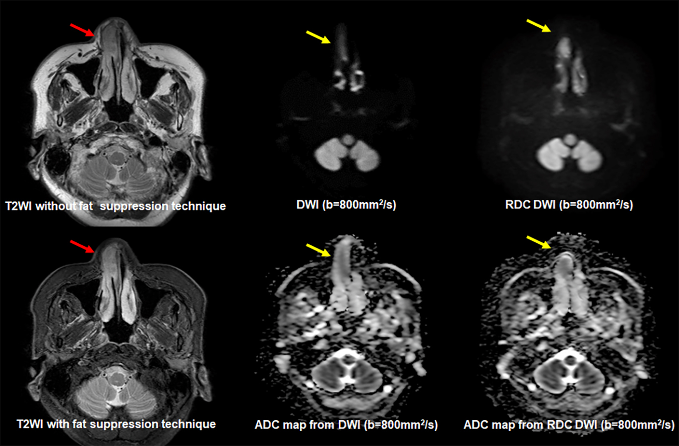
Figure 1. 73-year-old female patient with nasal cancer
On T2WI with and without fat suppression, nasal cancer (red arrow) demonstrates as high signal intensity with nasal cavity stenosis. When applied RDC technique, nasal cancer demonstrates without any distortion on DWI and ADC map and well matched with T2WIs (yellow arrows).
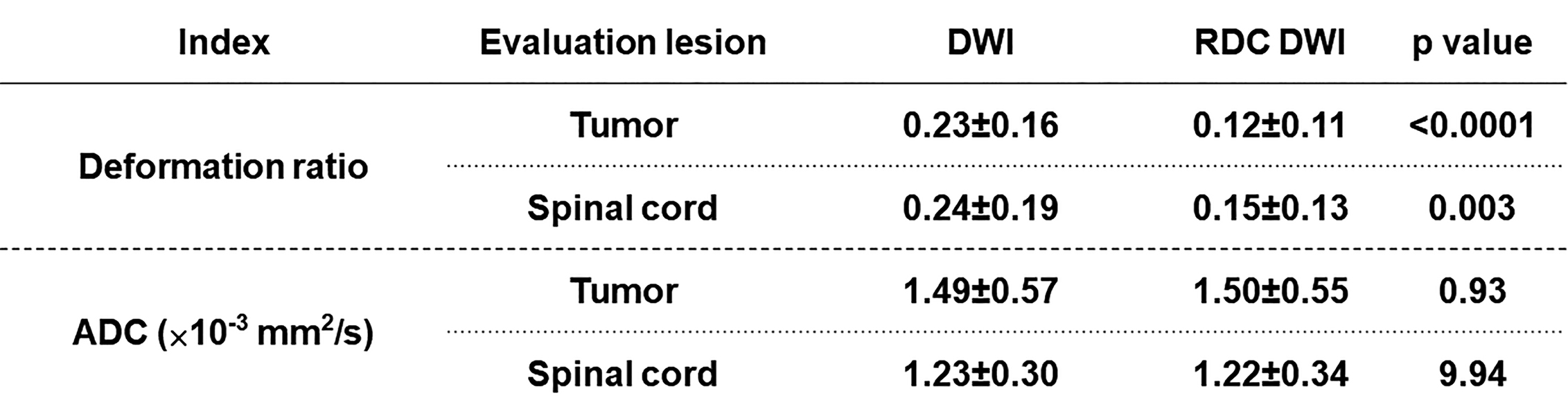
Figure 2. Comparisons of DRs and ADCs of tumor and spinal cord between RDC DWI and DWI.
There were significant differences of DR and ADC at tumor and spinal cord between DWI and RDC DWI (p<0.05).
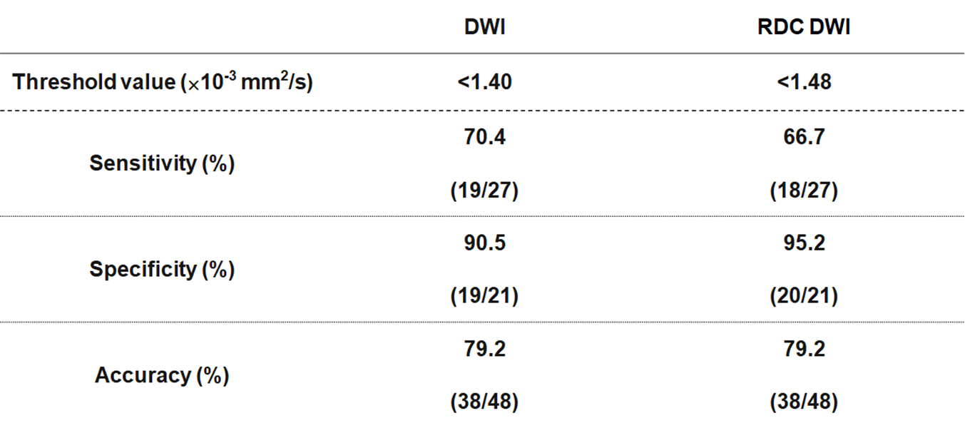
Figure 3. Diagnostic performance of each DWI with
feasible threshold value.
Sensitivity,
specificity and accuracy had no significant differences between DWI and RDC DWI
(p>0.05).
DOI: https://doi.org/10.58530/2023/1329