1320
Multi-channel CAscaded Multi-scale WAvelet with iterative REfinement (CAMWARE) network for accelerated whole-brain vessel wall imaging1University of Southern California, Los Angeles, CA, United States, 2University of California, Los Angeles, Los Angeles, CA, United States, 3Siemens Medical Solutions, Los Angeles, CA, United States
Synopsis
Keywords: Vessel Wall, Atherosclerosis
3D MR vessel wall imaging (VWI) is a non-invasive imaging modality for directly assessing intracranial arterial wall diseases. A typical intracranial VWI protocol requires 6-12 minutes per scan to obtain adequate spatial coverage and resolution. Such a long scan time hinders widespread use of VWI in clinical settings. We have developed a multi-channel application-ready intracranial vessel-dedicated CAscaded Multi-level WAvelet REfine (CAMWARE) network that enables a VWI scan within 4 minutes. The proposed network achieved significant improvement in vessel wall delination over conventional compressed sensing reconstruction.Introduction
3D MR vessel wall imaging (VWI)1 is a non-invasive, “looking-beyond-the-lumen” imaging method that can directly probe intracranial arterial wall diseases. However, the needs for large spatial coverage and submillimeter spatial resolution mandate a long acquisition 2-5 Recently, convolutional neural networks (CNN) have shown promising results on imaging acceleration in a wide variety of MR 6-8 In this work, we propose a real-world application-ready intracranial vessel-dedicated CAscaded Multi-level WAvelet REfine (CAMWARE) network that enables a whole-brain VWI scan within 4 min.Methods
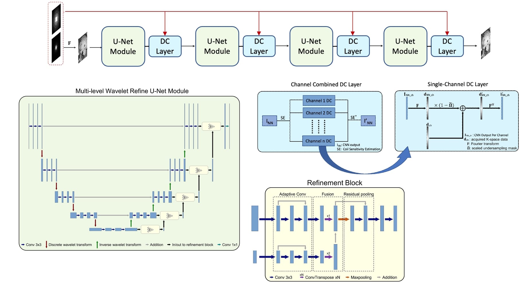
A multi-channel compatible CAMWARE network was designed. Similar to previous studies for CNN-based MR imaging acceleration, an unconstrained optimization problem can be formulated as:$$\arg \min _{\boldsymbol{I}}\left|\boldsymbol{I}-\mathrm{C}\left(\boldsymbol{I}_{\boldsymbol{u}} \mid \boldsymbol{\Theta}\right)\right|_2^2+\lambda|\boldsymbol{\Omega} \boldsymbol{F} \cdot \mathbf{S E} \cdot \boldsymbol{I}-\boldsymbol{d}|_2^2$$ where $$$\boldsymbol{I}$$$ denotes complex-valued MR images, $$$\boldsymbol{d}$$$ represents undersampled measurements in k-space, $$$\boldsymbol{I}_{\boldsymbol{u}}$$$ is the zero-filled reconstruction of $$$boldsymbol{d}$$$, $$$\mathrm{C}\left(\boldsymbol{I}_{\boldsymbol{u}} \mid \boldsymbol{\Theta}\right)$$$ is a mapping function conditioned on CNN parameter $$$\boldsymbol{\Theta}$$$, and $$$\boldsymbol{\Omega}$$$ and $$$\boldsymbol{F}$$$ represent undersampling pattern and Fourier encoding operators, respectively. $$$\mathbf{S E}$$$ represents the coil sensitivity estimation.To solve the above problem, we incorporated the data fidelity in the learning stage by introducing a combination of multi-coil data consistency (DC) layer7 , which replaces the k-space values of the reconstruction with the originally acquired values, at the end of the CNN. CAMWARE adopts a cascaded structure of U-Net and multi-channel Data Consistency (DC) Layer (Figure 1). To enhance the network performance in showing the vessel wall structures, we modified U-Net in two aspects: (a) Pooling and deconvolution operations, which are adopted in conventional U-Net to enlarge the receptive field and alter the resolution of feature maps, are replaced by discrete wavelet transform and inverse wavelet transform, respectively9 . (b) Multi-path refinement building block is added to fuse coarse features with fine-grained low-level features to restore sharp vessel wall boundaries10 . Four “CNN + DC” blocks are concatenated, with each additional block serving as an extra step to reduce the learning errors of the former block11 .
Data preparation:A total of 77 whole-brain VWI datasets, including 47 pre-contrast and 30 post-contrast scans, were acquired on a 3T system (Skyra, Siemens) with a standard 20-channel head-neck coil. The imaging protocol included: FOV=248x229x132 mm3, matrix size=448x414x240, 0.55 mm isotropic resolution, GRAPPA acceleration of 2x in the phase-encoding direction, 12.1 min acquisition time. Coil-separated complex-valued images reconstructed directly on the scanner were saved. A variable density Poisson-disc undersampling mask was retrospectively applied to the k-space data of each coil image generated by Fourier transforming the complex-valued images to simulate undersampled k-space which corresponded to an acceleration factor 6.5 or a 4-min acquisition. Coil sensitivity masks were estimated using ESPIRIT. The learning target of the network is then synthesized using the coil sensitivity mask.
Results
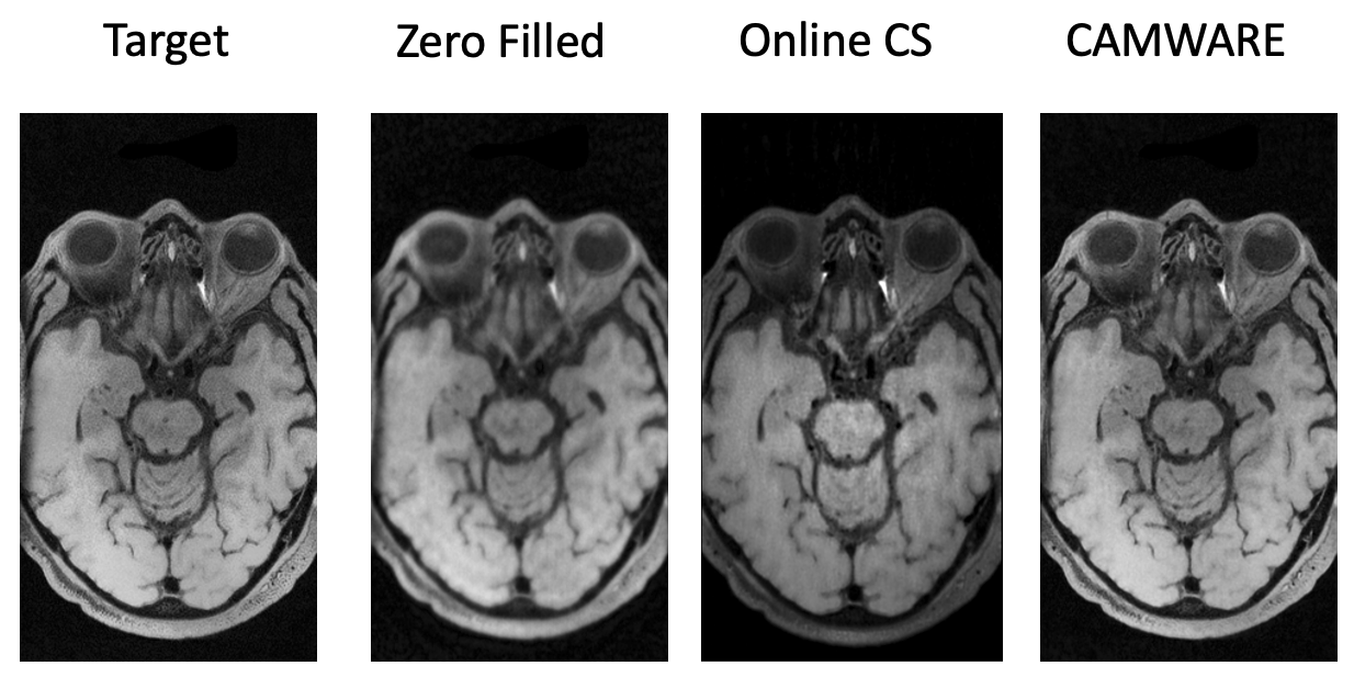
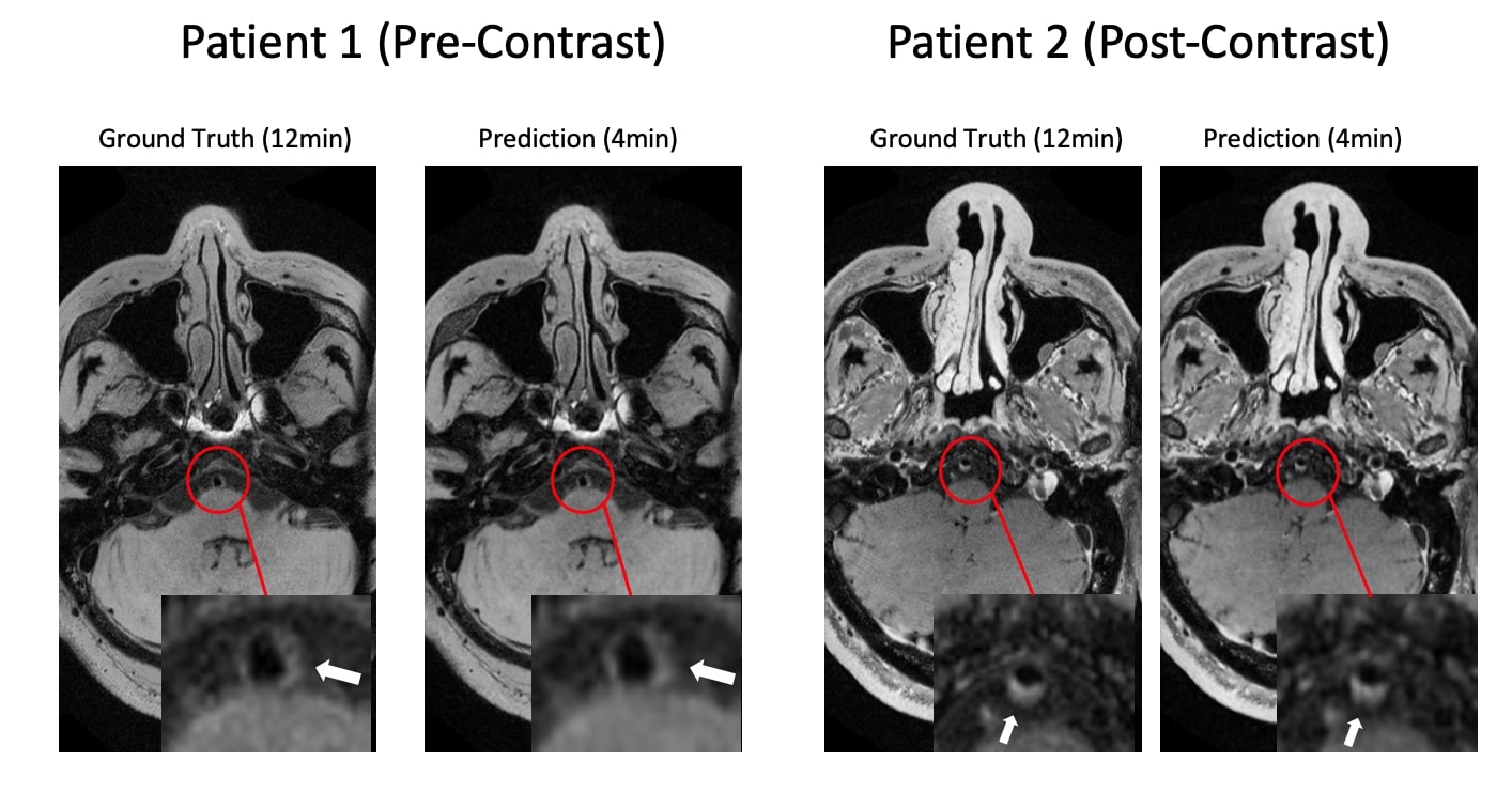
The qualitative comparisons as well as corresponding error maps are shown in Figure 2. The CAMWARE model achieved an average SSIM of 0.84 and PSNR of 30.21. CAMWARE faithfully reconstructed images with visually sharper delineation than the zero-filled reconstruction and commercial CS reconstruction. Figure 3 shows vessel wall images from two ischemic stroke patients. The pre-contrast plaque and post-contrast wall enhancement, both on the basilar artery are depicted accurately by the proposed model (4 min) with respect to the ground-truth (12 min).Discussion
In this work, we proposed a multi-channel compatible CAMWARE network that can reconstruct high-quality 0.55 mm isotropic-resolution whole-brain vessel wall images with 6.5x undersampled in k-space data. The proposed approach outperforms the state-of-the-art CS method. Our major innovations include: (a) preserving low-frequency details by incorporating discrete wavelet transform; (b) refining low-resolution features with fine-grained features; (c) cascading four “CNN + DC” blocks to let each additional cascade acts as an extra step to reduce the learning errors of the former block; (d) The incorporation of the sensitivity estimation in the data consistency layer renders the proposed structure ready for real-world applications. A recently optimized and widely used whole-brain VWI protocol requires 8 min per scan. With the novel deep learning method, a whole-brain VWI scan can further be accelerated to 4 min, thus allowing for complete VWI investigation (both pre- and post-contrast scans) within 10 min.Conclusion
In this work, we proposed a novel intracranial vessel dedicated multi-channel compatible CAMWARE network that holds the potential to enable 4-min whole-brain VWI.Acknowledgements
This work is supported by NIH/NHLBI R01 HL147355.References
1. Mandell, D. M., Mossa-Basha, M., Qiao, Y., Hess, C. P., Hui, F., Matouk, C., ... & Mikulis, D. J. (2017). Intracranial vessel wall MRI: principles and expert consensus recommendations of the American Society of Neuroradiology. American Journal of Neuroradiology, 38(2), 218-229.
2. Qiao, Y., Steinman, D. A., Qin, Q., Etesami, M., Schär, M., Astor, B. C., & Wasserman, B. A. (2011). Intracranial arterial wall imaging using three‐dimensional high isotropic resolution black blood MRI at 3.0 Tesla. Journal of Magnetic Resonance Imaging, 34(1), 22-30.
3. Zhang, L., Zhang, N., Wu, J., Zhang, L., Huang, Y., Liu, X., & Chung, Y. C. (2015). High resolution three dimensional intracranial arterial wall imaging at 3 T using T1 weighted SPACE. Magnetic resonance imaging, 33(9), 1026-1034.
4. Fan, Z., Yang, Q., Deng, Z., Li, Y., Bi, X., Song, S., & Li, D. (2017). Whole‐brain intracranial vessel wall imaging at 3 T esla using cerebrospinal fluid–attenuated T1‐weighted 3 D turbo spin echo. Magnetic resonance in medicine, 77(3), 1142-1150.
5. Yang, Q., Deng, Z., Bi, X., Song, S. S., Schlick, K. H., Gonzalez, N. R., ... & Fan, Z. (2017). Whole‐brain vessel wall MRI: A parameter tune‐up solution to improve the scan efficiency of three‐dimensional variable flip‐angle turbo spin‐echo. Journal of Magnetic Resonance Imaging, 46(3), 751-757.
6. Sriram, A., Zbontar, J., Murrell, T., Zitnick, C. L., Defazio, A., & Sodickson, D. K. (2020). GrappaNet: Combining parallel imaging with deep learning for multi-coil MRI reconstruction. In Proceedings of the IEEE/CVF Conference on Computer Vision and Pattern Recognition (pp. 14315-14322).
7. Schlemper, J., Caballero, J., Hajnal, J. V., Price, A. N., & Rueckert, D. (2017). A deep cascade of convolutional neural networks for dynamic MR image reconstruction. IEEE transactions on Medical Imaging, 37(2), 491-503.
8. Eun, D. I., Jang, R., Ha, W. S., Lee, H., Jung, S. C., & Kim, N. (2020). Deep-learning-based image quality enhancement of compressed sensing magnetic resonance imaging of vessel wall: Comparison of self-supervised and unsupervised approaches. Scientific Reports, 10(1), 1-17.
9. Liu, P., Zhang, H., Lian, W., & Zuo, W. (2019). Multi-level wavelet convolutional neural networks. IEEE Access, 7, 74973-74985.
10. Lin, G., Milan, A., Shen, C., & Reid, I. (2017). Refinenet: Multi-path refinement networks for high-resolution semantic segmentation. In Proceedings of the IEEE conference on computer vision and pattern recognition (pp. 1925-1934).
11. Quan, T. M., Nguyen-Duc, T., & Jeong, W. K. (2018). Compressed sensing MRI reconstruction using a generative adversarial network with a cyclic loss. IEEE transactions on medical imaging, 37(6), 1488-1497.
Figures