1313
Deep learning-based acceleration of compressed sensing non-contrast-enhanced coronary MRA in patients with suspected coronary artery disease1West China Hospital, Sichuan University, Chengdu, China, 2Affiliated Hospital of North Sichuan Medical College, Nanchong, China, 3Clinical Science, Philips Healthcare, Chengdu, China
Synopsis
Keywords: Vessels, Cardiovascular, Deep Learning
This study aims to investigate the feasibility of a compressed sensing artificial intelligence (CSAI) framework for non-contrast-enhanced coronary MRA. The image quality and the diagnostic performance of CSAI coronary MRA in patients with suspected CAD were fully evaluated using coronary computed tomography angiography (CTA) as the non-invasive clinical reference standard. The results shows that all recruited patients completed coronary MRA with high image quality and diagnostic performance within short scan time. Therefore, we conclude that the CASI coronary MRA could be a robust and safe non-invasive alternative for excluding significant disease in patients with suspected CAD.Introduction
Coronary artery disease (CAD) remains a major public health issue and one of the most frequent causes of death in countries 1. Three-dimensional (3D) whole-heart coronary magnetic resonance angiography (MRA) has shown significant potential for visualization and diagnosis of CAD without radiation exposure or the need for intravenous contrast, making it a compelling alternative. Despite the early promise, the clinical application of coronary magnetic resonance angiography (MRA) remains limited due to the long scan time and inferior image quality. Compressed sensing (CS) leverages the fact that MR images can be compressed in some domain, restoring the missing k-space data through an iterative reconstruction algorithm 2. Previous studies demonstrated that CS produced higher overall image quality than conventional parallel accelerated techniques in coronary MRA 3. However, traditional CS-based techniques exploit structured sparsity as an image first and then solve a sparsity-regularized optimization problem in an iterative fashion. They generally have the challenge of choosing optimal sparsity transforms and tuning parameters 4. More recently deep learning-based methods have been proposed to further improve CS-based reconstruction quality by learning optimal reconstruction parameters 5. A novel convolutional neural networks (CNN) that integrates and enhances the conventional CS approach, which ensures data consistency and incorporates domain-specific prior knowledge in extremely fast computational times 6, promising to further advance the field of coronary MRA reconstruction. Thus, this study aims to propose a novel non-contrast compressed sensing artificial intelligence (CSAI) coronary MRA methods and to evaluate its diagnostic performance for patients with suspected CAD using the coronary CTA as a non-invasive clinical reference standard.Methods
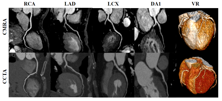
This study was approved by the institutional ethics committee. The MR examinations were performed on a 3T MR scanner (Ingenia Elition, Philips Healthcare) using a 16-channel body matrix coil combined with a 12-channel spine matrix coil. Fifty-six consecutive patients (60±10 years, 29 males) with suspected CAD were finally included in this study and underwent non-contrast CSAI coronary MRA. The details of CSAI coronary MRA using a free-breathing balanced SSFP sequence with a 3D non-selective pulse were listed: TR/TE = 2.4/1.2 ms; flip angle= 10°; field of view= 280×280×240 mm3; Acquisition voxel size= 1.5×1.5×1.5mm3; Reconstruction voxel size= 0.75×0.75×0.75mm3; A spectral presaturation with inversion recovery (SPIR) fat saturation and a T2 preparation (duration=30 ms) were performed. Data acquisition was accelerated by employing a CSAI framework with a factor of 6. The image quality of coronary segments was rated by two observers in consensus on a 5-point scale (1, non-diagnostic; 5, excellent). Images with a score ≥3 were considered to be satisfactory and acceptable for diagnosis. Significant coronary stenosis was defined as luminal diameter reduction of ≥50% in a reference diameter greater than 1.5 mm using an intention-to-read approach. Sensitivity, specificity, positive (PPV) and negative predictive values (NPV) of CSAI coronary MRA for the detection of significant CAD were calculated. Curved planar reformation (CPR) and volume rendering (VR) images were used for image display.Results and Discussion
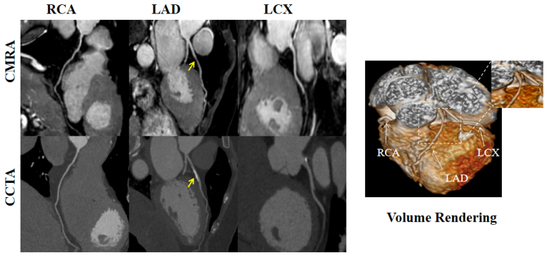
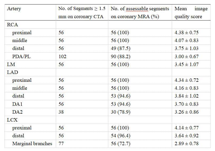
The mean image acquisition time was 8.3±2.5 min. Twenty-four (42.9%) had significant CAD on coronary CTA. The image quality of 56 patients with successful whole-heart coronary MRA was shown in Table 1. Overall, 718/777 (92.4%) of coronary MRA segments were deemed diagnostic. The sensitivity, specificity, positive predictive value, negative predictive value and diagnostic accuracy were as follows: per patient (91.7%, 81.3%, 85.7%, 78.6% and 92.9%), per vessel (82.5%, 92.2%, 89.9%, 76.7% and 94.4%) and per segment (76.8%, 97.7%, 96.1 %, 74.1% and 98.0%) respectively. Typical example images from selected patients with suspected CAD with both coronary MRA and coronary CTA were shown in Fig.1 and Fig.2.Conclusion
In conclusion, we have investigated a novel non-contrast-enhanced, radiation-free, whole-heart coronary MRA framework with compressed sensing artificial intelligence image reconstruction framework, which could provide excellent image quality and diagnostic performance within clinical feasible time for patients with suspected CAD. Especially, the coronary MRA has the better visualization in calcified segments than coronary CTA.Acknowledgements
No acknowledgements found.References
1 Tsao C W, Aday A W, Almarzooq Z I, et al. Heart Disease and Stroke Statistics—2022 Update: A Report From the American Heart Association[J]. Circulation, 2022,145(8).
2 Feng L, Benkert T, Block K T, et al. Compressed sensing for body MRI[J]. Journal of Magnetic Resonance Imaging, 2017,45(4):966-987.
3 Akçakaya M, Basha T A, Chan R H, et al. Accelerated isotropic sub-millimeter whole-heart coronary MRI: Compressed sensing versus parallel imaging[J]. Magnetic Resonance in Medicine, 2014,71(2):815-822.
4 Fuin N, Bustin A, Kustner T, et al. A multi-scale variational neural network for accelerating motion-compensated whole-heart 3D coronary MR angiography[J]. Magn Reson Imaging, 2020,70:155-167.
5 Bustin A, Fuin N, Botnar R M, et al. From Compressed-Sensing to Artificial Intelligence-Based Cardiac MRI Reconstruction[J]. Frontiers in Cardiovascular Medicine, 2020,7.[6] Foreman S C, Neumann J, Han J, et al. Deep learning–based acceleration of Compressed Sense MR imaging of the ankle[J]. European Radiology, 2022.
Figures