1299
Age-related alterations in soma and neurite fraction obtained from high-gradient diffusion MRI data across the lifespan1Department of Radiology, Athinoula A. Martinos Center for Biomedical Imaging, Massachusetts General Hospital, Charlestown, MA, United States, 2Department of Neurology, Massachusetts General Hospital, Harvard Medical School, Boston, MA, United States, 3MS Center Amsterdam, Anatomy and Neurosciences, Amsterdam Neuroscience, Amsterdam UMC location VUmc, Amsterdam, Netherlands
Synopsis
Keywords: Neurodegeneration, Aging
We studied alterations in soma and neurite signal fractions with age based on Soma And Neurite Density imaging (SANDI) in 43 healthy adults across the lifespan using multi-shell dMRI measurements acquired on the MGH Connectome scanner. We observed decreases in soma fraction with age in all cortical lobes, especially in the frontal lobe. The neurite fraction predominantly decreased with age in the genu of corpus callous. These results suggest a regionally selective aging effect on changes in compartmental composition within the brain, potentially reflecting alterations in microstructure associated with neurodegeneration.
Introduction
The aging human brain is characterized by progressive alterations in brain tissue microstructure including a defined sequence of myelination in development followed by demyelination in mid-to-late life, as well as gray matter changes such as neuronal loss, reduction in synaptic density, and cortical thinning1,2. Noninvasive imaging biomarkers to distinguish between normal and pathologic brain aging predisposing to neurodegeneration and dementia are currently lacking. Soma and Neurite Density Imaging (SANDI) is a biophysical model of diffusion MRI (dMRI) sensitive to the soma size and the soma/neurite volume fractions in the brain3. In this study, we sought to detect age-related changes in cortical and white matter microstructure obtained from SANDI applied to data acquired on the MGH Connectome scanner in healthy individuals across the lifespan.Methods
Forty-three cognitively healthy subjects (36.7±14.5 years, ranging from 22 to 72 years; 25 females) were scanned on a 3T Connectom MRI scanner with maximum gradient strength of 300mT/m using a 64-channel head coil. Diffusion-weighted images (DWIs) were acquired using a pulsed gradient spin-echo echo-planar-imaging sequence at two diffusion times (Δ=19, 49ms) with a fixed gradient duration (δ=8ms). Other parameters TE/TR=77/4000ms; 2mm isotropic voxel size, GRAPPA=2, SMS=2. A total of 16 shells were acquired with 8 b-values per diffusion time ranging from 0.05 to 6ms/μm2 for Δ=19ms and from 0.2 to 17.8ms/μm2 for Δ=49ms4. b=0 images were acquired every 16 DWIs. 32 gradient directions were sampled for b-values<2.3ms/μm2 and 64 directions otherwise. T1-weighted anatomical images were acquired using 3D MEMPRAGE: TE/TR=1.15/2530ms, TI=1100ms, GRAPPA=3, 1mm isotropic voxel. Cortical thickness measurements, gray matter segmentation, and corpus callosum subdivisions were obtained using FreeSurfer on T1-weighted images (Figure 1). After preprocessing the DWIs, the SANDI model was fitted to directionally averaged (spherical mean) DWIs for the two diffusion times separately using the SANDI-AMICO package3,5. We evaluated the correlations between variables using Pearson’s correlation coefficients. We corrected for multiple comparisons using the false discovery rate (fdr).Results
Overall cortical thickness was negatively correlated with age (r=-0.60, P<0.001) (Figure 2). Soma fraction, fis, decreased with age for all lobes at Δ=19ms (fdr P<0.04) (Figure 3). In the frontal lobe, parietal lobe, and cingulate gyrus, the neurite fraction, fin, and extracellular volume fraction, fec, increased significantly with age (Figure 3). Among the cortical lobes, the frontal lobe showed the strongest correlation between age and all three volume fractions. In addition, fin in the corpus callosum showed a significant decrease with age, especially in the genu of corpus callosum (r=-0.51, fdr P=0.003). Furthermore, cortical thickness significantly correlated with both fis and fin in the frontal lobe, parietal lobe, and cingulate gyrus (Figure 4). In corpus callosum, only the mid-body showed a significant correlation between cortical thickness and fis (r=0.40, fdr P=0.03). In the comparison of two diffusion times, correlations between age and fis in cortical lobes and between age and fin in corpus callosum were stronger at Δ=19ms than those at Δ=49ms (Figure 5).Discussion and Conclusion
We observed decreased cortical fis with age in a region-specific manner. The fis decrease with age was particularly significant in the frontal lobe, consistent with previous results showing regionally selective relationships between age and diffusion metrics in white matter6 and potentially related to neuronal degeneration in the frontal lobe with aging. The marked decrease in fin with age in the genu of corpus callosum may reflect preferential degeneration of connected white matter fibers in the frontal lobe6. The correlation between age and fin showed opposite trends in cortical areas and corpus callosum.At shorter diffusion time (Δ=19ms), the correlations between age and fis and fin are stronger than those at longer diffusion time (Δ=49ms). This is potentially due to the non-negligible exchange effect at longer diffusion time7-9, whereas SANDI model assumes no exchange between compartments at all time scales3. This is also manifested by the inconsistency between the measured signals and the predicted signals by SANDI fitting at high b-values at longer diffusion time (Δ=49ms) (data not shown). To reduce the signal sensitivity to the exchange, stronger gradients may be needed to access shorter diffusion times.
In this study, we observed the cortical thickness decrease with age as well as correlations between cortical thickness and microstructural parameters in the brain gray matter. The cortical thickness was highly correlated with cortical fis and fin , suggesting possible relationships between cortical atrophy and neuronal alterations with age. These findings merit further investigation of microstructural changes in the brain and their relationship to neurodegeneration in larger cohorts of individuals across the lifespan.
Acknowledgements
This work was supported by NIH under the award number: DP5OD031854, R01NS118187, P41EB015896, P41EB030006, U01EB026996, S10RR023401, S10RR019307, S10RR023043, K99AG073506.References
1. Pannese E. Morphological changes in nerve cells during normal aging. Brain Structure & Function. 2011;216(2):85–9.
2. Salat DH, Buckner RL, Snyder AZ, et al. Thinning of the cerebral cortex in aging. Cereb Cortex. 2004;14(7):721–30.
3. Palombo M, Ianus A, Guerreri M, et al. SANDI: a compartment-based model for non-invasive apparent soma and neurite imaging by diffusion MRI. NeuroImage. 2020;215:116835.
4. Huang SY, Tian Q, Fan Q, et al. High-gradient diffusion MRI reveals distinct estimates of axon diameter index within different white matter tracts in the in vivo human brain. Brain Structure and Function. 2020;225(4):1277-91.
5. https://github.com/daducci/AMICO
6. Fan Q, Tian Q, Ohringer NA, et al. Age-related alterations in axonal microstructure in the corpus callosum measured by high-gradient diffusion MRI. NeuroImage. 2019;191:325-336.
7. Olesen JL, Østergaard L, Shemesh N, et al. Diffusion time dependence, power-law scaling, and exchange in gray matter. NeuroImage. 2022;251:118976.
8. Jelescu IO, de Skowronski A, Geffroy F, et al. Neurite Exchange Imaging (NEXI): A minimal model of diffusion in gray matter with inter-compartment water exchange. NeuroImage. 2022;256:119277.
9. Lee HH, Olesen J, Tian Q, et al. Revealing diffusion time-dependence and exchange effect in the in vivo human brain gray matter by using high gradient diffusion MRI. In Proceedings of the 31st Annual Meeting of ISMRM 2022 (Vol. 2).
Figures
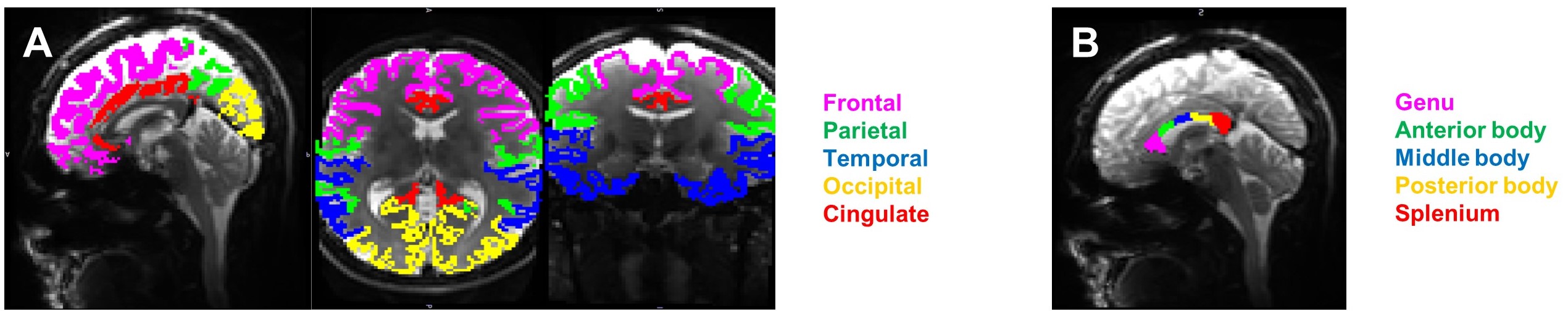
Figure 1. The cortical lobe ROIs (A) and corpus callosum ROIs (B) from Freesurfer

Figure 2. The correlation between age and cortical thickness. r value is Pearson’s correlation coefficient.
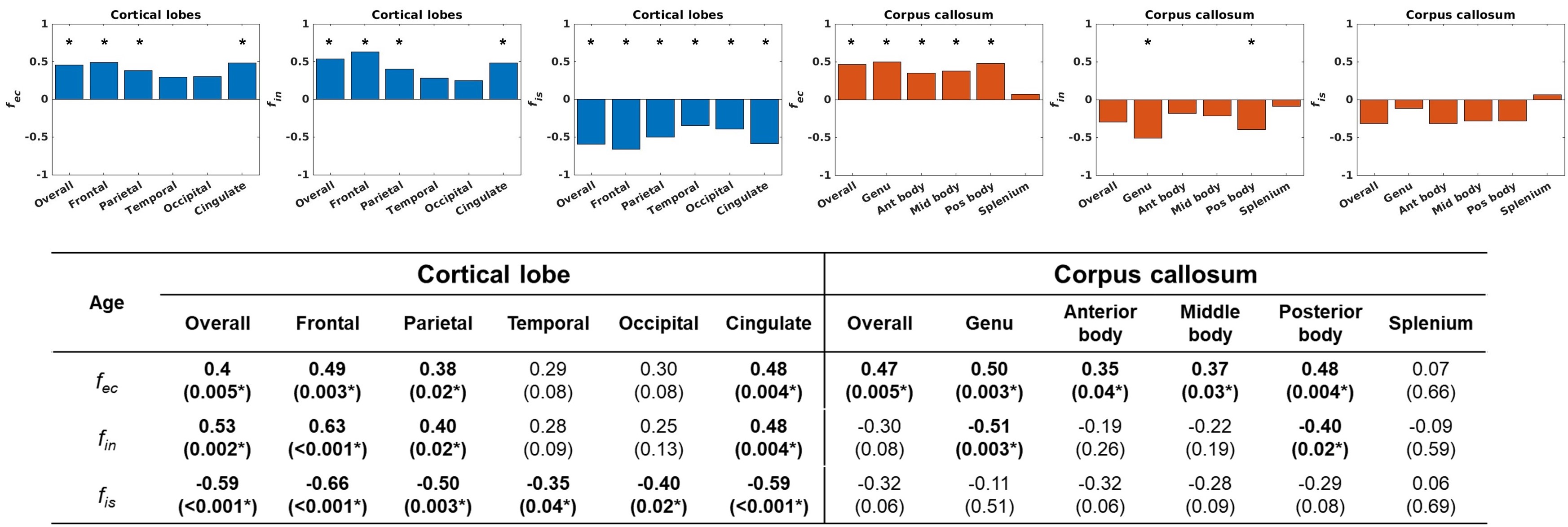
Figure 3. The correlation between age and volume fractions in the cortical lobes and the subdivisions of the corpus callosum at Δ=19ms. The bar graph shows Pearson’s r values. In the table, the data is Pearson’s r value and the data in parentheses is the P value after correcting fdr. *: fdr P-value< 0.05.
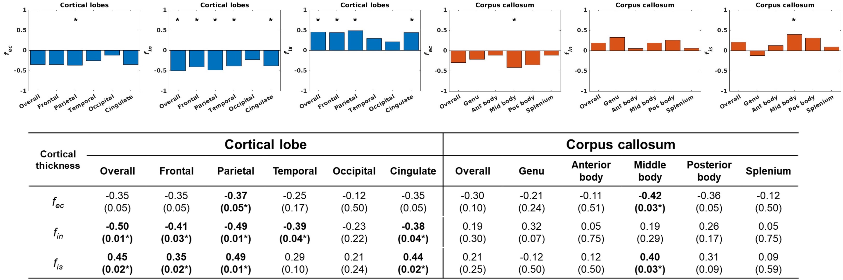
Figure 4. The correlation between cortical thickness and volume fractions in the cortical lobes and the subdivisions of the corpus callosum at Δ=19ms. The bar graph shows Pearson’s r values. In the table, the data is Pearson’s r value and the data in parentheses is the P value after correcting fdr. *: fdr P-value< 0.05.
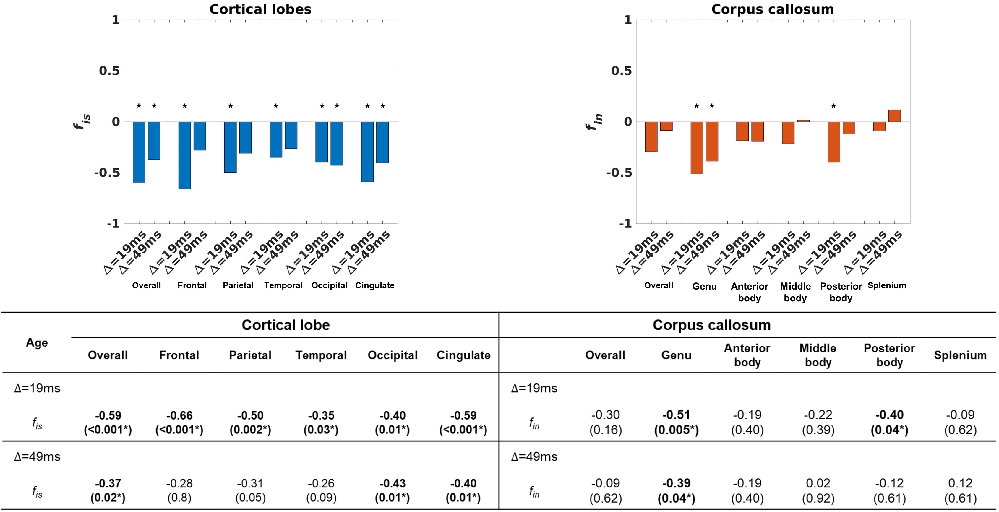
Figure 5. The correlation between age and soma volume fraction in the cortical lobes and between age and neurite volume fraction in the subdivisions of the corpus callosum at two different diffusion times. The bar graph shows Pearson’s r values. In the table, the data is Pearson’s r value and the data in parentheses is the P value after correcting fdr. *: fdr P-value< 0.05.