1274
Resting-state fMRI study of vigilance level under endogenous modulation based on ReHo and fALFF in humans under normal entrained conditions
Hanqi Xing1, Zhiwei Wu1, Mengya Ma1, Ziyang Song1, Yang Song2, Yunzhu Wu2, Yue Chang1, and Hui Dai1
1The First Affiliated Hospital of Soochow University, Suzhou, China, 2MR Scientific Marketing, Siemens Healthcare, Shanghai China., Shanghai, China
1The First Affiliated Hospital of Soochow University, Suzhou, China, 2MR Scientific Marketing, Siemens Healthcare, Shanghai China., Shanghai, China
Synopsis
Keywords: fMRI (resting state), Brain, sleep
Time-dependent neuromodulation mechanisms in human cognitive-behavioral tasks are not yet fully established. The present study aimed to analyze the changes in resting-state functional magnetic resonance imaging (rs-fMRI) blood oxygen level dependent (BOLD) signals over the 24-h day and their correlation with vigilance level. We recruited 20 healthy volunteers to be scanned at six-time points 24 hours a day. Compared to 9:00, 13:00, 17:00, and 21:00h, thalamic BOLD signals increased and vigilance level decreased at 1:00 and 5:00h. We speculate that the BOLD signals increased in the thalamus may represent a compensatory mechanism for maintaining relative vigilance level.Purpose
Whether neuronal activities in the brain maintain a constant level of activation throughout the day is not yet understood. It is also unclear the neural mechanism of time-dependent modulation in human cognitive-behavioral tasks [1]. The objectives of this study were to analyze the changes in fractional amplitude of low-frequency fluctuation (fALFF) and regional homogeneity (ReHo) of rs-fMRI over the 24-h day and their correlation with vigilance level.Methods and Materials
We recruited 20 healthy volunteers. All participants met the following inclusion criteria: (1) no history of major diseases or sleep disorders. (2) no cross-meridian travel, no shift work, and no irregular sleep-wake patterns during the 2 months before the experiment. (3) have a regular sleep schedule. Participants underwent six fMRI scanning sessions at six-time points (9:00, 13:00, 17:00, 21:00, 1:00, and 5:00) on the scanning day. After the scanning session, participants performed a 10-minute psychomotor vigilance task (PVT). In the PVT task, the longer the reaction time (RT), the worse the vigilance performance.All neuroimaging data were collected with a 3T MR scanner (MAGNETOM Skyra, Siemens Healthcare, Erlangen, Germany) using an 8-channel head coil at the Department of Radiology, the first affiliated hospital of Soochow University. Participants underwent rs-fMRI and sagittal high-resolution T1WI scanning. The rs-fMRI images were acquired using an echo-planar imaging sequence with the following parameters: TR = 2000 ms, TE = 30 ms, FOV = 256mm×256mm, matrix = 64 × 64, slice number = 33, slice thickness = 4 mm. High-resolution 3D sagittal T1WI was acquired using a spoiled gradient echo sequence with the following parameters: TR = 2300 ms, TE = 2.98 ms, FOV = 256mm × 256mm, slice thickness = 1 mm.
The fMRI data pre-processing was performed using Data Processing Assistant for Resting-State fMRI Advanced edition (DPARSFA) [2] software. The main steps include (1) Removing the first 10 time points of all data, slice timing, and realignment. (2) T1-weighted images were coregistered to the mean functional image. (3) Spatial normalization to the standard Montreal Neurological Institute (MNI) brain space. The fractional amplitude of low-frequency fluctuations(fALFF)mapping was calculated, which measures the relative contribution of the low-frequency range (0.01- 0.1 HZ) to the entire frequency range of the signal oscillations. Regional homogeneity (Reho) analysis was performed to assess the time series similarity of each voxel to its neighboring voxels using Kendall's coefficient concordance(KCC). In addition, spatial smoothing (FWHM = 6 mm) was also performed before the fALFF calculation, but after ReHo.
Group statistical analyses were performed using Data Processing and Analysis of Brain Imaging (DPABI) and IBM SPSS. A one-way repeated measures ANOVA was used on behavioral data and ReHo/fALFF at six-time points to assess changes in vigilance levels and neural activity in the whole brain. The mean values of fALFF/Reho were extracted from the thalamus at six-time points. Then, using SPSS software we performed a repeated measures ANOVA with subsequent pairwise comparisons between all time points for RTs and fALFF/ReHo. Spearman correlation analysis was performed to examine for a linear relationship between fALFF/ReHo in the thalamus and reaction time at the same time.
Results
A whole-brain, repeated measures ANOVA (P< 0.001 at the voxel level and P < 0.05 at the cluster level,GRF corrected) revealed that the main influence of time effect on fALFF in the left middle temporal gyrus, right thalamus, and left superior parietal lobule and Reho in left middle temporal gyrus, bilateral thalamus, right rolandic operculum, left cerebellum. Post-hoc two-tailed dependent t-tests with Fisher least significant difference test (Fisher LSD) correction (P < 0.05) based on fALFF averages in the right thalamus and Reho averages in the bilateral thalamus showed that fALFF/Reho significant increase at both 01:00 and 05:00h compared to 9:00, 13:00, 17:00 and 21:00h. A repeated measures ANOVA showed a significant effect of time effect on the 10th and 90th percentiles of RT (F=6.9, P=0.001; F=3.23, P=0.035). At both the 10th and 90th percentiles of RTs, participants had the worst vigilance levels at 01:00 and 05:00h compared to 9:00, 13:00, 17:00, and 21:00h. Decreased vigilance levels coincided with increased rs-fMRI BOLD signals in the thalamus at 01:00 and 05:00h. The present study shows a positive linear relationship between fALFF/Reho in the thalamus and 10th percentile RTs at the same time of day. This relationship between vigilance levels and resting-state BOLD signals may point to a compensatory mechanism for maintaining relative vigilance level.Discussion and conclusions
In this study, we investigated the influence of time-effects on resting-state fALFF/Reho signals during the 24-hour cycle and explored the neural basis of time-effect-dependent changes in vigilance level through the association of rs-fMRI data with PVT performance. This study first reveals the time dependence of this relationship between endogenous resting-state BOLD fALFF/Reho signals and vigilance performance. Our study found reaction times and resting-state neural activities in the thalamus are largely consistent during the 24-hour cycle, and we speculate that fALFF/Reho increases in the thalamus may have a direct role in the maintenance of vigilance performance. These results demonstrate that increased sustained thalamic neural activity near melatonin-producing offset areas during the subjective late night and early morning hours is a mechanism for maintaining sustained attention.Acknowledgements
This work was partially supported by the National Natural Science Foundation of China (grant number 81971573), the Suzhou Gusu Medical Youth Talent (grant number GSWS2020019) and the Project of Invigorating Health Care through Science, Technology and Education, Jiangsu Provincial Medical Youth Talent (grant number QNRC2016709).References
1. Cordani, L., et al., Endogenous modulation of human visual cortex activity improves perception at twilight. Nat Commun, 2018. 9(1): p. 1274.
2. Chao-Gan, Y. and Z. Yu-Feng, DPARSF: A MATLAB Toolbox for "Pipeline" Data Analysis of Resting-State fMRI. Front Syst Neurosci, 2010. 4: p. 13.
Figures
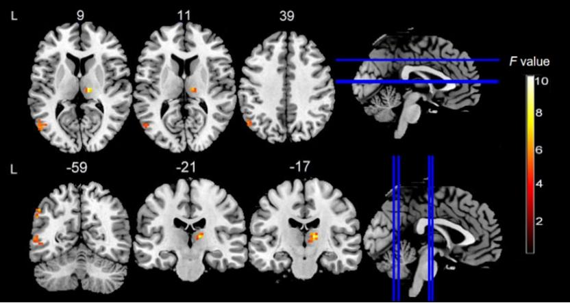
Significant effect of time effect(9:00,13:00,17:00,21:00,1:00,5:00) for resting-state BOLD fALFF(transverse and coronal).
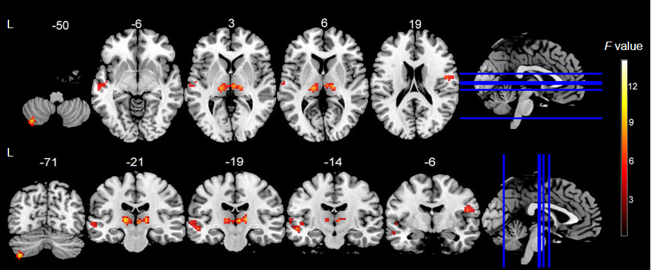
Significant effect of time effect(9:00,13:00,17:00,21:00,1:00,5:00) for resting-state ReHo(transverse and coronal).
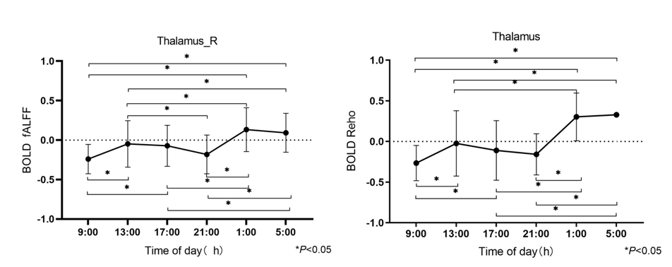
Thalamic resting-state BOLD fALFF and Reho trends throughout the day .
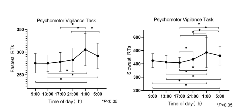
The 10th (fastest RTs) and 90th (slowest RTs) percentiles of RTs trends throughout the day.
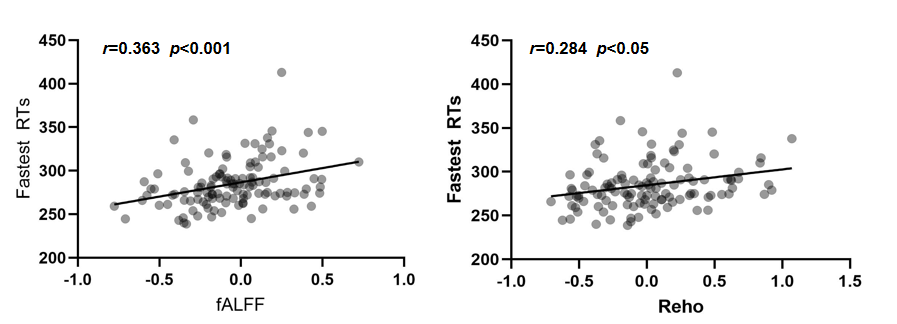
BOLD fALFF and Reho is positively associated with the 10th percentiles of RTs.
DOI: https://doi.org/10.58530/2023/1274