1239
RF shimming in the spinal cord at 7T1NeuroPoly Lab, Institute of Biomedical Engineering, Polytechnique Montreal, Montreal, QC, Canada, 2Mila - Quebec AI Institute, Montreal, QC, Canada, 3Functional Neuroimaging Unit, Centre de recherche de l'Institut universitaire de gériatrie de Montréal, Montreal, QC, Canada, 4Centre de recherche du CHU Sainte-Justine, Université de Montréal, Montreal, QC, Canada
Synopsis
Keywords: Parallel Transmit & Multiband, Spinal Cord
Spinal cord MRI at 7T suffers from Tx inhomogeneity. RF shimming is therefore potentially useful in this region. Here, we evaluate for the first time the effect, both on signal intensity and signal homogeneity, of multiple RF shimming algorithms implemented in the open-source ‘Shimming Toolbox’, and compare the results with vendor solutions.Introduction
Recently, multiple dedicated spinal cord (SC) coils with parallel transmit (pTx) capability have been developed by research groups and commercial vendors [1–5]. To fully exploit the improvements in signal intensity and homogeneity offered by pTx coils, the amplitudes and phases of each transmit (Tx) channel have to be adjusted for each patient during a radiofrequency (RF) shimming step. While scanner vendors offer RF shimming algorithms, these are opaque to the user, necessitating the development of open-source RF shimming algorithms.Here, we demonstrate for the first time the impact of four RF shimming algorithms implemented in the open-source Shimming Toolbox [6] on signal intensity and homogeneity in the cervical SC and compare them with vendor solutions.
Methods
Acquisition:One subject (21, M) was scanned using a custom 8Tx/20Rx cervical SC array [5] and a Siemens 7T Terra system.
To calculate RF shim weights, two scans were acquired. The magnitude and phase of each transmit field channel was mapped using a vendor-provided sequence using a pre-saturated TurboFLASH readout with sagittal orientation, 2×2×3.66mm resolution, FOV=240×388×84mm, covering cervical levels C1-T4. B0 shim gradients were calculated on a rectangular volume around the SC, and kept constant throughout the session. A 2D GRE scan with with axial orientation, 0.6×0.6mm2 in-plane resolution, 50 slices of 3mm slice thickness, TR/TE=600/2.86ms, FA=15° was acquired without RF shimming (“CP mode”), and was used for segmenting the SC during RF shim weight calculation as described below. Once RF shim weights were calculated, six more GRE scans were acquired with different RF shim weights (all other image parameters were kept the same). In “Vendor: Volume Specific”, the B0 shim volume was defined as the region of interest (ROI) for RF shimming, and the shim weights were calculated by the scanner. In “Vendor: Patient Specific”, shim weights were likewise calculated by the scanner, but the ROI was defined as the whole FOV. For all other "Custom" RF shimming approaches, the weights were calculated as described below:
RF shim weight calculation:
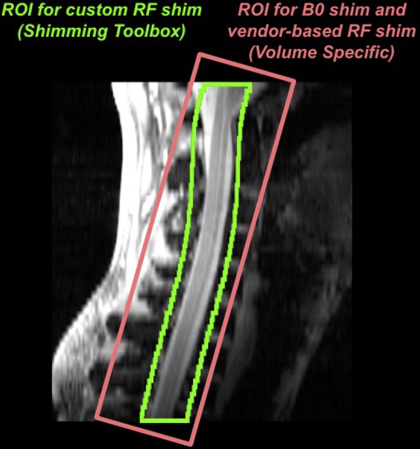
The SC was segmented on the GRE scan using the Spinal Cord Toolbox [7]. A cylindrical mask of 25mm diameter was derived from this segmentation and used to define the ROI for RF shim weight calculation in ST (Figure 1). Four RF shimming algorithms were implemented in ST. Each algorithm results in a set of complex weights (w) that minimize the following expressions:
For "phase-only shimming", the coefficient of variation (CV) of the phase of each transmit channel was minimised within the ROI, without taking the magnitude into account:
$$w_{\operatorname{phase-only}_{\text{}}}=\operatorname*{argmin}_{w}\left\{\frac{\sigma\!\left(\left| B_1^+(w) \right|\right)}{\mu\!\left(\left| B_1^+(w) \right|\right)} \right\}$$
where: $$$B_1^+(w) = \sum_{n}^{N_{Tx}}\frac{1}{\sqrt{N_{Tx}}}{B_1^{+}}_{n}e^{i{w}_{n}}$$$ and $$${B_1^+}_{n}$$$ is the measured $$${B_1^+}$$$ map of Tx channel n, and $$${\sigma\!\left(\left| B_1^+(w) \right|\right)}$$$ and $$${\mu\!\left(\left| B_1^+(w) \right|\right)}$$$ are the standard deviation and the mean of the shimmed $$${B_1^+({w})}$$$ map , respectively. Crucially, only the phase of the $$${B_1^+}$$$ maps is adjusted (via $$${e}^{i{w}_{n}}={e}^{i{\phi}_{n}}$$$) during the shim weight calculation, while the amplitude is left unmodified.
For "CV reduction", the CV of both the phase and magnitude of each transmit channel within the ROI was taken into account:$$w_{\operatorname{CV_{red}}_{\text{}}}=\operatorname*{argmin}_{w}\left\{\frac{\sigma\!\left(\left| B_1^+(w) \right|\right)}{\mu\!\left(\left| B_1^+(w) \right|\right)} \right\}$$
where $$$B_1^+(w)=\sum_{n}^{N_{Tx}}\frac{1}{\sqrt{N_{Tx}}}{A_{n}}{B_1^{+}}_{n}e^{i{\phi}_{n}}$$$ and $$$w_{n}=A_{n}\mathrm{e}^{i{\phi}_{n}}$$$. Thus, $$$w_{\operatorname{CV_{red}}_{\text{}}}$$$ weights affect both the phase (via $$${e}^{i{\phi}_{n}}$$$) and the magnitude (via $$$A_{n}$$$) of each transmit channel.
For "Target 20nT/V", a target $$${B_1^+}$$$ amplitude (“t”) of 20 nT/V was used to calculate the shim weights: $$w_{\operatorname{target}_{\text{}}}=\operatorname*{argmin}_{w}\left\{\sum_{n}^{N_{voxels}} \left(\left| B_1^+(w) \right|-t\right)^2 \right\}$$
For "SAR efficiency", the local SAR, derived from VOPs, is taken into account to increase the signal within the confines of SAR,$$w_{\operatorname{SAR_{eff}}_{\text{}}}=\operatorname*{argmin}_{w}\left\{ \operatorname{}SAR_{eff}(w)\right\}$$ $$\operatorname{SAR_{eff}(w)}=\frac{\mu (\left| B_1^+(w)\right|)}{\sqrt{max(\operatorname{SAR_{local}}(w))}}$$
$$$w_{\operatorname{SAR_{eff}}_{\text{}}}$$$ and $$$w_{\operatorname{target}_{\text{}}}$$$ affected both the magnitude and phase of each transmit channel.
Analysis:
For all RF shimming scenarios, the GRE scans were coregistered to the PAM50 template[8], the signal along the SC between cervical levels C1-T1 was extracted, and the CV of this signal was computed.
Results
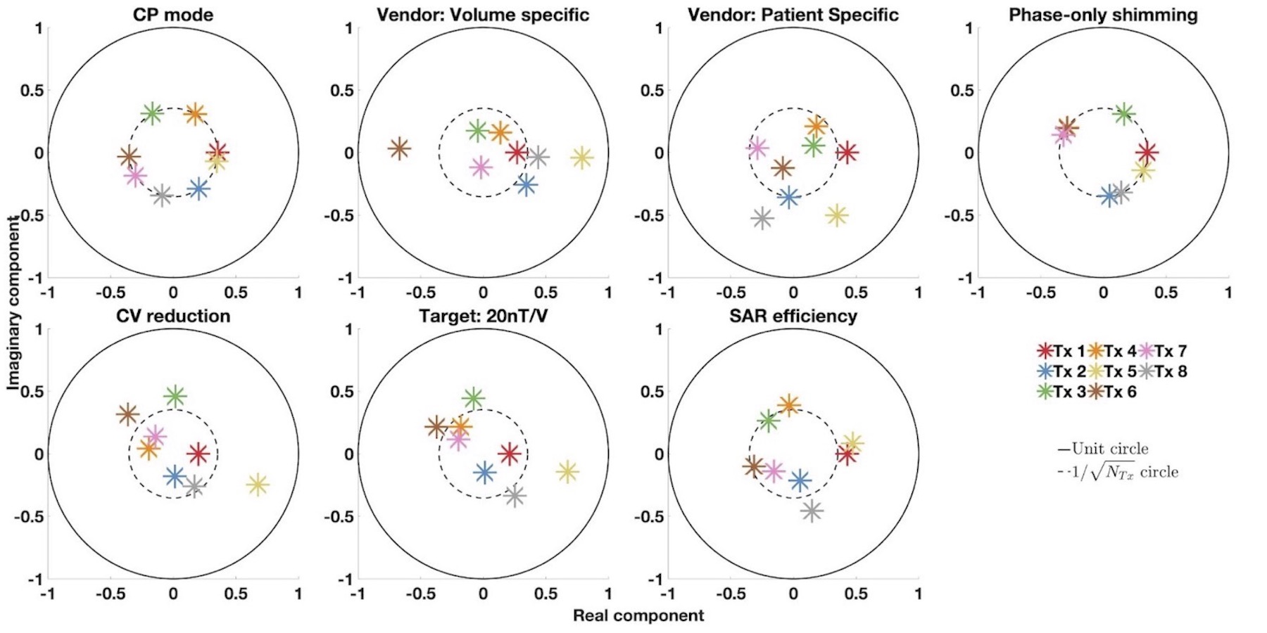
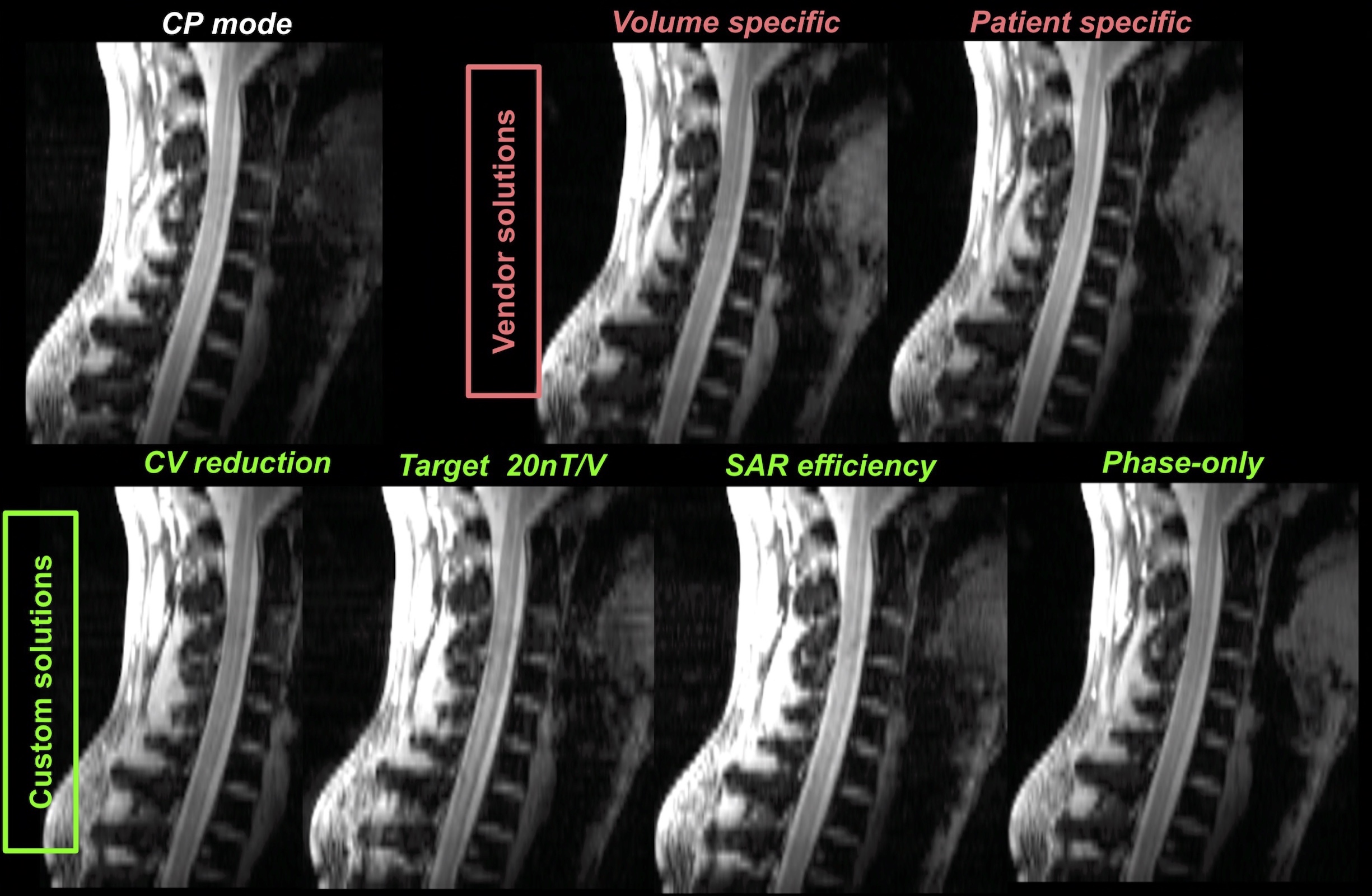
Figure 2 shows the complex shim weights for each RF shimming scenario. Vendor-computed shim weights for either scenario differ noticeably from the custom shim weights.Reflecting on the variation of shim weights, the signal intensity and homogeneity along the sagittal midline varies across all RF shimming scenarios, as seen in Figure 3.
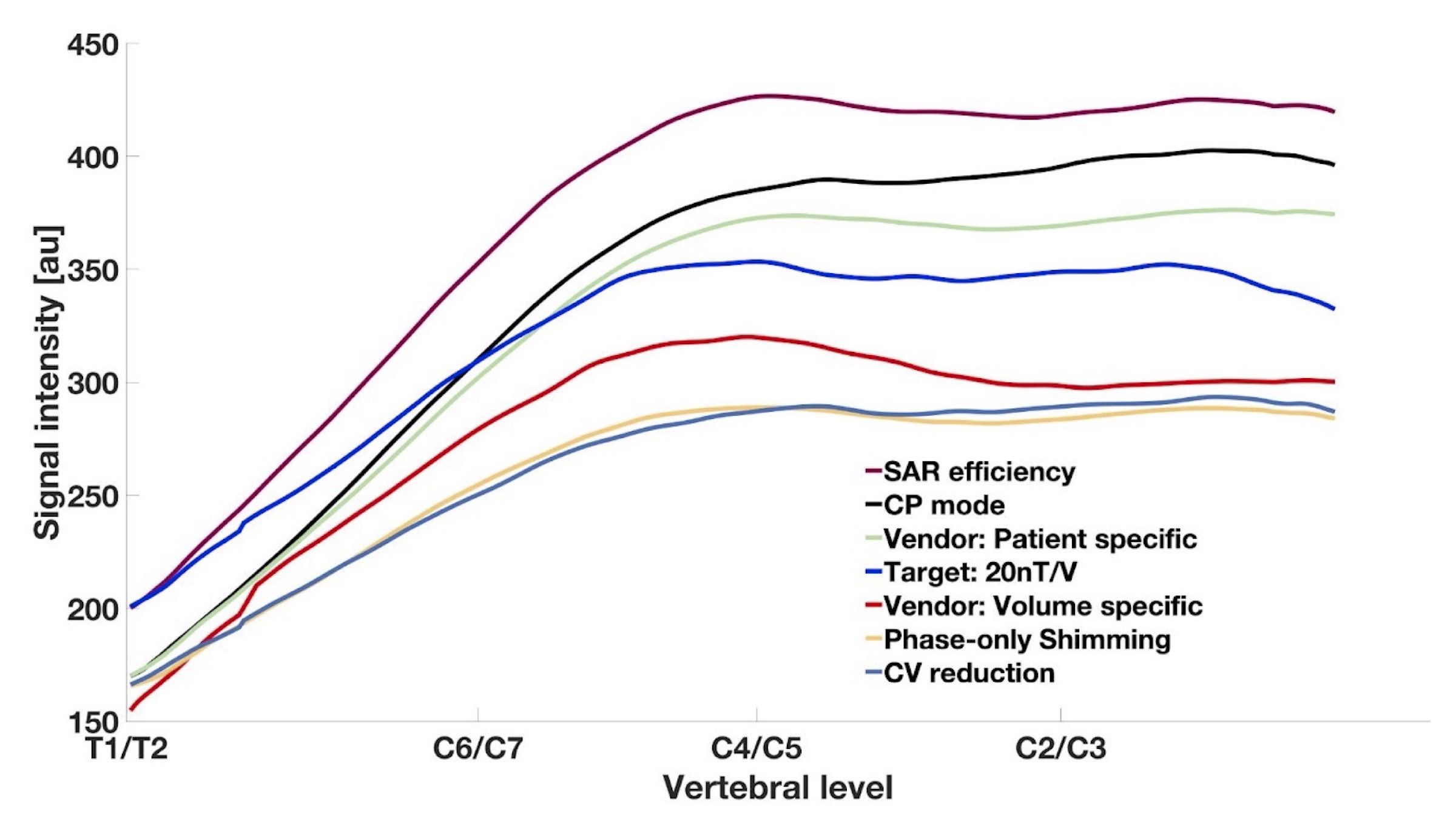
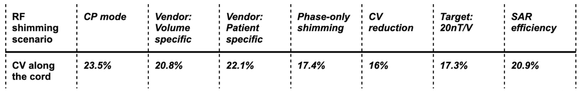
Along the SC, signal intensity was decreased for all RF shim scenarios apart from SAR efficiency, as seen in Figure 4. Correspondingly, CV along the cord was decreased for most RF shim scenarios, with vendor-provided shimming being outperformed by 3 out of the 4 custom algorithms.
Discussion
Our work highlights the importance of open-source RF shimming methods as implemented in the Shimming Toolbox. Depending on the algorithm used, RF shimming can result in an overall increase of signal intensity, at the cost of homogeneity, or in a more homogeneous excitation profile at the cost of lower average signal. Thus, the RF shimming algorithm should be tailored to the application. Notably, vendor-provided approaches are limited to whole-FOV optimisation or rectangular ROIs, rendering them ill-suited for patients with excessive lordosis.Future work will focus on generalising our findings across multiple subjects and across more modalities for anatomical and functional imaging.
Acknowledgements
Funded by the Canada Research Chair in Quantitative Magnetic Resonance Imaging [950-230815], the Canadian Institute of Health Research [CIHR FDN-143263], the Canada Foundation for Innovation [32454, 34824], the Fonds de Recherche du Québec - Santé [28826], the Natural Sciences and Engineering Research Council of Canada [RGPIN-2019-07244], the Canada First Research Excellence Fund (IVADO and TransMedTech), the Courtois NeuroMod project and the Quebec BioImaging Network [5886, 35450], and MITACS Accelerate Fellowship.References
1. Sigmund EE, Suero GA, Hu C, McGorty K, Sodickson DK, Wiggins GC, Helpern JA. High-resolution human cervical spinal cord imaging at 7 T. NMR Biomed. 2012;25: 891–899.
2. Zhao W, Cohen-Adad J, Polimeni JR, Keil B, Guerin B, Setsompop K., Serano P., Mareyam A., Hoecht P., Wald LL. Nineteen-channel receive array and four-channel transmit array coil for cervical spinal cord imaging at 7T. Magn Reson Med. 2014;72: 291–300.
3. Rietsch SHG, Brunheim S, Orzada S, Voelker MN, Maderwald S, Bitz AK., Gratz M., Ladd ME., Quick HH. Development and evaluation of a 16-channel receive-only RF coil to improve 7T ultra-high field body MRI with focus on the spine. Magn Reson Med. 2019;82: 796–810.
4. Lopez Rios N, Topfer R, Foias A, Guittonneau A, Gilbert KM, Menon RS, Wald LL., Sotckmann JP., Cohen-Adad J. Integrated AC/DC coil and dipole Tx array for 7T MRI of the spinal cord. Proc Intl Soc Mag Reson Med 27
5. May MW, Hansen S-LJD, Kutscha N, Multani GK, Mahmutovic M, Poniatowski M, Gumbrecht R, Kimmilgen R, Adriany M, Chhang Y, Guerin B, Triantafyllou C, Wald LL, Kiel B A Patient-Friendly 16ch Tx / 64ch Rx Array for Combined Head and Neck Imaging at 7 Tesla. Proc Intl Soc Mag Reson Med 29
6. D’Astous A, Cereza G, Papp D, Gilbert K, Stockmann J, Alonso-Ortiz E, Cohen-Adad J Shimming Toolbox: An open-source software toolbox for B0 and B1 shimming in Magnetic Resonance Imaging, Magn Reson Med 2022, DOI: 10.1002/mrm.29528 [in prod]
7. De Leener B, Lévy S, Dupont SM, Fonov VS, Stikov N, Louis Collins D, Callot V, Cohen-Adad J. SCT: Spinal Cord Toolbox, an open-source software for processing spinal cord MRI data. Neuroimage. 2017;145: 24–43.
8. De Leener B, Fonov VS, Collins DL, Callot V, Stikov N, Cohen-Adad J. PAM50: Unbiased multimodal template of the brainstem and spinal cord aligned with the ICBM152 space. Neuroimage. 2018;165: 170–179.
Figures