1237
Hybrid Active and Passive Local Shimming (HAPLS) Targeting Ultra-high-order B0 Spherical Harmonic (SH) Terms in Two-Region MRI
Zhi Hua Ren1, Jason Stockmann2,3, Andrew Dewdney4, and Ray F. Lee1
1Zuckerman Mind Brain Behavior Institute, Columbia University, New York, NY, United States, 2Athinoula A. Martinos Center for Biomedical Imaging, Massachusetts General Hospital, Boston, MA, United States, 3Harvard Medical School, Boston, MA, United States, 4Siemens Healthcare GmbH, Erlangen, Germany
1Zuckerman Mind Brain Behavior Institute, Columbia University, New York, NY, United States, 2Athinoula A. Martinos Center for Biomedical Imaging, Massachusetts General Hospital, Boston, MA, United States, 3Harvard Medical School, Boston, MA, United States, 4Siemens Healthcare GmbH, Erlangen, Germany
Synopsis
Keywords: Shims, Shims
An MRI scanner equipped with global shim systems fails to reach state-of-the-art when shimming two isolated ROIs simultaneously for two reasons: non-optimal spherical harmonic based shimming routine, and significant high-order B0 inhomogeneities, even though the two-area shimming can be essential in scan scenarios, such as bilateral breasts or dyadic brains. To address these challenges, a hybrid active and passive local shimming (HAPLS) technique is proposed to shim two isolated areas in one FOV simultaneously. Both the simulation and experimental results validated that HAPLS can complementarily address the bifocal and high-order inhomogeneities, and locally improve in vivo B0 homogeneity as well.Introduction
An MRI scanner equipped with global shim systems fails to reach state-of-the-art when shimming two isolated ROIs simultaneously for two reasons: non-optimal spherical harmonic-based shimming routine, and significant high-order B0 inhomogeneities, even though the two-area shimming can be essential in scan scenarios, such as bilateral breasts or dyadic brains1-4. To address these challenges, a hybrid active and passive local shimming (HAPLS) technique is proposed to simultaneously shim two isolated regions of interest areas within the whole FOV.Methods
All simulations, optimization, and experimental validation were performed on a 3T Siemens Prisma scanner. The top three dominant SH terms of the B0 field within 50-cm DSV (Figure 1(a)) are all ultra-high-order ones – a(12,0), a(14,0), and a(8,0), and they are at 12.3, 5.84, and 1.94 (in ppm), respectively. Shimming two isolated regions (Figure 1(b)) simultaneously is challenging because: (1) the background B0 field in both areas contains up to 14th order SH field inhomogeneities; (2) the scanner’s built-in SH-based in vivo shim routines are designed for one volume, so it cannot effectively shim two off-center volumes simultaneously and optimally as shown in Figure 2. The peak-to-peak (pk-pk) ppm for the two targeted imaging regions are 3.39 ppm and 8.92 ppm, respectively. To address the challenge of shimming two-region MRI, a hybrid active and passive local shimming (HAPLS) technique is proposed to shim two isolated regions within one large FOV simultaneously by taking advantage of both PS and AS.Local Passive Shimming
A local passive shimming (PS) system is constructed by optimized bilateral 20x11 ferromagnetic chip arrays to compensate for the magnet’s significant high-order B0 inhomogeneities at the boundary of the manufacturer’s specified homogeneous volume, thus locally improving the available FOV. The optimized PS chip arrays were affixed surrounding the two imaging regions with a CNC-machined acetal holder and 3D-printed fixtures as shown in Figure 3 (a-b). For the optimization of PS, the shim field generated by the PS chips was calculated by a current model5-6, and the thickness of the chip stack was taken as variables to optimize the standard deviation (SD) of the B0 field by a genetic algorithm in MATLAB.
Local Active Shimming
The active shimming (AS) is used to homogenize the center frequencies of two regions which may be shifted differently after applying PS. Furthermore, AS will deal with the residual lower-order and the additional subject-specific field inhomogeneities. The local AS consists of 40-channel DC loops powered by 64-channel current amplifiers as shown in Figure 3(c-d)7-9. The volumetric field maps of each shim coil were acquired by the double-echo gradient-echo field mapping sequence, and then used for the optimization of the current distribution of AS. The optimization of AS is conducted on the B0 field after PS, where the high-order inhomogeneities have been largely removed. The SD across two regions was minimized to get the optimal current distribution by a nonlinear constrained optimization algorithm (fmincon) in MATLAB.
Results
The optimized distributions of the PS chip array, and the optimized current distribution of AS were shown in Figure 3(a,f), respectively. The experimental setup to perform HAPLS was shown in Figure 3(e), and the experimental field maps are shown in Figure 4(a-b). Compared to B0 field in Figure 2(a), the high-order inhomogeneities in transversal xy-planes were greatly reduced after PS. Compared to no shim, the RMS decreased from 0.473 to 0.255 ppm by 46.1% with HAPLS, and the nonlinearity of the BZ field along the x-axis was significantly reduced as shown in Figure 4(c). The volume ratio containing MR voxels within a 0.5-ppm frequency span increased from 64.3% to 81.3% by 26.3% as shown in Figure 4(d).Discussion
To demonstrate the capability of the proposed HAPLS method for shimming two brains in dyadic fMRI, the 3D B0 maps of two brains were acquired, and then applied HAPLS to them. The PS is the same as that shown in Figure 3(a), and the AS was re-optimized. The B0 field within two brains before and after applying simulated HAPLS is shown in Figure 5. With HAPLS, the RMS within two brains decreased from 0.461 ppm to 0.211 ppm by about 54.2%, and the 95% pk-pk ppm was reduced from 1.54 ppm to 0.87 ppm by 44%. Due to that the 40-ch AS coil array is more confined on +x and -x regions, the regions close to the isocenter of the scanner were not improved too much. In the dual-head AC/DC coil array we are building, both local FOVs will be 360-degree surrounded, and a better shim performance will be expected.Conclusion
The hybrid active and passive local shimming was proposed to shim two-region MRI studies with high-order B0 field inhomogeneities where conventional SH shimming is unable to achieve the required improvements. The PS focuses more on locally improving the high-order field inhomogeneities which are an intrinsic property of the scanner type, whilst the AS concentrates mainly on the residual low-order terms and magnetic susceptibility effects. Both the simulation and experimental results validated the feasibility of the proposed method, and suggested that the HAPLS technique will locally improve in vivo B0 homogeneity as well.Acknowledgements
The authors would like to thank for the grants from NSF 1926789 and NIH U24EB028984, also thank the support from Bernd Stoeckel.References
- Lee RF, Dai W, Jones J. Decoupled circular-polarized dual-head volume coil pair for studying two interacting human brains with dyadic fMRI. Magn Reson Med. 2012;68(4):1087-1096. doi:10.1002/mrm.233132.
- Lee RF. Dual logic and cerebral coordinates for reciprocal interaction in eye contact. PLoS One. 2015;10(5):1-23. doi:10.1371/journal.pone.01217913.
- Hancu I, Govenkar A, Lenkinski RE, Lee SK. On shimming approaches in 3T breast MRI. Magn Reson Med. 2013;69(3):862-867. doi:10.1002/mrm.243074.
- Ren ZH, Stockmann JP., Dewdney A, Lee RF. Hybrid Active and Passive Local Shimming (HAPLS) for Two-region Magnetic Resonance Imaging (MRI). In: In Proceedings of the 30th Annual Meeting of ISMRM; 2021:3327.5.
- Ren ZH, Mu WC, Huang SY. Design and Optimization of a Ring-Pair Permanent Magnet Array for Head Imaging in a Low-Field Portable MRI System. IEEE Trans Magn. 2019;55(1):1-8. doi:10.1109/TMAG.2018.28766796.
- Ren ZH, Gong J, Huang SY. An Irregular-Shaped Inward-Outward Ring-Pair Magnet Array with a Monotonic Field Gradient for 2D Head Imaging in Low-Field Portable MRI. IEEE Access. 2019;7:48715-48724. doi:10.1109/ACCESS.2019.29098347.
- Current driver: Current driver - RF Coil Lab. https://rflab.martinos.org/index.php?title=Current_driver:Current_driver. Accessed April 6, 2022.8.
- Stockmann JP, Witzel T, Keil B, et al. A 32-channel combined RF and B0 shim array for 3T brain imaging. Magn Reson Med. 2016;75(1):441-451. doi:10.1002/mrm.255879.
- Stockmann JP, Wald LL. In vivo B0 field shimming methods for MRI at 7 T. Neuroimage. 2018;168(June 2017):71-87. doi:10.1016/j.neuroimage.2017.06.013
Figures
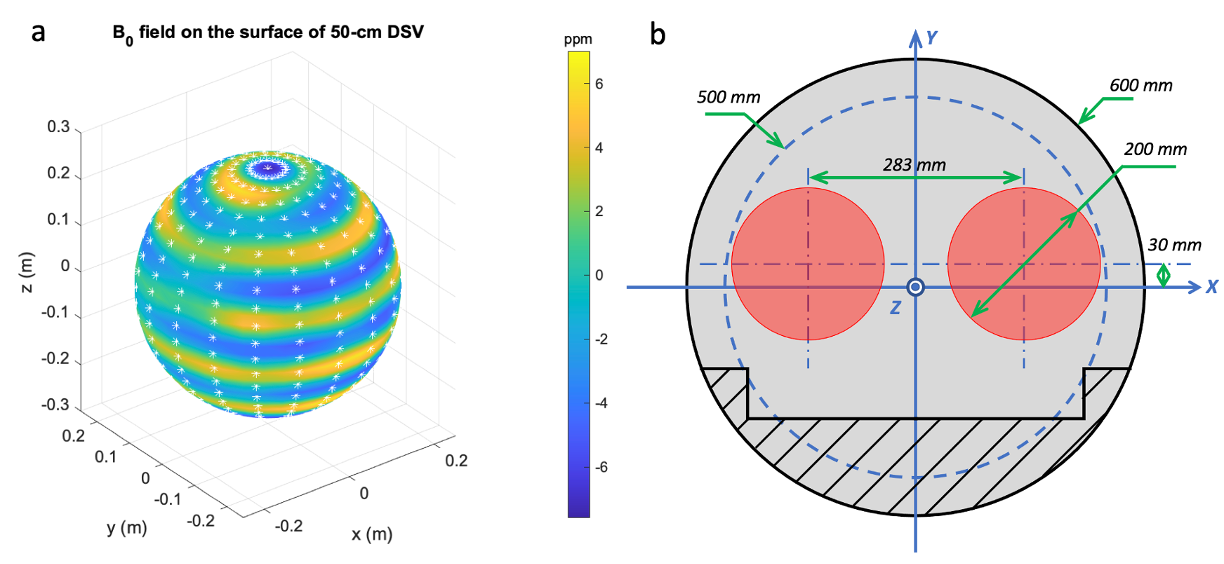
Figure 1 (a) The plot of the magnetic field on the surface of the 50-cm DSV, and white star signs depict the positions of the field probe magnetometer of Siemens Array Shim Device used to take the measurement. (b) The location and dimension of bi-focal regions targeted for shimming two heads at the same time. The left region (-x side) and the right region (+x side) will be referred to as sub-FOV1 and sub-FOV2 in this paper, respectively.
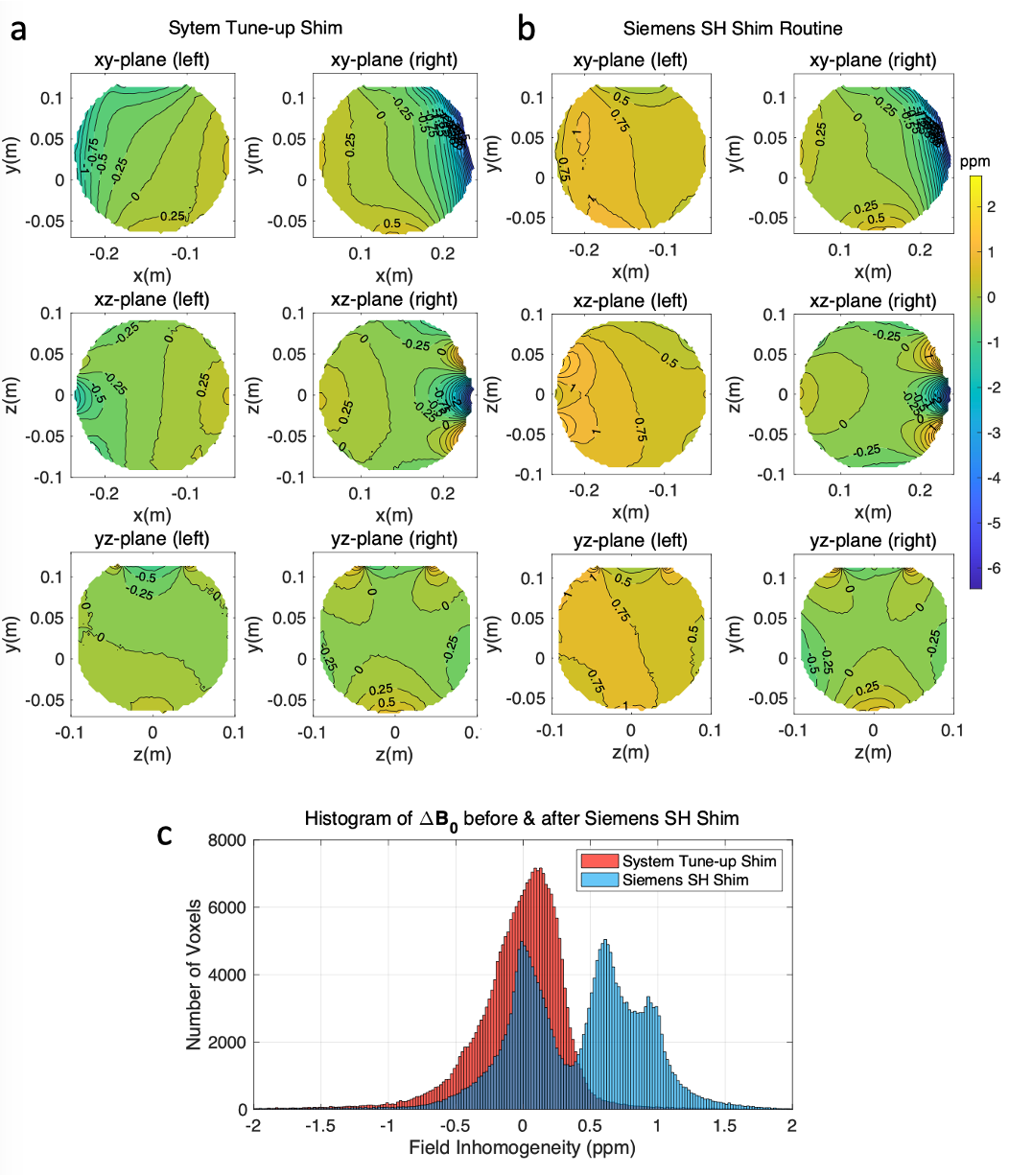
Figure 2 Acquired B0 field maps inside the paired 20-cm home-made spherical phantoms with system tune-up shim setting and after automatic SH shimming are shown in (a) and (b), respectively. The automatic SH shimming cannot handle localized high-order inhomogeneities, and only focused on the left region (-x). (c) The histogram plot of the ∆B0 field distribution with system tune-up shimming and automatic SH shimming. As can be seen, the center frequencies of the two regions were separated by about 60 Hz after automatic SH shimming.
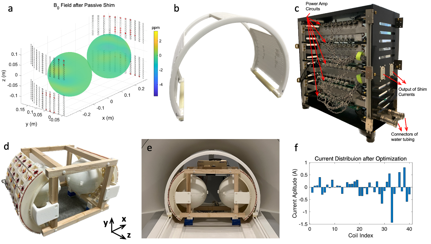
Figure 3 (a) B0 field on the spherical surface of two sub-FOVs after PS is shown. The optimized chip distribution (red cuboids) is also illustrated, and transparent chip blocks depict that no chips are there based on optimization results. (b) The assembly of the PS fixture with shim chips affixed. (c) The 64-channel shim current supply system. (d) The 40-channel bilateral AS coils with home-made phantom filled with CuSO4 solution. (e) The setup in the scanner to perform HAPLS. (f) The current distribution for 40-channel active shim coils after optimization.
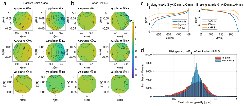
Figure 4 The field map within two phantoms with PS and with HAPLS are shown in (a) and (b), respectively. (c) BZ field along the x-axis @ y = 30 mm in both sub-FOVs is shown. (d) Histogram plot of field inhomogeneity within both sub-FOVs before and after applying HAPLS.
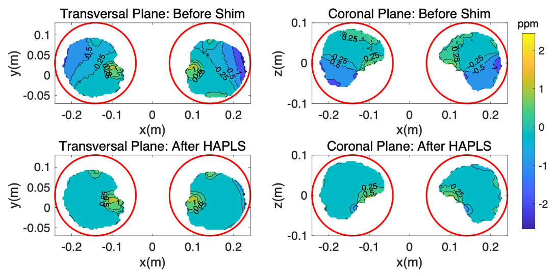
Figure 5. The B0 field in two brains within two targeted imaging regions before and after applying HAPLS. The red circles depict two targeted 20-cm imaging regions.
DOI: https://doi.org/10.58530/2023/1237