1236
Flexible use of AC/DC coil for eddy-currents and concomitant fields mitigation with applications in diffusion-prepared non-Cartesian sampling1Department of Radiology, Stanford University, Stanford, CA, United States, 2Department of Electrical Engineering, Stanford University, Stanford, CA, United States, 3Athinoula A. Martinos Center for Biomedical Imaging, Massachusetts General Hospital and Harvard Medical School, Charlestown, MA, United States, 4Stanford Center for Cognitive and Neurobiological Imaging, Stanford University, Stanford, CA, United States, 5GE Healthcare, Milwaukee, WI, United States, 6BARNLabs, Muenzkirchen, Austria, 7Harvard Medical School, Charlestown, MA, United States
Synopsis
Keywords: Shims, Shims
This work provides a demonstration that the AC/DC shim-array can be flexibly used to correct undesirable eddy-current and concomitant fields effect in MRI acquisitions, with simulation results showing its effectiveness at mitigating eddy-current induced phase in diffusion-prepared acquisition and in mitigating concomitant fields in non-Cartesian trajectories, such as spiral. Phantom and invivo experiments were also performed on a 46-channel AC/DC shim-array to demonstrate high-fidelity multi-shot 3D diffusion-prepared acquisition without need for SNR-zapping amplitude stabilizer.Introduction
Local multi-coil shimming (AC/DC shim-array)(1,2) have been shown to provide improved B0 homogeneity to mitigate EPI distortions for structural, functional and diffusion MRI applications(3–5). This dual-purpose AC/DC shim-array provides both high spatial-order B0 field control dynamically at a high temporal resolution as well as good parallel-imaging capability. In this work, we proposed the flexible use of the AC/DC shim-array for eddy-currents and concomitant fields corrections in MRI acquisitions. We demonstrated its feasibility in applications of a cardiac-gated diffusion-prepared multi-shot 3D-acquisition and 2D-spiral sampling trajectory.Methods
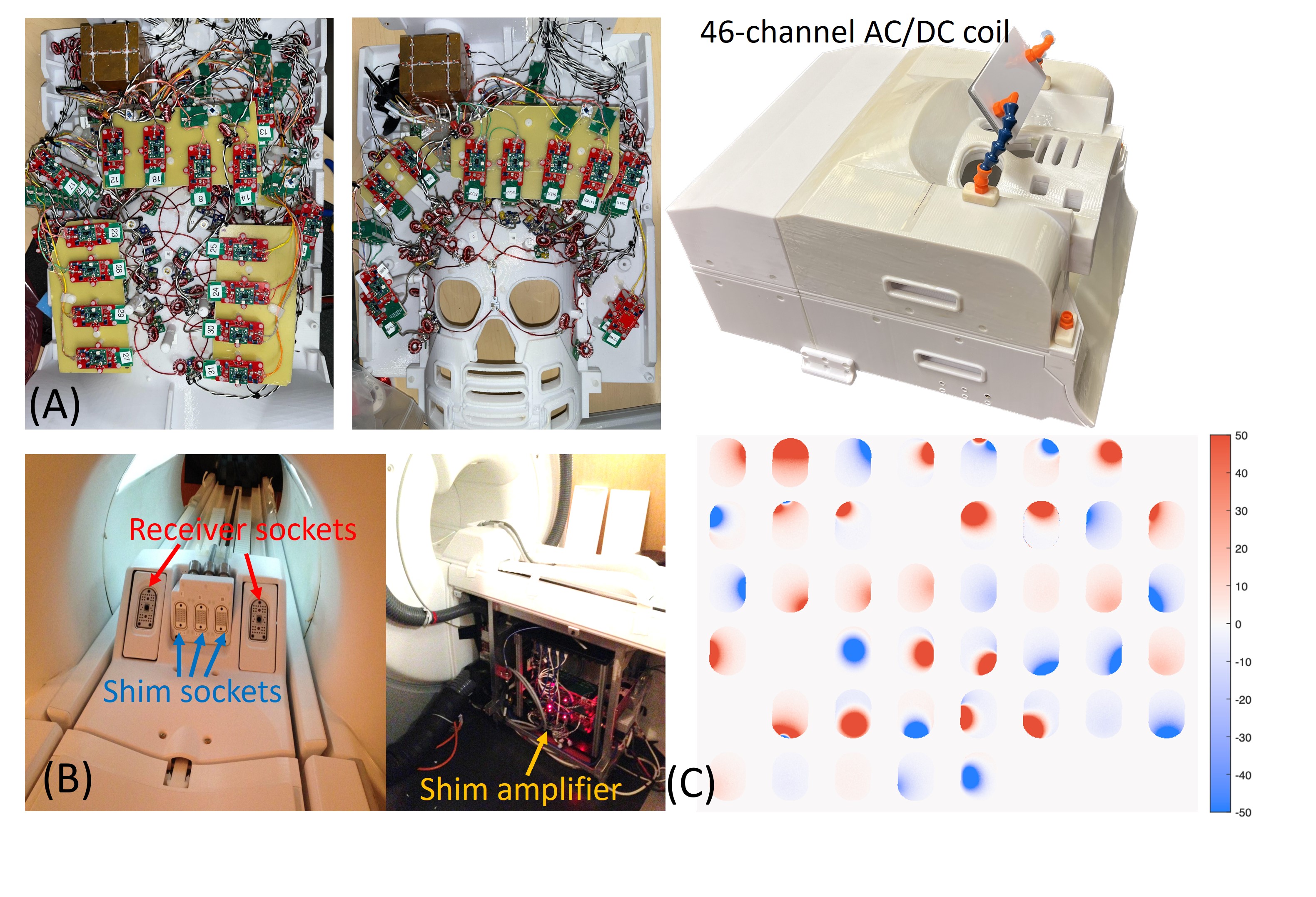
AC/DC shim-array: Figure1 shows coil elements, receiver and shim sockets, and shim amplifier of the 46-channel AC/DC shim-array. The shim amplifier was placed underneath the patient table as a convenient way without the need to modify the MRI scanner substantially. Figure1(C) is the calibration B0maps on a single slice.Eddy-currents correction using AC/DC shim-array in diffusion-prepared acquisition: Diffusion-prepared (DP) acquisition could achieve high SNR with short diffusion-preparation time and are compatible with various readouts(6,7) However, with the tip-up pulse, shot-to-shot phase variations from physiological noise and eddy-currents will cause undesirable magnitude variations. A magnitude stabilizer prior to the tip-up pulse was proposed to mitigate the magnitude variations at a cost of losing half of the signals(8). Therefore, in this work, we synergistically used the AC/DC shim-array to correct the eddy-currents and M1-compensated diffusion-preparation(9) with cardiac gating rather than magnitude stabilizer to minimize physiological noise-induced shot-to-shot magnitude variations, enabling robust diffusion MRI with full signal level.
The eddy-currents correction includes:
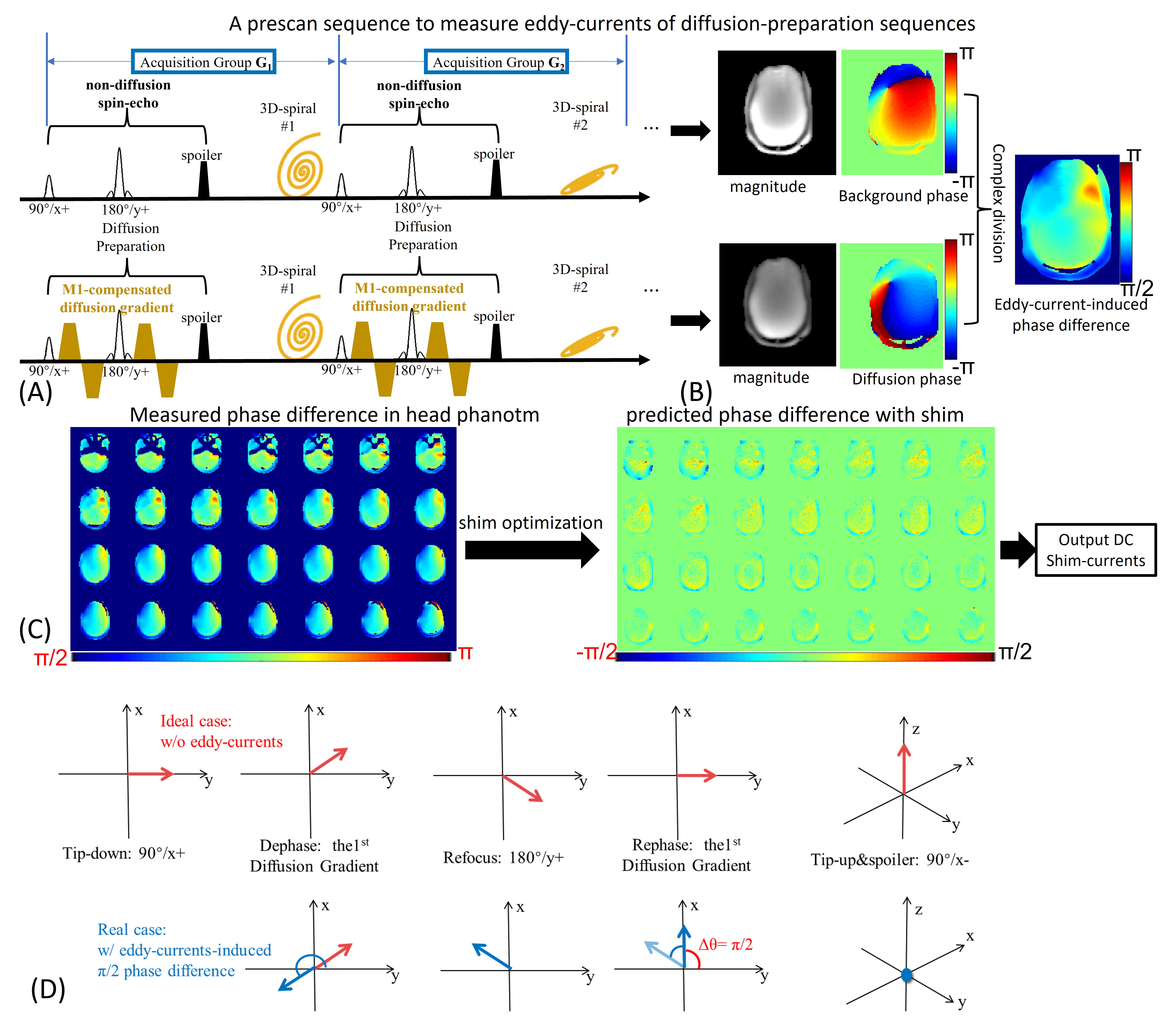
(i) A one-time eddy-current-induced phase characterization: A spin-echo diffusion acquisition was implemented as a fast prescan to measure phase differences between b=0&600s/mm2 in a phantom, which correspond to an estimation of the eddy-current-induced phase in a DP sequence, as shown in Figure2(A).
(ii) The extracted phase difference from (i) was set as the input of DC-shim currents optimization. The optimal DC shim currents of the AC/DC shim-array were then computed using the calibration B0 map basis, to create opposite phase-maps to compensate for the eddy-current-induced phase differences(Figure2(B)&(D)).
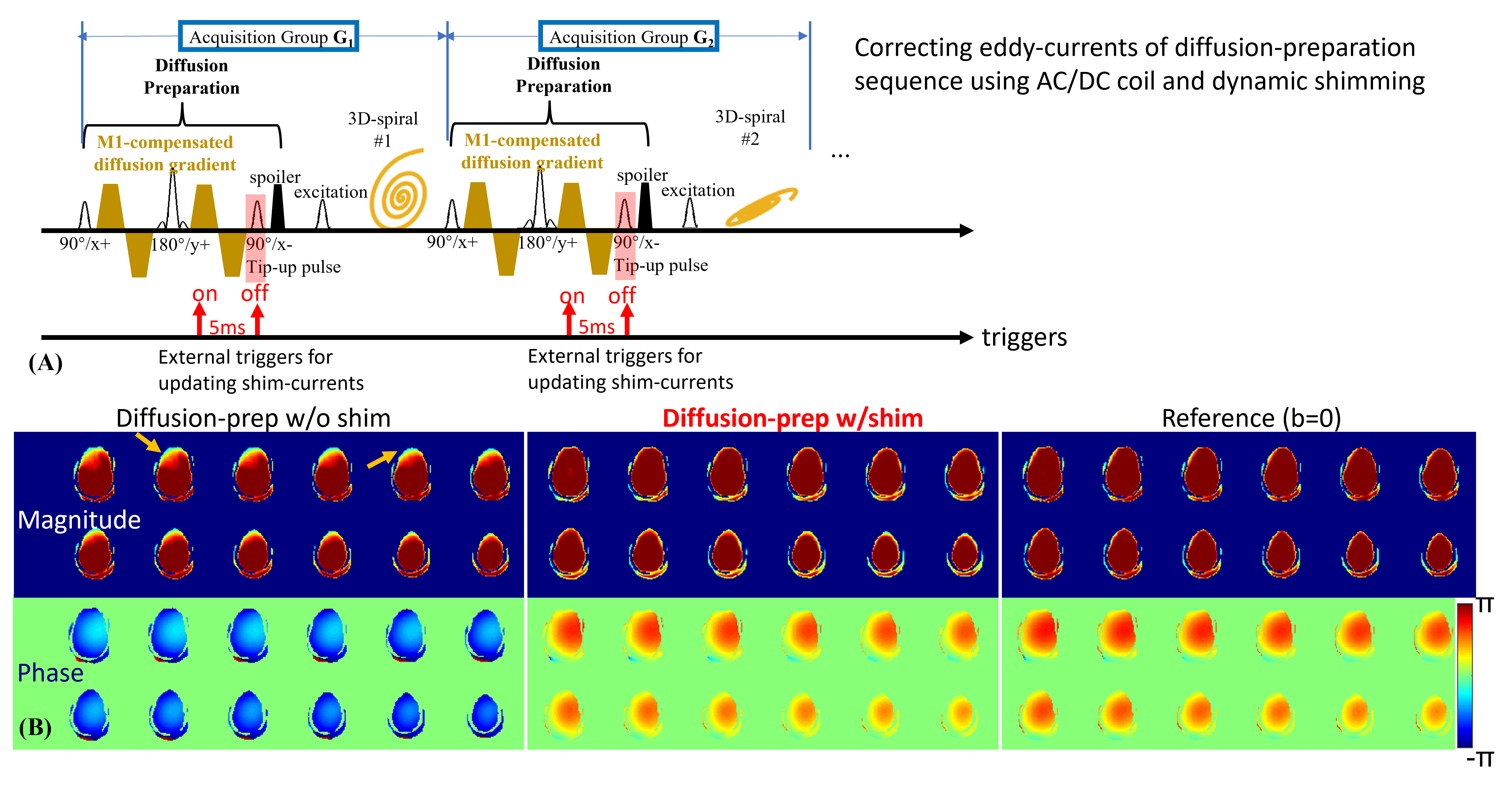
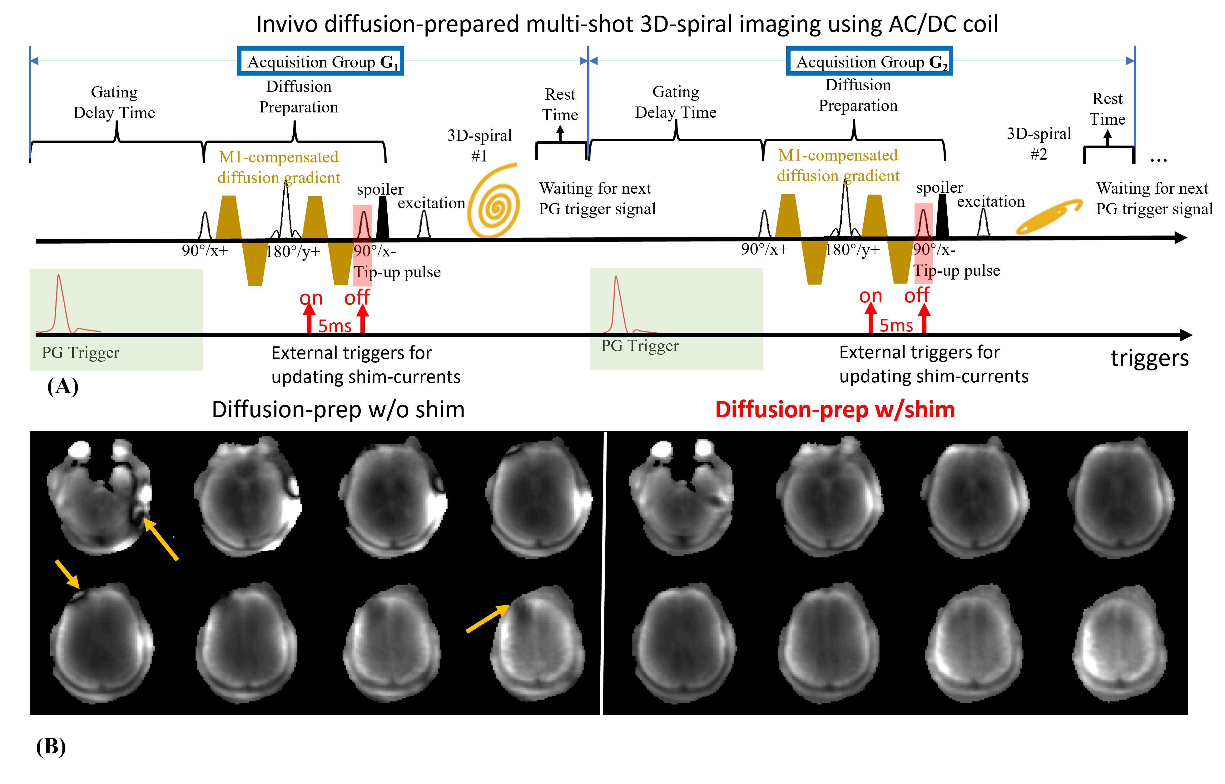
(iii) The calculated shim currents are applied during the diffusion-encodings of the DP sequence that has the same diffusion encodings as the prescan in(i). The shim currents update every TR through external triggers of the sequence(Figure3(A)), which enables compensation of differing eddy-currents from different diffusion-directions. For invivo diffusion acquisition, cardiac-gating was implemented to minimize cardiac-pulsation-induced phase instability during the DP(Figure4(A)).
To validate the proposed method, both phantom and invivo datasets were acquired.FOV:240mm, 64-shot-3D spiral-projection trajectories were sampled(matrix size: 96×96×96). Three diffusion-directions with b=600s/mm2 were acquired with TR/DP-time=500/50ms. The shim duration=5ms during the diffusion-encoding. All the experiments were scanned on a 3T GE UHP scanner.
Concomitant field correction using AC/DC shim-array in non-Cartesian sampling: The Concomitant fields from gradient encoding can cause additional phase-accrual during the readout which resulted in image blurring/artifacts, particularly in acquisitions on high-performance gradient systems and low-field scanner(10,11) where such fields can be large. From Maxwell's equations, the concomitant field Bc is approximated to the second order of Taylor-series expansion of B, which can be expressed as:
$$B_c≈(\frac{G_z^2}{8B_0})(X^2+Y^2)+ (\frac{G_x^2+G_y^2}{2B_0})(Z^2)-(\frac{G_xG_y}{2B_0})(XZ)-(\frac{G_yG_z}{2B_0})(YZ)$$
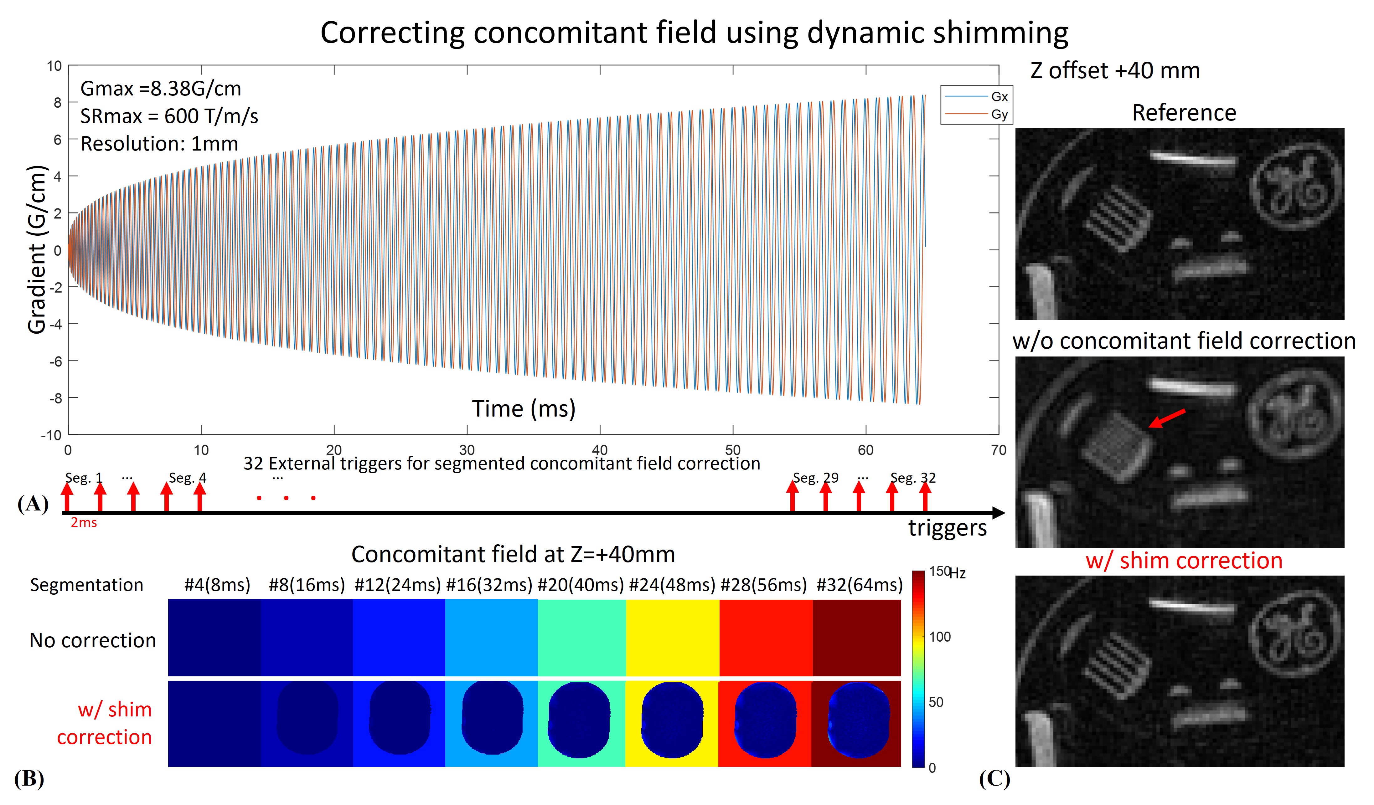
where (Gx,Gy,Gz) are the time-dependent gradient fields and (X,Y,Z) are spatial coordinates. Using this equation, we simulate the concomitant field of a 64ms single-shot 2D-spiral trajectory at 3T, as shown in Figure5(A):FOV=220mm, Gmax=8.38G/cm, maximum slew-rate = 600T/m/s, resolution:1mm. Considering the temporal switch limits of the AC/DC shim array(12), we uniformly divide the 64ms trajectory into 32 segments and calculate the concomitant field every 2ms. Similar to eddy-current correction, we create opposite phase-accrual to compensate the concomitant fields every 2ms.
Results
Figure2(C) shows the prescan measured phase-difference between b=0&diffusion acquisitions, and the predicted phase difference after shimming. With the shim optimization, the eddy-current-induced phase differences were minimized to zero.Figure3(B)&4(B) shows DP images with and without shim correction on a head-shaped phantom and a healthy volunteer, respectively. Without eddy-currents correction, the bias of eddy-current-induced phase differences causes signal loss, as indicated in yellow arrows in DP images in Figs3(B)&4(B), while shim-corrected DP images show higher signals in these regions without signal dropout. With eddy-currents correction, the DP phantom images in Figure3(B) also show a similar phase compared to the reference b=0 images, demonstrating the eddy-current-induced phase differences were compensated by shim currents.
Figure5(B) shows the concomitant field maps at Z=40mm. The phase from concomitant fields accumulates across long readout and caused image blurring as shown in Figure5(C). With masked shim correction, the phase accrual in the region of interest (inside the phantom/brain) shown in Figure5(B) was compensated, which resulted in sharper images in Figure 5(C).
Discussion and conclusion
In this work, we propose flexible usage of the AC/DC shim-array for eddy-current and concomitant field corrections. We demonstrated eddy-current mitigation in a diffusion-prepared acquisition, to facilitate high-fidelity 3D-invivo diffusion-MRI. This technique is also applicable to other efficient sampling schemes, such as fast-spin-echo and GRASE. We also demonstrated on a 2D-spiral simulation that the AC/DC shim-array can solve the image-blurring due to strong concomitant fields at low-field MRI scanners or high-performance gradient-systems. Future work will explore correction on more complex trajectories, where undesirable fields from eddy-current and concomitant fields obtained using field probes could provide a high-fidelity target for mitigation.Acknowledgements
This study is supported in part by GE Healthcare and NIH fundings: R01-EB020613, R01-EB019437, R01-MH116173, U01-EB025162, U24EB028984, R01EB028797, and P41EB030006.References
1. Stockmann JP, Witzel T, Keil B, et al. A 32-channel combined RF and B0shim array for 3T brain imaging. Magn. Reson. Med. 2016;75:441–451 doi: 10.1002/mrm.25587.
2. Han H, Song AW, Truong T-K. Integrated parallel reception, excitation, and shimming (iPRES). Magn. Reson. Med. 2013;70:241–247 doi: 10.1002/mrm.24766.
3. Juchem C, Umesh Rudrapatna S, Nixon TW, de Graaf RA. Dynamic multi-coil technique (DYNAMITE) shimming for echo-planar imaging of the human brain at 7 Tesla. Neuroimage 2015;105:462–472 doi: 10.1016/J.NEUROIMAGE.2014.11.011.
4. Kim T, Lee Y, Zhao T, Hetherington HP, Pan JW. Gradient-echo EPI using a high-degree shim insert coil at 7 T: Implications for BOLD fMRI. Magn. Reson. Med. 2017;78:1734–1745 doi: 10.1002/mrm.26563.
5. Liao C, Stockmann J, Tian Q, et al. High-fidelity, high-isotropic-resolution diffusion imaging through gSlider acquisition with B1+ and T1 corrections and integrated ΔB0/Rx shim array. Magn. Reson. Med. 2020;83:56–67 doi: 10.1002/mrm.27899.
6. Van AT, Cervantes B, Kooijman H, Karampinos DC. Analysis of phase error effects in multishot diffusion-prepared turbo spin echo imaging. Quant. Imaging Med. Surg. 2017;7:238–250 doi: 10.21037/qims.2017.04.01.
7. Lu L, Erokwu B, Lee G, et al. Diffusion-prepared fast imaging with steady-state free precession (DP-FISP): A rapid diffusion MRI technique at 7 T. Magn. Reson. Med. 2012;68:868–873 doi: 10.1002/mrm.23287.
8. Gao Y, Han F, Zhou Z, et al. Multishot diffusion-prepared magnitude-stabilized balanced steady-state free precession sequence for distortion-free diffusion imaging. Magn. Reson. Med. 2019;81:2374–2384 doi: 10.1002/mrm.27565.
9. Alexander AL, Tsuruda JS, Parker DL. Elimination of eddy current artifacts in diffusion-weighted echo-planar images: The use of bipolar gradients. Magn. Reson. Med. 1997;38:1016–1021 doi: 10.1002/mrm.1910380623.
10. Cheng JY, Santos JM, Pauly JM. Fast concomitant gradient field and field inhomogeneity correction for spiral cardiac imaging. Magn. Reson. Med. 2011;66:390–401 doi: 10.1002/mrm.22802.
11. Du YP, Zhou XJ, Bernstein MA. Correction of concomitant magnetic field-induced image artifacts in nonaxial echo-planar imaging. Magn. Reson. Med. 2002;48:509–515 doi: 10.1002/mrm.10249.
12. Xu J, Stockmann J, Bilgic B, et al. Multi-frequency wave-encoding (mf-wave) on gradients and multi-coil shim-array hardware for highly accelerated acquisition. In: Int. Soc. Mag. Res. Med. ; 2020. p. 618.
Figures