1227
Rapid In Vivo Prostate Microstructure MRI using Diffusion-Relaxation Correlation Spectrum Imaging with Random Matrix Theory-Based Denoising
Zhaohuan Zhang1, Shu-Fu Shih1, Kyunghyun Sung1, Steven Raman1, and Holden H. Wu1
1Department of Radiology, University of California, Los Angeles, Los Angeles, CA, United States
1Department of Radiology, University of California, Los Angeles, Los Angeles, CA, United States
Synopsis
Keywords: Prostate, Microstructure
Diffusion-Relaxation Correlation Spectroscopic Imaging (DR-CSI) has shown promises for quantifying prostate microscopic tissue compartments for prostate cancer characterization, but in vivo DR-CSI faces challenges such as lower signal-to-noise ratio (SNR), and signal averages could lead to prolonged scan time. This work investigated the combination of DR-CSI with random matrix theory-based denoising to take advantage of the large number of TE-b values contrast encodings to improve SNR, and enables rapid in vivo prostate microstructure MRI in <6min at 3T.Introduction
Microstructural MRI has the potential to improve the diagnosis and characterization of prostate cancer (PCa)1-5. Multi-dimensional spectral MRI approaches6-8, such as Diffusion-Relaxation Correlation Spectroscopic Imaging (DR-CSI)6, were developed to probe tissue microstructure without pre-assuming the number of tissue compartments and properties. DR-CSI was recently validated and showed promising capabilities for quantifying microscopic tissue compartments in PCa using ex vivo 3T MRI compared to whole-mount histopathology9. The feasibility of in vivo prostate DR-CSI is also being actively investigated10-11.For in vivo DR-CSI, challenges regarding the low signal-to-noise ratio (SNR) using a body array coil, as well as managing the time to acquire multiple combinations of TE and b-values (TE-b) to encode diffusion-relaxation information, need to be addressed. A common strategy to maintain SNR for diffusion-weighted MRI (DWI) is through signal averaging (e.g., up to 10 averages for higher b-values12). However, this would lead to prolonged scan times of 12~30 min for DR-CSI and increased motion sensitivity. An alternative strategy is to acquire a fewer number of unique encodings to balance scan time against averages but requires protocol design optimizations11,13.
Recently, random matrix theory (RMT)-based denoising techniques14-15 demonstrated encouraging results in reduction of thermal noise for diffusion MRI by exploiting the redundancy of noise statistics across multiple dimensions of space and diffusion/relaxation/directional contrast encodings. DR-CSI datasets, which can consist of large numbers of TE-b encodings, could potentially take advantage of RMT denoising to improve SNR instead of relying primarily on signal averaging.
Therefore, the purpose of this study was to investigate the feasibility of combining DR-CSI with RMT denoising using a protocol that sampled multiple unique TE-b encodings (no averaging) within a clinically practical scan time (<6 min) for in vivo prostate microstructure mapping at 3T.
Methods
Acquisition: In an IRB-approved study, four male subjects (age: 61~65 years old) with clinical suspicion of PCa (patient demographics in Fig.1 ) were scanned at 3T (Prisma, or Vida; Siemens) with a body array coil only, including standard T2-weighted (T2w) turbo-spin-echo (TSE) and single-shot spin-echo echoplanar imaging (ss-SE-EPI) DWI, and a DR-CSI sequence (based on ss-SE-EPI) that acquired a total of 72 encodings (24 TE-b combinations x 3 directions, single average) in 5min50s (Fig.1A). Special care was taken to ensure the DR-CSI directional TE-b images (instead of trace-averaged images) were output from the scanner for subsequent processing. Parameters are listed in Fig.1 . The bmax value of 600 s/mm2 was chosen to (1) adapt to the higher tissue diffusivities in vivo, estimated by the body vs. room temperature difference from a previous ex vivo DR-CSI protocol (bmax~1200-15009,15), and (2) avoid violating the Gaussian diffusion model that DR-CSI assumes for its exponential basis functions, due to more pronounced intra-compartmental diffusion kurtosis effects at b>600 s/mm2 for in vivo prostate4,16.RMT denoising14: All acquired DR-CSI TE-b encoding magnitude images (24x3=72) were reconstructed using GRAPPA on the scanner, exported offline, and stacked for joint denoising using Marchenko-Pastur distribution (MP)-informed Principal Component Analysis14 , (MP-PCA) (Fig.2) with a spatial patch size of [5,5,5] voxels and optimal shrinkage of singular values.
DR-CSI Microstructure Modeling: From the denoised directional TE-b images, 3 scan-trace averages were calculated and used for DR-CSI model fitting (Fig.2). Voxel-wise T2-diffusivity (D) spectra were calculated for all voxels within the prostate using non-negativity and spatial total variation constraints6,9. Signal component fraction maps (fA, fB, fC) were generated by integrating the area under each individual spectral peak (A, B, and C, which reflect epithelium, stroma, and lumen9) observed on T2-D spectra for each subject.
Results
DR-CSI with RMT denoising reduced thermal noise in TE-b images compared to standard reconstruction (example in Fig. 3). In prostates, DR-CSI consistently resolved three T2-D spectral peaks in prostate tissue. Fig 4A shows the averaged T2-D spectrum of an example slice with area under peak A in the region (D=350·10-6 mm2/s, T2=80 ms), peak B in (D=1350·10-6 mm2/s, T2=40 ms), and peak C in (D=3500·10-6 mm2/s, T2≥150 ms). Corresponding maps (fA, fB, fC) are shown in Fig.4B. Fig.4C showed the region-specific T2-D spectra and fA, fB and fC measurements in different anatomical spatial locations. Lastly, Fig.5 showed DR-CSI T2-D spectral signal component fraction maps fA, fB and fC across apex, mid-gland and base in three representative subjects.Discussion
The relative positions of three distinct in vivo prostate DR-CSI peaks shared similarities with the three peaks resolved at 3T ex vivo DR-CSI9, showing the promise of DR-CSI for resolving prostate sub-voxel tissue microscopic compartments in vivo. Apparent improvement in DR-CSI image quality, especially at longer TE and higher b-value, were achieved with RMT denoising. Unlike some of the previous studies using endorectal coil to boost SNR, our DR-CSI protocol only used body array coil and showed encouraging performance resolving tissue compartments. The scan time (5 min 50 s) is similar to that of clinical prostate DWI protocols, and is among the shortest compared to existing prostate microstructural MRI sequences1-5 .Conclusion
This work developed rapid in vivo prostate microstructure MRI at 3T in <6 min using DR-CSI with RMT denoising, which demonstrated feasibility for successfully resolving three major T2-D spectral peaks in prostate tissues.Acknowledgements
This work was supported in part by the NIH/NCI (R01 CA248506), the UCLA Department of Radiological Sciences, the UCLA Jonsson Comprehensive Cancer Center, and Siemens Medical Solutions USA. The authors thank the clinicians, study coordinators, and MRI technologists at UCLA. The authors also acknowledge the use of open source MPPCA MATLB code from the NYU Biophysics MRI group.References
[1] Panagiotaki E, Walker-Samuel S, Siow B, et al. Noninvasive quantification of solid tumor microstructure using VERDICT MRI. Cancer Res 2014;74(7):1902–1912.[2] Sabouri S, Chang SD, Savdie R, et al. Luminal Water Imaging: A New MR Imaging T2 Mapping Technique for Prostate Cancer Diagnosis. Radiology 2017;284(2):451–459.
[3] Chatterjee A, Bourne RM, Wang S, et al. Diagnosis of Prostate Cancer with Noninvasive Estimation of Prostate Tissue Composition by Using Hybrid Multidimensional MR Imaging: A Feasibility Study. Radiology 2018;287(3):864–873.
[4] Lemberskiy G, Fieremans E, Veraart J, Deng FM, Rosenkrantz AB, Novikov DS. Characterization of prostate microstructure using water diffusion and NMR relaxation. Front Phys. 2018 Sep;6:91.
[5] Wu D, Jiang K, Li H, et al. Time-dependent diffusion MRI for quantitative microstructural mapping of prostate cancer. Radiology 2022;303(3):578–587.
[6] Kim D, Doyle EK, Wisnowski JL, Kim JH, Haldar JP. Diffusion-relaxation correlation spectroscopic imaging: A multidimensional approach for probing microstructure. Magn Reson Med 2017;78(6):2236–2249.
[7] Benjamini D, Basser PJ. Magnetic resonance microdynamic imaging reveals distinct tissue microenvironments. Neuroimage 2017;163:183–196.
[8] Jan Martin, Alexis Reymbaut, Manuel Schmidt, Arnd Doerfler, Michael Uder, Frederik Bernd Laun, Daniel Topgaard,
Nonparametric D-R1-R2 distribution MRI of the living human brain, NeuroImage, 2021; 245; 118753
[9] Zhang Z, Wu HH, Priester A, et al. Prostate microstructure in prostate cancer using 3-T MRI with diffusion-relaxation correlation spectrum imaging: validation with whole-mount digital histopathology. Radiology 2020;296(2):348–355
[10] Zhang Z and Wu HH. Towards in vivo prostate microstructure mapping using diffusion-relaxation correlation spectrum imaging. ISMRM 2020.
[11] D. Kim, J. Chen, J. Xiao, B. P. Lee, K. King, J. P. Haldar, N. A. Terrault, Z. Fan.
Diffusion-Relaxation Correlation Spectroscopic Imaging (DR-CSI) in In-Vivo Body: Feasibility Study on Liver and Prostate.
AAPM, 2022.
[12] Maurer MH, Heverhagen JT. Diffusion weighted imaging of the prostate-principles, application, and advances. Transl Androl Urol. 2017 Jun;6(3):490-498.
[13] Zhang Z, Afshari Mirak S, Hosseiny M, Azadikhah A, Bajgiran A, Priester A, Sung K, Sisk AE, Reiter RE, Raman S, Enzmann DR, Wu HH. A Data-Driven Sequential Backward Selection Framework to Accelerate Diffusion-Relaxation Prostate Microstructure Mapping. ISMRM 2022.
[14] Jelle Veraart, Dmitry S. Novikov, Daan Christiaens, Benjamin Ades-aron, Jan Sijbers, Els Fieremans, Denoising of diffusion MRI using random matrix theory, NeuroImage, Volume 142,2016.
[15] Gregory Lemberskiy, Steven Baete, Jelle Veraart, Timothy M Shepherd, Els Fieremans, and Dmitry S Novikov. Achieving sub-mm clinical diffusion MRI resolution by removing noise during reconstruction using random matrix theory. Proceedings of ISMRM 2019.
[16] Rosenkrantz AB, Padhani AR, Chenevert TL, Koh DM, De Keyzer F, Taouli B, Le Bihan D. Body diffusion kurtosis imaging: Basic principles, applications, and considerations for clinical practice. J Magn Reson Imaging. 2015 Nov;42(5):1190-202.
Figures
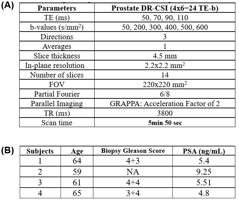
Fig 1: (A) Detailed parameters for the evaluated in vivo prostate DR-CSI protocol. (B) Patient demographic information. The most recent biopsy Gleason Score (GS) and prostate-specific antigen (PSA) level were reported.
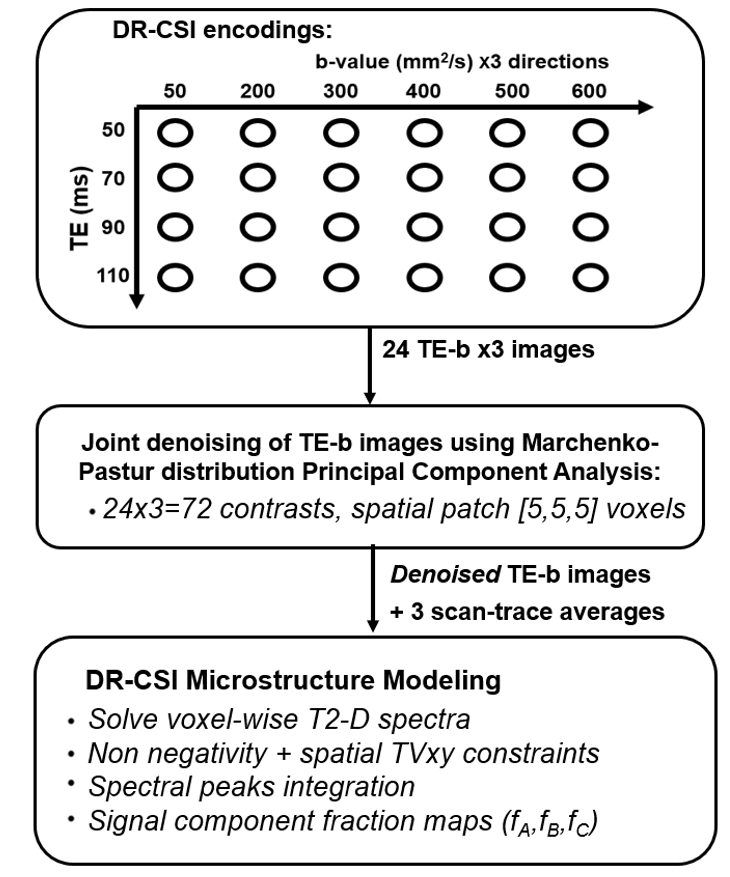
Fig 2. Pipeline for DR-CSI acquisition, RMT denoising, and reconstruction of T2-D spectral signal components for prostate microstructure mapping. TVxy: total variation in x and y.
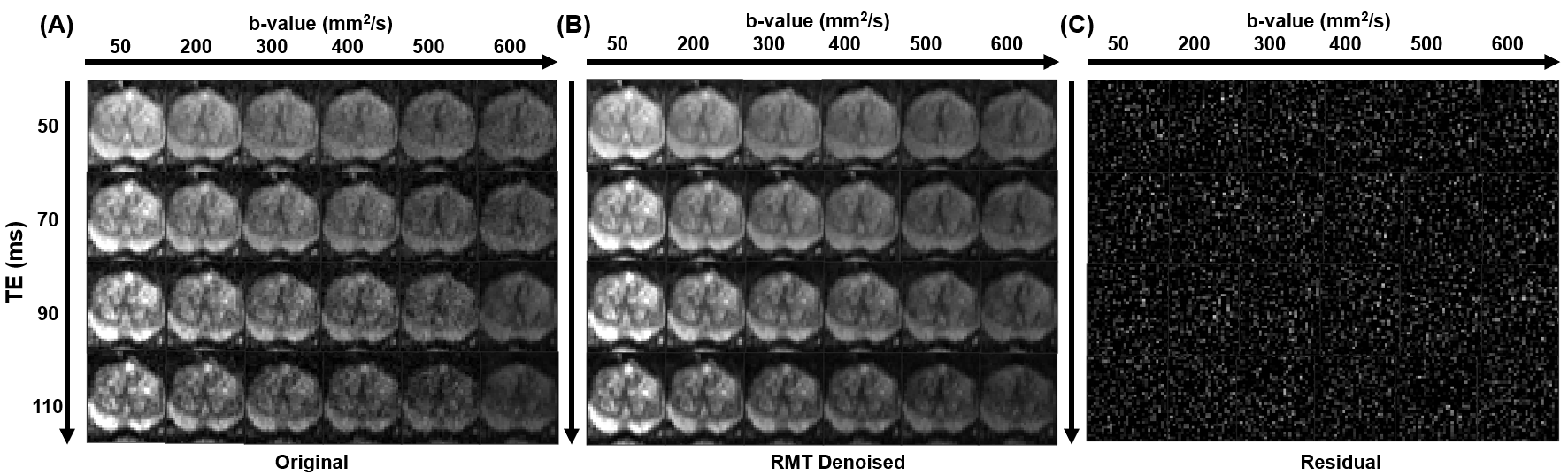
Fig 3: (A) Original DR-CSI images (cropped to focus on the prostate) with 24 TE-b encodings (z-direction shown, no average) from one representative subject. (B) RMT-denoised DR-CSI images. (C) Subtracted noise residuals (original – RMT-denoised), which displayed no anatomical details and appeared uniformly in terms of variance across all encodings. All images are displayed using the same window/level.
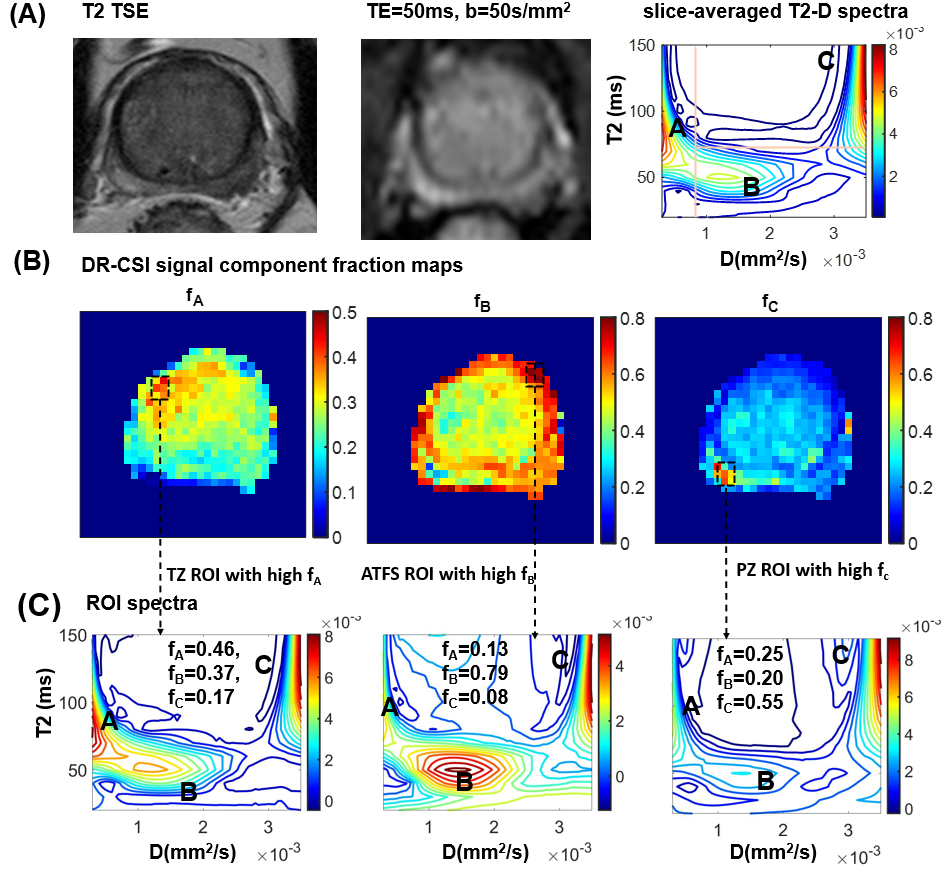
Fig 4: (A) T2w TSE MRI, DR-CSI (TE=50 ms, b=50 s/mm2 in x), and the averaged DR-CSI T2-D spectrum of the same slice from one subject. The T2-D spectrum exhibits three distinct spectral peaks A, B, and C, reflecting effects of underlying microscopic tissue compartments. Definitions of area under the peaks are delineated by orange lines. (B) DR-CSI T2-D spectral signal component fraction maps fA, fB, and fC. (C) Region of interest (ROI) specific T2-D spectra from spatial locations in the transition zone (TZ), anterior fibromuscular stroma (ATFS), and peripheral zone (PZ).
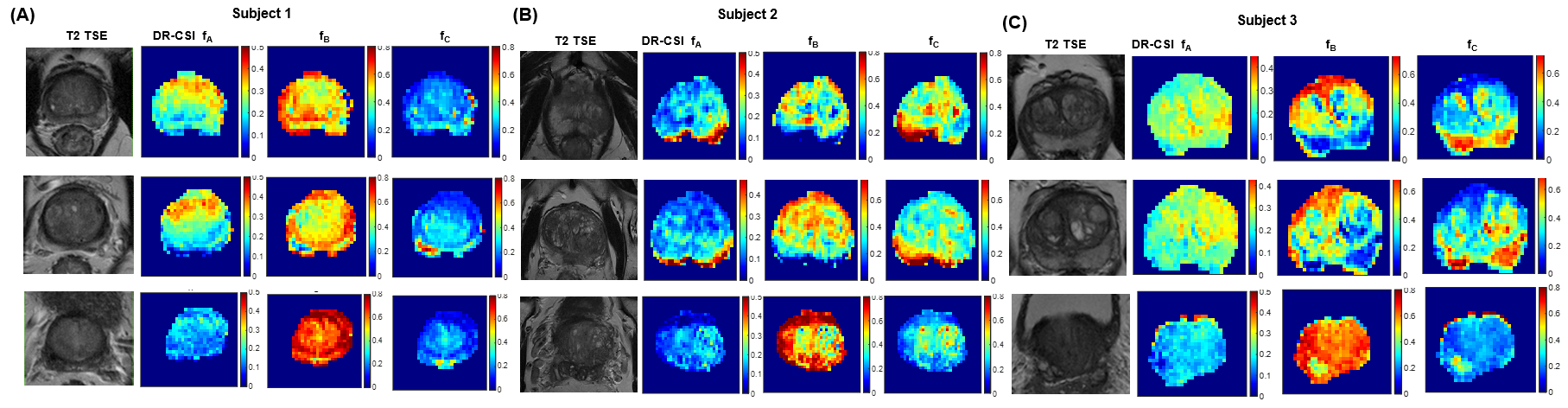
Fig 5: (A-C) T2w TSE MRI and DR-CSI T2-D spectral signal component fraction maps across slices in the prostate apex, mid-gland, and base from three representative subjects. A consistent trend of increase of fB and decrease of fC from prostate apex toward base (third rows) were observed in all subjects, which was consistent with known variations in prostate tissue composition (less glandular tissues). Anterior prostate exhibited a higher fB than posterior, which was consistent with the presence of denser muscle tissue in the prostate anterior fibromuscular stroma region.
DOI: https://doi.org/10.58530/2023/1227